Tuberculosis (TB), which is caused by Mycobacterium Tuberculosis, one of the oldest recorded human afflictions, is still one of the biggest killers among the infectious diseases.
Apart from this, TB is the most common cause of death in human immunodeficiency virus (HIV) positive patients, since both (TB &HIV) are more destructive together than either alone.
A total of 30 million people worldwide are infected with HIV and one third of them are co-infected with TB, which account for up to ~33% mortality per year.
Furthermore, the prevalence of multi-drug-resistance (MDR) TB in HIV infected patients, have rises up to 9%, since the last global drug resistance survey.
The global threat of XDR tuberculosis has great significance for the public health field. For one thing, its very existence is a reflection of weaknesses in tuberculosis management, which should minimize the emergence of drug resistance.
"A total of 30 million people worldwide are infected with HIV and one third of them are co-infected with Mycobacterium tuberculosis, which account for up to ~33% mortality per year"
What is Mycobacterium tuberculosis
Mycobacterium tuberculosis are rod or slightly curved rod, approximately 1-10 µm long and 0.3-0.6 µm in diameter, occurring singly, in pair or as small clumps.
Mycobacterium tuberculosis is a slow-growing, facultative intracellular pathogen that can survive and multiply inside macrophages and other mammalian cells.
It is non-motile, non-spore forming, aerobic and weakly gram positive. It shares number of genes with other members of the Mycobacterium genus.
It has a unique cell wall, due to the dominant presence of mycolic acids that make up more than 50% of its dry weight. It is distinguished from other bacilli by its acid fastness upon Zeihl Neelsen staining.
How Mycobacterium tuberculosis grows in culture?
The bacilli grow slowly with a generation time of 15-20 hours in vitro. The colonies appear in about three weeks and may some times takes up to eight weeks.
The optimum temperature for the growth of M. tuberculosis is 37°C with the optimum pH 6.4-7.0 and growth does not occur below 25°C or above 40°C.
Several solid and liquid media have been defined for the cultivation of M. tuberculosis bacilli.
The solid media most widely used for routine culture is Lowenstein Jensen (L-J) media and Middle brook 7H10 and Middle brook 7H9 for drug susceptibility testing (Recommended by the International Union against Tuberculosis).
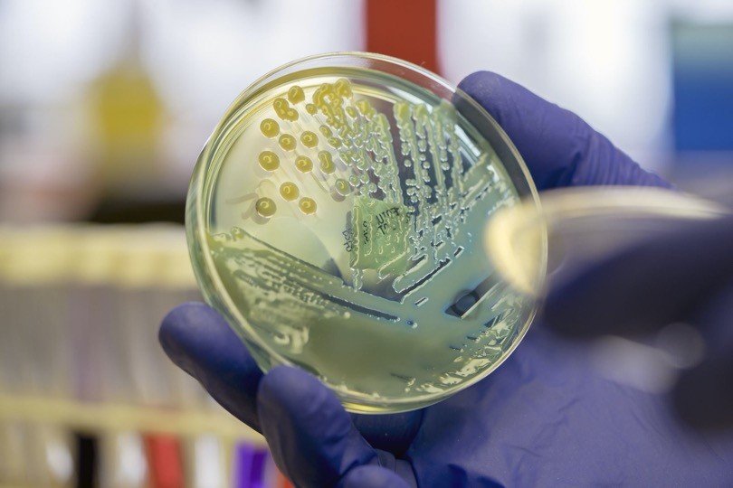
On solid media, M. tuberculosis form opaque, dry colonies with a wrinkled or nodular surface. Colonies are creamy white, becoming yellowish or buff colored on further incubation, while in liquid media M. tuberculosis grows in the bottom and form serpentine cords or duster.
Global epidemiology of Mycobacterium tuberculosis
According to WHO surveys (2006) of the effectiveness of surveillance system, and death registration, there were an estimated 8.9 million new cases of TB in 2004. Based on data from 175 countries, the prevalence of MDR among new cases and among the previously treated cases are 3.1% and 19.3%, respectively.
When including other ten countries which reported data on combined drug resistance only, the global prevalence of MDR in all cases of TB has been reported to be 4.6%.
"XDR-TB is more expensive and difficult to treat than MDR-TB and outcomes for patients are worse, therefore understanding the magnitude and distribution of XDR-TB is important"
XDR-TB is more expensive and difficult to treat than MDR-TB and outcomes for patients are worse, therefore understanding the magnitude and distribution of XDR-TB is important.
Data from this report indicate that XDR-TB is widespread with 45 countries having reported at least one case.
The high proportions of XDR-TB among MDR-TB as well as the large overall burden suggest a significant problem within the countries of the Soviet Union.
Japan and republic of Korea in a previous study, has also shown a high proportion of XDR-TB among MDR.
South Africa reported a moderate proportion of XDR-TB among MDR-TB cases.
However, the underlying burden of MDR-TB is considerable and 44% of TB patients are estimated to be co-infected with HIV (WHO Report, 2008).
Among 17,690 M. tuberculosis isolates included in the WHO surveillance for drug resistant M. Tuberculosis in the network of TB laboratories 20% were MDR and 2% XDR.
In addition, population based data on drug susceptibilities of M .tuberculosis isolates obtained from USA (for 1993-2004), Latvia (for 2000-2002) and South Korea (2004) shows that 4%, 19% and 15% of MDR-TB cases, respectively, could be classified as XDR-TB.
Thus, the distribution of XDR-TB has been found to be worldwide (WHO Report, 2008).
Classification of Mycobacterium tuberculosis
Mycobacterium tuberculosis belongs to family Mycobacteriaceae and it is the only genus in this family.
The majority of the species that comprise the genus Mycobacterium are non-pathogenic environmental bacteria related closely to the soil bacteria Streptomyces and Actinomyces.
However a few species are highly pathogenic, including M. tuberculosis complex, M. leprae, and M. ulcerans, the causative agents of tuberculosis, leprosy and Buruli ulcers, respectively.
"The Mycobacterium tuberculosis complex consists of M. tuberculosis, along with M. bovis, M. canettii, M. africanum, and M. microti"
The Mycobacterium tuberculosis complex consists of M. tuberculosis, along with M. bovis, M. canettii, M. africanum, and M. microti, which are closely related organisms (share >99% identity at the nucleotide level for some loci) and are pathogenic.
However, they differ significantly in morphology, biochemistry, host range, and disease patterns in experimental animals.
Mycobacterium tuberculosis is the causative agent of tuberculosis in humans, although in part of Africa, M. africanum causes more cases of tuberculosis than M. tuberculosis.
In contrast, infection by M. canettii appears to be rare. M. bovis has a broad host range, including humans and cattle, and was a major cause of human tuberculosis prior to pasteurization of milk. M. microti is a pathogen of voles but is avirulent in humans and mice.
Evolution of Mycobacterium tuberculosis
The recent studies into the evolution of Mycobacterium tuberculosis have indicated that Mycobacterium tuberculosis is actually more closely related to a common founder strain that is M. bovis. This implies that either humans infected cattle with M. tuberculosis, resulting in the divergence of M. bovis, or the two strains evolved in parallel from a founder strain that infected both humans and cattle.
The ancestor of M. tuberculosis most likely arose from the M. tuberculosis-like species of the M. tuberculosis complex that are found today in central Africa.
This might indicate that humans were exposed to tuberculosis-mediated selection pressure much earlier, and more comprehensively, than was previously assumed. However, M. africanum and M. microti represent intermediates in this new evolutionary scenario.
Natural Habitats of Mycobacterium tuberculosis
The genus mycobacterium includes obligate parasites, saprophytes and opportunistic pathogens. Most of the species are free living in soil and water, but the major ecological niche for other such as M. tuberculosis complex and M. leprae is diseased tissues of human and other warm-blooded animals.
Cell wall composition of Mycobacterium tuberculosis
M. tuberculosis possesses a polysaccharide cell wall that resembles gram positive bacteria in having disaccharide linker between peptidoglycan and polysaccharide.
The cell wall of M. tuberculosis contains 3 classes of mycolic acids: α-, keto-, and methoxymycolates. α-Mycolic acid has 2 cyclopropane rings, which are in the cis configuration, while the keto- and methoxymycolates have1 ring each in either cis or trans configuration.
In addition to trehalose, mycolates are linked to arabinogalactan which is covalently attached to the peptidoglycan layer. The pathogenic strains of M. tuberculosis posses “cord factor”, which are highly toxic in mice.
"The cell wall of M. tuberculosis contains 3 classes of mycolic acids: α-, keto-, and methoxymycolates. α-Mycolic acid has 2 cyclopropane rings"
Recent studies suggest that cord factor is made up of trehalose dimycolate (TDM), which are composed of a non-reducing sugar trehalose linked to mycolic acids.
Share
Required 'Candidate' login to applying this job. Click here to logoutAnd try again