o The bacterial plasma membrane and everything inside of it is called the protoplast.
o A protoplast isn’t a complete bacterium.
o Protoplast: Plant, bacterial or fungal cell with the cell wall removed using either mechanical or enzymatic means.
o Surrounding the protoplast is the bacterial envelope.
o The component of the envelope, adjacent to the plasma membrane is the cell wall.
o One of the functions of the cell wall is to prevent the protoplast from bursting.
o Most bacteria (prokaryote) are hypertonic (hypotonic solution) to their environment (this means that the aqueous solution of their cytosol contains more particles than the aqueous solution surrounding them.
o The resulting osmotic pressure causes a net movement of water into of the cell).
o Compare isotonic where the cytosol contains the same amount of particles and hypotonic (hypertonic solution) where the cytosol contains less particles.
o The cell wall is strong and able to withstand high pressure.
o As the cell fills with water and the hydrostatic pressure builds, it eventually equals the osmotic pressure and the filling stops.
o If the cell wall is removed, the plasma membrane cannot withstand the pressure.
o Osmosis describes the movement of water.
Peptidoglycan
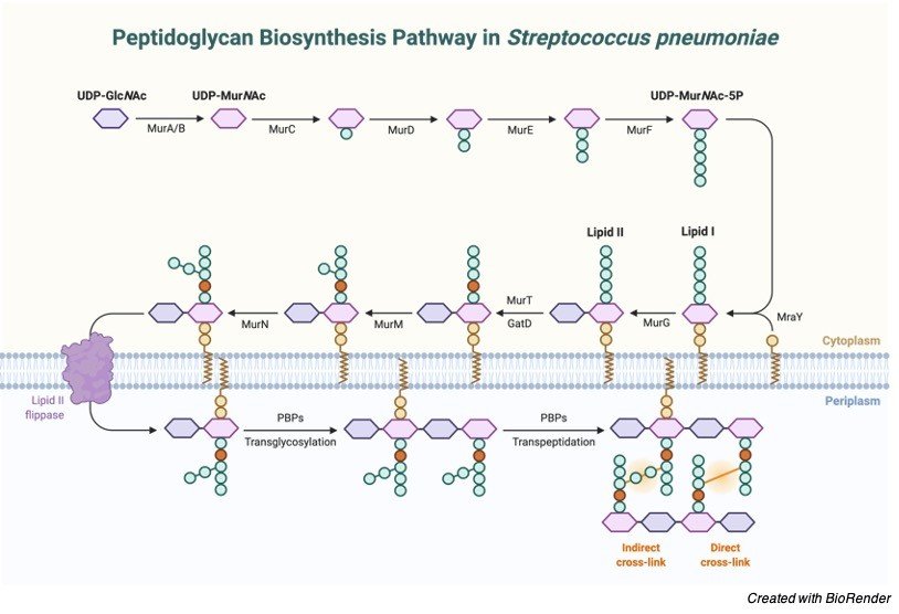
o Peptidoglycan, also known as murein, is a polymer consisting of sugars and amino acids that forms a mesh-like layer outside the plasma membrane of bacteria, forming the cell wall.
o The polymers are crosslinked by an interbridge of more amino acids.
o It is porous, so it allows large molecules to pass through.
Structure and Synthesis of Peptidoglycan
o Archaea don’t have peptidoglycan cell walls.
o Peptidoglycan is more elastic than cellulose, which as we recall is carbohydrate formed by plants and contains Beta linkages (only bacteria eat Beta linkages and is part of the plants cell wall).
o Some antibacterial drugs such as penicillin interfere with the production of peptidoglycan by binding to bacterial enzymes which make the crosslinks.
"One of the functions of the cell wall is to prevent the protoplast from bursting"
oLysozyme, an enzyme produced naturally by humans, attacks the crosslinks as well.
o In both cases the cell wall is disrupted and the cell lyses killing the bacterium.
o Don’t confuse this with a lysosome which are organelles containing digestive enzymes
o Capsules: This type of surface layer is composed primary of polysaccharides.
o If the layer is strongly adhered to the cell wall, it is called a capsule; if not, it is called a slime layer.
o Not all bacteria have this.
o These layers provide resistance to phagocytosis and serve as antigenic determinants.
o One method of classification of bacteria is according to the type of cell wall that they possess.
o A staining technique, called gram staining, used to prepare bacteria for viewing under the light microscope, stains two major cell walls differently
Gram Positive Bacteria
o The first type is called gram-positive bacteria.
o Gram-positive bacteria are those that are stained dark blue or violet by Gram staining.
o This is in contrast to Gram- negative bacteria, which cannot retain the crystal violet stain, instead taking up the counterstain and appearing red or pink.
o Gram-positive organisms are able to retain the crystal violet stain because of the high amount of peptidoglycan in the cell wall.
o Thick peptidoglycan layer.
Gram Positive Cell Wall
o Stain blue/violet.
o The space between the peptidoglycan layer and the plasma membrane is known as the periplasmic space and it contains proteins that help the bacteria acquire nutrition.
Gram Negative Bacteria
o Gram-negative bacteria appear red or pink in color when gram stained.
o Many species of Gram-negative bacteria are pathogenic, meaning that they can cause disease in a host organism.
o This pathogenic capability is usually associated with certain components of Gram-negative cell walls, in particular the lipopolysaccharide (also known as LPS or endotoxin) layer.
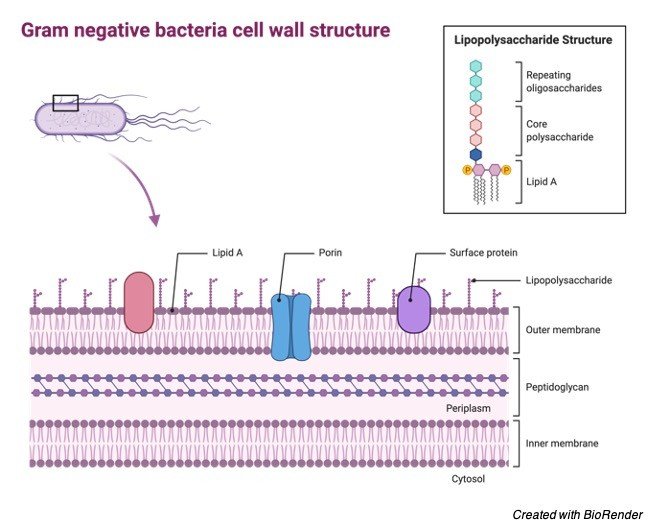
Gram Negative Cell Wall
o The following characteristics are displayed by Gram-negative bacteria:
1. Thin peptidoglycan layer (which is present in much higher levels in Gram-positive bacteria)
2. Outer membrane containing lipopolysaccharide (LPS) (can form a protective barrier from antibodies and many antibiotics) outside the peptidoglycan layer, this outer membrane is also more permeable than the inner, even allowing molecules the size of glucose to pass through
3. Porins exist in the outer membrane, which act like pores for particular molecules
4. A lipoprotein in the outer membrane called Braun’s lipoprotein points inward toward the cell wall and attaches covalently to the peptidoglycan
5. There are two spaces between the layer of peptidoglycan and the two membranes
6. Stain red/pink
7. The periplasmic space is the space between the two membranes.
o Some gram-negative bacteria possess fimbrie or pili (not to be confused with the sex pilus discussed below).
o Fimbriae are short tentacles that can attach a bacterium to a solid surface.
o They are NOT involved in cell motility.
"Outer membrane is also more permeable than the inner, even allowing molecules the size of glucose to pass through"
o Bacterial flagella are long, hollow, rigid, helical cylinders made from a globular protein called flagellin; these shouldn’t be confused with eukaryotic flagella which are made up of microtubules.
o They rotate counterclockwise.
o When they are rotated clockwise, the bacterium tumbles.
o This tumbling acts to change the orientation of the bacterium allowing it to move forward in a new direction.
o The movement of a bacterium toward or away from a particular stimulus is called taxis.
o Such stimuli include chemicals (chemotaxis) and light (phototaxis).
o The flagellum is propelled using the energy from a proton gradient rather than by ATP.