Apoptosis also known as cell suicide was discovered in the year 1972 by 3 scientists, Wyllie, Kerr and Currie. Its called cell suicide because the cell decides to kill itself as the cell has completed its function and, in the process, it will contract and the remaining broken pieces are engulfed with any effects to the other process.
Apoptosis is the planned death of a cell where 50-70 million cells die every day within a person. Thus, could also be called as cellular death as it is a highly specialized type of process for the extrusion of the cells within the body.
Why Apoptosis?
Along with the various organelles, cells also has a program fitted in it so that its aware when it has to initiate the apoptosis. This helps them to remove unwanted and harmful cells. Thus, apoptosis plays a pivotal role in processes such as cell cycle development, immune system operation, development of embryo and death of cell via chemicals.
The other roles of apoptosis in various fields are such as to segregate tissue in its respective classes. It also plays role in deleting cancerous cells, where the error in the cells can’t be resolved thus, undergoing apoptosis.
It is also seen to function when the antigen i.e the foreign particle is excluded so that it can make way for the necessary cells required, which could further prevent various sorts of diseases. A major motive of apoptosis is to maintain a healthy balance between the worn-out cells and the new cells.
Mechanism of Apoptosis
The mechanism of cell death process is highly organized, consisting of multiplexes steps and is energy dependent with 4 pathways to achieve the breakdown of the fragments, which is the ultimatum of apoptosis.
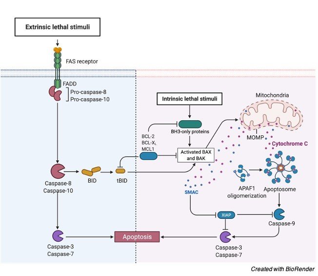
i. Extrinsic Pathway
For the apoptosis to begin, ligand and the receptor belonging to the tumor necrosis family initiate a transmembrane receptor mediated interaction. The TNF family has a death domain which allows the signal to reach the signaling pathway from the surface of the cell, with the death domain consisting of 80 amino acids loaded with cysteine.
There are 2 models in the extrinsic pathway and they are FasL /FasR and TNF ligand/TNFR 1 model both of them will interact by attaching the receptor to the respective ligand. Once the receptor is bound to the ligand, it will trigger cytoplasmic adapter proteins bringing the death domains.
The FasL FasR interaction will activate the FADD and the interaction between the TNF ligand and TNFR will activate RIP and FADD. Thus, the FADD will attach to procaspase 8 due to dimer formation of death domain and procaspase 8 will be switched on along with a death inducing signaling complex (DISC) being initiated. Once caspase 8 is triggered apoptosis process will start to proceed.
Apoptosis Pathway Diagram
ii. Intrinsic Pathway
In this pathway the signal will reach inside the cell and thus the name intrinsic/ intracellular which could be either positive or negative signals. Positive signals could be free radicals, toxins, radiations and others whereas negative signals could be lack of certain necessary factors such as hormones or growth factors which could initiate the apoptosis process.
The positive signals will release 2 various proteins in the cytosol from the transmembrane space due to the unlocking of the mitochondrial permeability transition pore. The first protein group poses cytochrome c which attaches to Apaf-1 (Apoptotic protease activating factor -1) and pro-caspases 9 leading to the establishment of apoptosome. After which the procaspase is nicked into its working form caspase 9 and then again nicked to form caspase 3 by the apoptosome.
There are also other proteins in the first group that initiate apoptosis and are HtrA2/Omi and SMAC (second mitochondria derived activator of caspases). The second group of protein would come into picture only when the cell has decided to die and thus leading to proteins secretion from mitochondria and will be broken into fragments and condensed.
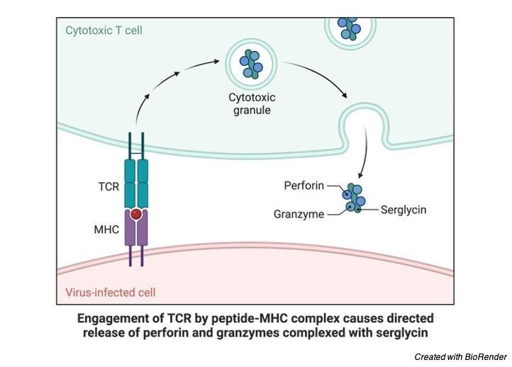
iii. Perforin Pathway
In this pathway, the toxic effect on the infected or cancerous cell is removed with the help of a molecule called perforin which secretes granules which are of 2 type, granzyme A and B which translocates the granule to the required cell and the pathway. Granzyme A will activate caspase and initiate apoptosis. Granzyme B will activate procaspase 10 at aspartate residues and nick factors such such as ICAD (Inhibitor of Caspase Activated Dnase).
To release cytochrome c granzyme B will use mitochondrial pathway for enlargening the death signal. Granzyme A nicks the Dnase when it’s in the vicinity of the cell to inhibit the apoptosis of tumor cell. Further granzyme will inhibit Dnase and nick the SET complex and this complex will further reassure that the DNA is intact and the inactivation of this complex will result in apoptosis and the DNA would be left that way.
iv. Execution Pathway
At the end of the intrinsic and extrinsic pathways begins the execution pathway. This pathway begins by triggering the caspase that will further trigger the proteases which will break proteins and endonucleases will break the nuclear material. Caspase 8, 9 or 10 will activate Caspase 3 which will further activate Caspase activated Dnase (CAD) which will further shrinken up the chromatid and degrade the DNA inside the nucleus.
Caspase 3 will cleave gelsolin, an actin binding protein to form apoptotic bodies. These apoptotic bodies will show the signs of phosphatidylserine on their outer coverings and the cell dies without any inflammatory reaction taking place.
Apoptosis Inhibitor
In order to prevent apoptosis from occurring, and the infected or dead cells to live, signaling pathways has to be inhibited. After which the inhibitors of apoptosis come into picture which are the anti-apoptotic protein factors called IAPs (Inhibitors of apoptosis) and Bcl-2. IAPs as their names suggest are the inhibitors prevent cell death and in human population this class poses 8 proteins, in which each one has a domain called the Baculovirus IAP Repeat which would link itself to the proteins involved along with caspases. Caspase 3 and Caspase 9 will attach to proteins such as XIAP to stop the process.
The other inhibitor called the Bcl – 2 is concerned with the movement of mitochondrial membrane. For the intrinsic pathway, Bcl-2, Bcl- x, BAG are some of the proteins stopping the cytochrome c and altering the mitochondrial membrane thus preventing the apoptosis to take place.
c- FLIP is a protein, which will attach to caspases and FADD to inhibit the extrinsic pathway of cell death.
The failure of the cells to apoptosis is the reason behind diseases such as myeloma, cancer and leukemia. It can further lead to mutation of the inhibitor protein XIAP causing a genetic disorder or even failure of immune system function.
Apoptosis Regulation
To regulate apoptosis, various proteins and other factors come together, however the mostly recruited one’s are Bcl-2 and IAP which has the authority to prevent or continue the cell death process.
In the extrinsic pathway, regulation takes place with the help of a protein called Toso which will not trigger caspase 8 as it has been blocked by the Fas. The intrinsic pathway of regulation requires Bcl-2 protein to regulate the movement of the mitochondrial membrane.
For the apoptosis regulation, cytochrome c is released from the mitochondria by the change in the mitochondrial membrane mobility. These proteins released along with Smac will regulate apoptosis by stop the IAPs. Thus, apoptosis takes place. To activate apoptosis, proteins like Puma and Noxa stop the anti-apoptotic protein.