A key function of macrophages is to engage and clear invading microorganisms.
A variety of receptors present on the plasma membrane of phagocytes mediate interactions with pathogens.
A novel formulation of nanoparticles/ drugs needs to be checked for the uptake by macrophages.
Macrophages are involved in evading the foreign particles and eliciting the immune response.
Thus, in vitro drug uptake is necessary before the formulation can move ahead and be used for in vivo studies.
Depending on the type of molecule and the mode of its uptake, endocytic process has been broadly classified into pinocytic, phagocytic and receptor mediated pathways.
However, further categorization of the processes has been done depending on whether or not the process utilizes clathrin, a molecule known to be very important for transport of transmembrane receptors involving coated pits.
Phagocytosis is engulfment of large particulate matter specially shown by macrophages and neutrophils.
The engulfment of particles in this process forms phagosomes. The vesicles in this case are usually larger than 250nm.
Similarly, pinocytosis is a process of “cell drinking”, here the particles taken up by the cell are usually very small in size.
Created with BioRender
About Current Cellular Drug Uptake Protocol
In the present experiment, we would study about uptake of fluorescent beads which are opsonized with IgG.
These beads are ~2μm in diameter; hence they would be taken up by phagocytosis route.
Opsonization with IgG enhances their phagocytic uptake.
Along with this formulation of iron particles coated with RBITC-BSA (Rhodamine B Isothiocyanate-Bovine Serum Albumin) would also be studied.
Since iron particles are very small is size ~25nm, they are usually taken up by pinocytosis/receptor mediated endocytosis.
To understand the mechanism of uptaking particles it needs to be checked at 37°C versus 4°C, competitive inhibition and also a saturation curve of receptors, which can give us the idea whether the uptake is receptor mediated or not.
Principle of Cellular Drug Uptake
Isolated rat peritoneal macrophages are used for in vitro drug uptake of particles which are in the range of nanometer to micrometers.
The particles which are in the range of nanometers (~25nm) are expected to be internalized by the process of pinocytosis/receptor mediated endocytosis, whereas those in micrometer (2μm) range are internalized by the process of phagocytosis.
Cellular Drug Uptake Requirement
1. Rhodamine B Isothiocyanate-Bovine Serum Albumin coated iron particles.
2. Opsonized fluorescent beads
3. Phosphate buffer saline
Procedure of Cellular Drug Uptake
1. Visualize rat peritoneal macrophages isolated from earlier experiment in inverted microscope. Assess for their morphology (oval, translucent) and size.
2. For opsonization of fluorescent beads, add 50 μl of beads suspension in 100 μl of rabbit serum. Put the mixture at 37 C shaker for two hours.
3. Treat the macrophages with RBITC-BSA-iron and opsonized Fluorescent beads.
4. After the treatment for respective duration, wash with Phosphate buffer saline for 3 times.
5. Add 500μl of PBS in each well/petridish and visualize in fluorescent microscopy.
Cellular Drug Uptake Analysis
Drug uptake could be studied quantitatively as well as semi-quantitatively.
To study the uptake quantitatively, after thorough washing, we need to scrap out all the cells, lyse them using hypotonic lysis buffer and then measure the fluorescence using fluorimeter.
More percent of drug uptaken by macrophages correlate to more fluorescence in lysed macrophages.
Evaluate results of the percentage of drug uptaken by comparing the fluorescent drug inside the cell per milligram of protein versus drug suspension added.
Semiquantitative estimation can be done using fluorescent microscope.
Checking out the fluorescent images as well as phase contrast gives us understanding of drug uptake inside the cell.
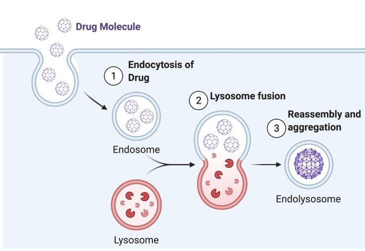
Internalization of drug inside the macrophages will form vesicles (endosomes).
Thus, more is the uptake of the drug per cell; we will visualize more endosomes per cell.
We can calculate the percentage of phagocytes out of the total number of cells with fluorescent phagocytic vesicles to assess the phagocytic uptake.
Assessment of florescent beads is to be done in blue filter and for nanoparticles in green filter using florescent microscope.