Chromatin is a nucleic acid (DNA or RNA) and protein combination (e.g. histones). It was discovered in 1882. Originally considered to be merely a colourful material in the nucleus, chromatin was subsequently shown to be proteins linked to DNA, and DNA was recognised as the bearer of genetic information. As a result, we may describe chromatin as a material made up of DNA and proteins (known as histones).
Histones
Histones are basic proteins with a positive charge that attach to DNA’s negatively charged phosphate units. Chromatin consists of two primary components: the cell’s DNA and related proteins. Histones are the proteins that are involved. Or, to put it another way, chromatin is made up of proteins known as histones.
Chromatin is a kind of DNA packing. It can wrap around itself or get harmed during cell division if it isn’t packed correctly. Cells are measured in micrometres, while DNA may be up to 3 metres long. Tight packing is necessary to accommodate such a long structure inside a micrometre cell.
Nucleosomes
Nucleosomes are formed when a DNA molecule wraps around histone proteins to form tight loops.
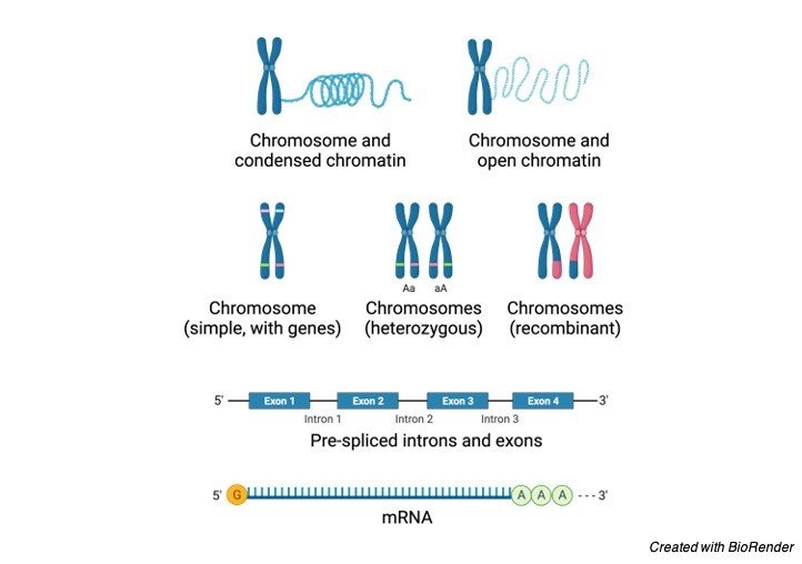
The nucleosomes coil and bundle together to create chromatin fibre, which is a kind of fibre. These chromatins, in turn, loop and fold around to form a chromosome with the aid of proteins. That is why a chromosome is said to carry a portion or all of an organism’s genetic material. Because of its tightly coiled shape, DNA is now protected after it is condensed into a chromosome.
Chromatin is also important for controlling the transmission of genetic information. A material composed of DNA or RNA and proteins, such as histones. During cell division, it condenses to create a chromosome.
Where is Chromatin Found?
Chromatin is present in the nucleus of eukaryotic cells. Here’s a diagram to help you visualise where it’s located within the cell nucleus.
Genes in Chromatin
The genes that are present in chromatin can be switched on or off. It indicates that in certain cells, one half of the gene is active (“switched on”) and the other is inactive (“switched off”). It’s chromatin, of course.
The researchers confirmed this by studying the on and off states of genes in chromatin using a fruit fly as a model organism. Their research revealed five distinct kinds of chromatin based on the presence of certain proteins.
Chromatin Structure
In eukaryotic chromatin, the histone proteins and DNA have almost the same mass (although there are also some cells with non-histone proteins instead). The nucleosome is a structural unit of chromatin, which is made up of DNA and proteins (histone and non-histone). This structure can be found throughout an organism’s genetic material. Below is a diagram showing chromatin packing into a higher-order structure.
"Beads on a String" Model of Chromatin
The initial degree of compaction for DNA inside the nucleus is provided by DNA and histone proteins. The nucleosome is chromatin’s fundamental structural unit. When DNA is wrapped around histones (the protein core) to create a “bead-like” structure, it is called a nucleosome. A nucleosome is the name of this bead-like structure. The “beads-on-a-string” type of chromatin is shown in figure 3 as the second from the top. The nucleosome is a 146-base-pair DNA molecule that is wrapped around eight proteins called histones. When DNA is wrapped around histones, a nucleosome is produced.
H1, H2A, H2B, H3, and H4 are the five distinct kinds of histones. When two H2A and H2B proteins join with H3 and H4 proteins, a histone core is formed. A nucleosome is formed by wrapping 145 base pairs of DNA twice around this protein structure. The length of linker DNA varies based on the gene activity of the species and can range from 10 to 95 base pairs. Every 200 base pairs, a nucleosome with a length of 10 nm appears.
When seen under a microscope, chromatin resembles beads strung on a string. Nucleosomes are the name of these beads. The nucleosome is made up of eight proteins called histones. The nucleosomes wrap themselves into a 30nm spiral to produce a solenoid. Additional histone proteins aid in the formation of the chromatin structure in this solenoid. Due to its more compact shape, chromatin condenses into chromosomes.
Chromatin and DNA
DNA is packaged in chromatin. To fit inside a nucleus, DNA and related proteins are compacted into chromatin.
How DNA is Assembled in Chromatin Structure?
The process of assembling DNA into chromatin involves multiple stages. H3e and H4 proteins are the first to bind to DNA, followed by H2A and H2B. A sub-nucleosomal particle with 146 base pairs of DNA is produced. The second phase is maturation, which involves ATP establishing a constant nucleosome core spacing. Folding of linker histones into a nucleofilament of 30 nm structure is the next stage. Further folding happens in the final phase, resulting in a greater amount of packing. The packing ratio is estimated to be around 7000.
Euchromatin vs Heterochromatin
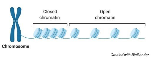
Heterochromatin and euchromatin are the two types of chromatin. Heterochromatin is extremely condensed and can not generally be transcribed, whereas euchromatin is less condensed and can be transcribed. Constitutive heterochromatin and facultative heterochromatin are two types of heterochromatin. Constitutive heterochromatin refers to the DNA sequences found in all of an organism’s cells. Highly repetitive DNA is linked to constitutive heterochromatin.
Similarly, not all cells have facultative heterochromatin. The gene that codes for beta-globin in animals, for example, is found in certain cells but not in blood cells. In eukaryotic cells, chromatin is a combination of proteins and DNA, as previously stated. Nuclear DNA is tightly compacted and wrapped around nuclear proteins to fit inside the nucleus, rather than existing as linear strands.
In the interphase nucleus, chromatin is divided into two types: euchromatin and heterochromatin. Euchromatin refers to a kind of chromatin that is structurally loose. In terms of transcription and replication, it is generally active. Allowing RNA and DNA polymerases to transcribe and replicate DNA, respectively, is permissive. Heterochromatin is the chromatin that is less active. It contains dormant genes and is smaller in size.
Chromatin Function
Initially, chromatin was thought to be the material that gave the cell nucleus its colour. It was later discovered that it is more than simply a colouring agent; it is also one of the most significant DNA expression controllers. The structure of chromatin is also crucial for DNA replication. DNA is packaged in chromatin and nucleosomes, resulting in a tightly closed structure that enzymes essential for transcription, replication, and repair of DNA can not access.
DNA structural packing is transcriptionally restrictive, allowing only a modest amount of gene expression. DNA may be copied and transcribed more easily when nucleosome structures are open or broken.
Some repressors and activators interact with RNA to regulate gene activity and modify the chromatin structure during the transcription process. Activators alter the structure of nucleosomes, causing RNA polymerase assembly to be stimulated. A comparable control of chromatin shape happens during replication, allowing the replication mechanism to be in place at the replication origin.
The control of gene expression is another function of chromatin. The genes can be made transcriptionally inactive by placing them near quiet heterochromatic chromatins using the location effect variegation method. Silent heterochromatin chromatins and genes can be separated by up to 1000 kilobase pairs. Because it causes phenotypic diversity, this phenomenon is referred to as epigenetic.
The extremely condensed structure of heterochromatin, according to scientists, inhibits DNA transcription. However, how the surrounding non-heterochromatic areas are impacted is still unknown. Proteins in chromatin can travel to nearby areas, causing a similar restrictive effect, according to the researchers. The researchers also speculated that heterochromatin may be found in compartments of the nucleus that are not accessible to transcription factors. As a result, transcription factors may not have direct access to chromatin in the nucleus.
DNA replication is influenced by chromatin structure. The euchromatin and other active regions of the genome, for example, replicate faster. The replication process is also sluggish in the heterochromatin and quiet regions surrounding it. Below are some of chromatin’s other key activities.
i. DNA Packaging
The primary purpose of chromatin is to compact lengthy DNA strands into a much smaller area. In comparison to where it resides, DNA has a relatively long linear length. DNA must be compressed in some way in order to fit comfortably and securely without tangling or harm. Condensing is the process of compacting DNA into the nucleus. The packing ratio refers to how tightly DNA is packed within a body. DNA has a packing ratio of around 7000. DNA is not packed directly into the structure of chromatin because of the high amount of compaction. Rather, there are numerous organisational hierarchies.
The nucleosome is initially packed by wrapping DNA around it. A packing ratio of 6 is obtained as a result of this. Both heterochromatin and euchromatin have the same packing. Wrapping the beads in a 30 nm fibre, which is also present in mitotic chromosomes and interphase chromatin, achieves the second degree of compaction. The packing ratio is increased from 6 to 40 with this wrapping. Compaction is done in the third stage by injuring the fibre into loops, domains, and scaffolds. In mitotic chromosomes, the packing ratio climbed to 10,000, whereas interphase chromatins had a packing ratio of 1000.
During metaphase, chromosomes are most compact. The DNA of eukaryotic cells must be evenly split into two daughter cells during cell division. The DNA is severely compressed during this phase, and the chromosome uncoils after the cell division is completed. When linear DNA is compared to the length of metaphase chromosomes, the packing ratio can be as high as 10,000:1. Phosphorylation of histone H1 results in this high amount of compaction.
ii. Transcription Regulation
Transcription is the process of genetic information being transferred from DNA to proteins. After that, the information is translated into RNA. The translation of RNA into functional proteins is the final step.
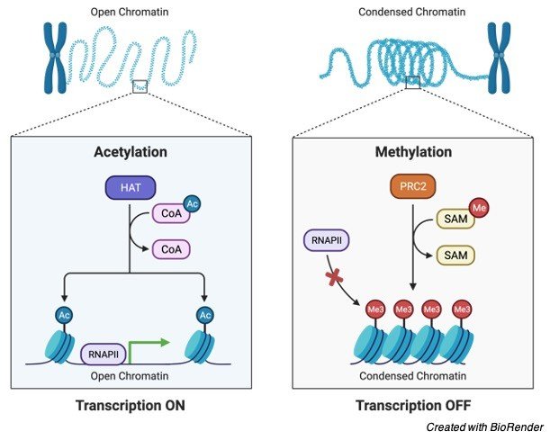
Chromatin regulates the transcription process. The transcription will halt if chromatin is reinforced and access to read the proteins is restricted. Heterochromatin is a densely packed form of chromatin in which proteins are unable to read the DNA. While euchromatin is not as densely packed, proteins can carry out the DNA description process. Similar to active and inactive chromatins, there are active and inactive chromatins that can contribute to transcriptional bursts or discontinuity.
The attachment and dissociation of transcription factor complexes in chromatin are other factors in transcription. This phenomenon is thought to be the cause of the significant diversity in gene expression among cells in an isogenic population.
iii. Chromatin and DNA Repair
The packaging of DNA into chromatin is essential for all DNA-based activities. Because of the dynamic organisation of proteins, chromatin can change its form and structure. Chromatin relaxation happens when DNA is damaged. This relaxes the proteins, allowing them to attach to DNA and repair it.
iv. Chromatin in Mitosis
Mitosis is a cell division process in which the resultant two cells (daughter cells) have the same chromosomal type and number as the parent nucleus. During the four stages of mitosis, chromatin plays a crucial role.
The chromatin fibres wrap around to create the chromosomes during this phase. At the centromere, two chromatids are joined to form a replicated chromosome.
Metaphase: During this phase, chromatin condenses to an excessive degree.
Anaphase: The spindle microtubules bring the two identical chromosomes to the cell’s end and separate them during this phase.
Telophase: The new chromosomes are split into their own nucleus during this phase. Uncoiling causes chromatin fibres to become less condensed at this stage. Two identical cells with the same number of chromosomes are generated.
Structure and Function of Chromatin
Chromatin is a macromolecule composed of DNA, RNA, and proteins. Its name, which literally means colourful substance, comes from the fact that it can be easily identified by staining. The nucleosome is chromatin’s fundamental structural unit. A DNA segment is wrapped around the histone protein cores of each nucleosome in chromatin. When this nucleic acid and protein combination becomes compact during cell division, it becomes a chromosome. Its responsibilities include packing DNA into a smaller volume so that it can fit within the cell, strengthening DNA to aid mitosis and meiosis, and regulating expression. In eukaryotic cells, chromatin is located in the nucleus, whereas in prokaryotic cells, it is found in the cytoplasm.
Chromatin, Chromosome, and Chromatid
Despite the fact that all three structures, chromatin, chromosomes, and chromatids, are made up of DNA and are found in the nucleus of the cell, they are each uniquely designated as follows:
Chromatin vs Chromosome
The primary distinction between chromatin and chromosomes is that chromatin is made up of DNA and histones packed into a fibre, whereas chromosomes are single-stranded forms of condensed chromatin. The fine fibre of chromatin serves as the foundation for the chromosomal structure. While the activities of chromatin have been defined, the function of chromosomes is critical for mutation, regeneration, cell division, variation, and inheritance. Furthermore, chromatin condenses to form a chromosome during cell division, and the chromosome is double-stranded with an X shape. The centromere is a region that connects the two strands to the centre of the cell.
Chromosomes Location in Cells
A eukaryotic cell’s chromosomes are found in the nucleus. In prokaryotes, such as bacteria, the chromosome is usually a single loop of stable chromosomal DNA in the nucleoid. Non-histone proteins are linked to prokaryotic DNA. There is no nucleus in viruses, so the chromosome appears as a small linear or circular DNA or RNA molecule, typically without any structural proteins, wrapped in an envelope or capsid around its head.
Chromatin vs Chromatid
Chromosomes are divided into two strands. A chromatid is the solitary strand of the chromosome. At the end of cell division, these chromatids split to form daughter chromosomes. Thus, chromatin is distinct from chromatid in that chromatin is made up mostly of DNA and associated proteins in the form of fibre, whereas chromatid is a chromosomal component. Yes, chromatin is present in the chromatid.
Chromatin vs Nucleosome
The nucleosome is a piece of DNA that wraps around the protein’s core. Chromatin is a DNA-protein complex that aids in the condensation of DNA for packing into the nucleus.