Gamete: Definition, Formation, Examples, and Facts
Gamete Definition
A gamete is a mature reproductive or sex cell with a haploid number of chromosomes (i.e., just one set of dissimilar chromosomes) and the ability to fuse with another haploid reproductive cell to produce a diploid zygote. The zygote is created by fusing (or merging) two gametes, namely a male and a female gamete. Fertilization is the process of gametes coming together to form a zygote.
What are Gametes?
Gametes are an organism’s reproductive cells. Gametes are also referred to as sex cells. Female gametes are called ova or egg cells, and male gametes are called sperm.
A mature haploid reproductive cell generated by gametogenesis and that which unites with another from the opposing sex at fertilisation to form a zygote that grows into a new person.
Gamete Etymology
From the Ancient Greek (gamet), which means “woman.” sex cell; reproductive cell are synonyms. One of the gametes is generally bigger and non-motile, as is customary. It’s also called a female gamete, ovum, or egg cell.
The other gamete cell is smaller in size and motile. It’s also known as a sperm cell or a male gamete. Each human gamete has 23 chromosomes, and when they fuse, a diploid zygote with 46 chromosomes is formed.
These reproductive cells are generated in the male and female gonads, or reproductive organs, in mammals. Male gametes are pollen in seed-bearing plants, whereas female gametes are contained in the plant’s ovules. In plants, however, the gamete may or may not be a haploid cell.
Types of Gametes
The gametes used in fertilisation might be identical (known as isogamy) or distinct (known as polygamy) (referred to as anisogamy).
i. Isogamy
Isogamy refers to gametes that have identical morphology, such as size and form. Heterogamy is another name for this situation. These gametes aren’t classified as either male or female. These gametes are denoted by the letters “+” or “-“.
Unicellular algae gametes, Chlamydomonas reinhardtii, and Carteria palmata are examples.
ii. Anisogamy
Anisogamy refers to gametes with morphological differences, such as size and form. Female and male gametes are the two kinds of gametes. The gamete with the smallest size is called a sperm or male gamete, whereas the gamete with the largest size is called an ova, egg, or female gamete. Additionally, these gametes can be both motile and non-motile.
In the instance of Polysiphonia, a red algae, both gametes are non-motile. A zygote is formed when a non-motile sperm joins a non-motile egg. Spermatia is a non-motile male gamete or sperm. This may also be found in flowering plants where both non-motile gametes are present in the gametophyte. Pollen is a non-motile male gamete found in plants.
iii. Oogamy
Oogamy occurs when one of the gametes, the male gamete or sperm, is motile while the other gamete, the egg or female gamete, is non-motile in humans and animals. Oogamy is a situation in which a big non-motile egg is fertilised or will fuse with a tiny motile sperm to produce a zygote.
Anisogamy (or heterogamy) is a type of sexual reproduction that involves female and male gametes of differing sizes. In contrast, isogamy is a kind of sexual reproduction in which both the male and female gametes are the same size.
Size of the Gametes
The size of the gametes can also be used to classify them. Gametes are classified as follows based on their size:
• Microgametes are tiny versions of gametes. These are motile, usually generated in vast numbers, and do not have nutrition storage. Consider sperm cells.
• Macrogametes are gametes that are larger than normal. These are non-motile, generated in small quantities, and contain a high amount of nutrient storage. Egg cells or ova are two examples.
Gamete Examples
Male and female gametes are created in their reproductive organs through a process known as “gametogenesis.” A diploid (2n) cell goes through meiosis to create four haploid (n) cells during gametogenesis.
In most cases, gametogonia is the first step in the gametogenesis process. The primordial germ cells give rise to gametogonia (PGCs). Mitosis is the mechanism by which these germ cells multiply. These cells are transported to the gonadal ridge in the late embryonic stage, where they are known as gametogonia. Following the development of gametogonia, subsequent gametogenesis results in the creation of an egg or sperm, depending on the individual’s sex.
Males and females have entirely distinct gametogenesis processes. Spermatogenesis is the gametogenesis that results in the creation of sperm, whereas oogenesis is the gametogenesis that results in the formation of an egg or ova.
What are some gamete examples?
A gamete is a haploid pair of chromosomes found in a reproductive cell or sex cell. It is generated by a germ cell that goes through gametogenesis, which is a gamete production process involving meiosis. Oogenesis is the gametogenesis that leads to the development of the female gamete. Spermatogenesis is the process of creating a male gamete.
Structure and Function: Sperm Cell
The sperm cell is the male reproductive cell or gamete. Anisogamy is a condition in which the structure of the male gamete differs from that of the female gamete. Sperm in animals, including humans, are tiny and motile. The flagellum is the motile organ found in sperm. Sperm cells have a finite lifespan and are unable to divide. Mammalian sperm have two different structures that are separated by a single membrane.

• The haploid nucleus, which is densely packed with DNA, is found in the head. A sperm cell also contains an acrosome, a thin, flattened sac-like structure, and a vacuole, in addition to the extremely compressed genetic material. The enzyme essential for breaching the ovary or egg cell is found in the acrosome. Exocytosis is the process through which the enzyme is released. The head is 5.1 m by 3.1 m in size.
• The tail directs the sperm towards the egg and finally enters it, ending up at the nucleus’s posterior end. This is also the sperm cell’s longest section. The tail is 50 metres in length. The tail travels at a rate of 1-3 millimetres each second.
The neck, which is rich in mitochondria, connects the two sections, namely the head and tail. Mitochondria are vital to the sperm cell because they supply all of the energy required for sperm motility. Mitochondria manufacture the ATP necessary for sperm motility. The neck also includes centrioles in addition to mitochondria. Sperm is a haploid gamete that has 23 chromosomes in humans.
Spermatogenesis
Spermatogenesis is a process that happens in male humans’ testes and begins only after they reach puberty. Spermatogenesis, on the other hand, is a continuous process that persists throughout a person’s lifespan once it begins (unlike oogenesis). Seminiferous tubules are tubular structures where spermatogenesis takes place.

Spermatogonia, or immature germ cells, are found at the basal lamina on the outside border of the seminiferous tubules. Mitosis is the mechanism by which these germ cells proliferate indefinitely. Some of these proliferating cells do not proliferate and become primary spermatocytes. These main spermatocytes then proceed through the first meiotic phase, in which each pair of homologous chromosomes contributes to cross-over, before going through division I of meiosis, which results in the production of two secondary spermatocytes with 22 duplicated autosomal chromosomes (it can be a duplicated X or a duplicated Y chromosome).
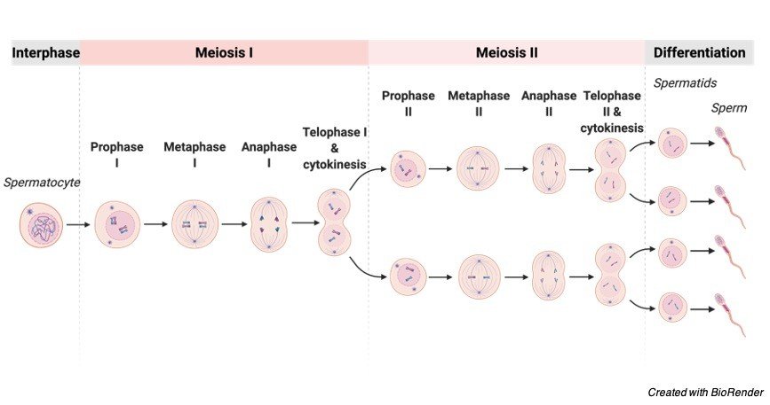
Phase II of meiosis occurs in these secondary spermatocytes, culminating in the creation of haploid spermatids, which are later differentiated to produce sperm. These sperm eventually make their way into the lumen of the seminiferous tubule. Later, sperm go to the epididymis, a coiled tube located above the testes where they develop and are stored in a liquid known as semen. This entire process takes around 70 days and may live in the female reproductive canal for over 5 days. Outside the body, however, they may only live for a few hours.
Sperm can be frozen for months or years and then thawed to preserve their ability to fertilise eggs. The fructose in the semen provides energy to the sperm for motility.
Sperm can’t swim backwards, which is an intriguing fact.
Ferns, cycads, and ginkgo trees all have flagellated sperm. Nematode sperm are amoeboid in nature. Rather than swimming, they move around by crawling.
Non-motile sperms, for example, non-motile sperms of Polysiphonia, a red alga, are dispersed by water currents after being discharged, relying on environmental circumstances for dispersal and finally reaching the egg cells. These non-motile sperm are carried by flies, butterflies, and insects.
Sperm have developed a number of important modifications that make them effective cells.
The following are some of these adaptations:
• Sperm have a streamlined shape and a tapered head, which aids in motility and agility.
• The energy for sperm movement is provided by condensed packing of mitochondria (almost 70 in number) in the neck region of the sperm.
• Sperm include certain basic amines that aid in the successful fertilisation of an egg by allowing them to establish an alkaline microenvironment even in the acidic vaginal canal.
• The sperm acrosome includes lysosomal enzymes (such as lysozyme) that aid sperm penetration into the egg during fertilisation.
Function of Sperm
The sperm’s job is to go to the egg and fuse or fertilise it to produce a zygote, transferring the male genetic material and centriole in the process (which eventually determines the microtubule cytoskeleton). The colour of the eyes, hair, and skin of the progeny is determined by the genetic content of the sperm. The X and Y chromosomes in sperm determine the sex of the offspring.
Structure and Function: Ovum (Egg Cell)
The egg cell, also known as the ovum, is a non-motile ovoid or spherical gamete generated in the female reproductive system’s ovaries. A fertilised egg is bigger than sperm. The average diameter of a human egg is around 0.1mm. It ranges from 1-2mm in fish and frogs. The biggest egg is the ostrich egg, which measures 170 x 135 mm. In humans, the egg or ova is a haploid gamete with 23 chromosomes.

The cytoplasm of an egg is called ooplasm. The egg’s cytoplasm is divided into two parts: the formative yolk and the nutritional yolk. Because the human egg has such a small amount of nutritious yolk, it is referred to as alecithal. The cytoplasm of avian eggs, on the other hand, is dense in nutritional yolk (which is made up of lipoproteins, pigment granules, and water).
The germinal vesicle, which houses the egg’s nucleus, and the germinal spot, a vacuole, are both found in the cytoplasm. The ovum’s nucleus is big, bloated with nucleoplasm, and positioned eccentrically. As a result, the human ovum has polarity, with animal and vegetable poles. The animal pole is the side of the ovum that contains the nucleus and polar body, whereas the vegetable pole is the opposite side. The cortex is a peripheral layer made up of microvilli and cortical granules that surround the cytoplasm.
Protective Membranes of Ovum
The zona striata or zona pellucida is a thick, translucent membrane that surrounds the ovum. The vitelline membrane is a thin layer that lies underneath the zona pellucida. The zona pellucida and the vitelline membrane are separated by a thin gap known as the perivitelline space. The corona radiata, which radiates from the egg surface, is the outermost layer, which lies above the zona pellucida.
Function of Ova
The major purpose of the ovum is to transport genetic material, which in a human ovum consists of 23 pairs of chromosomes, and to result in the creation of a zygote following fusion with the male gamete. It also offers the necessary conditions for sperm to fertilise the egg. The nutrients from the ovum are necessary for the zygote’s development after fertilisation.
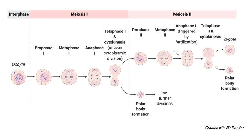
Oogenesis
The female reproductive organ known as the ovaries undergoes oogenesis, which is the process of ovum differentiation. The oogenesis process varies from species to species. An ovarium is formed in humans from germ cells that are present at the time of a female child’s birth. As a result, there are two major phases of oogenesis:
Pre-natal Phase
The ova are made up of oogonia, which is a kind of germ cell. Mitosis allows the oogonia to multiply and produce primary oocytes. At the moment of birth, all of the germ cells multiply to produce a significant number of primary oocytes (roughly 2 million). A
fter birth, no primary oocytes are generated (this is in contrast with spermatogenesis, wherein the primary spermatocytes are continuously formed at puberty). The primordial follicle is formed when initial oocytes are surrounded by follicular cells. Prior to the birth of the female offspring, the main oocyte starts the first meiotic division. However, until adolescence, this meiotic division is not completed and halts at the prophase in the diplotene stage.
In summary, the embryo initiates the first phase of meiotic division, which then enters a stop period for around 12 years until puberty occurs. It transmits the signal to restart meiosis at that point. Some oocytes have been seen to remain in the meiotic prophase for over 50 years. In addition, only 400 primary oocytes develop in a female’s lifetime out of a population of millions.
Post-natal Phase
The latent phase of the primary oocyte lasts until adolescence. The main oocyte undergoes maturation in the ovary after birth and before puberty. The primary oocyte remains inside the follicles during maturation, when it grows in size and forms the zona pellucida membrane.
The main oocyte restarts and completes meiosis before ovulation. The cytoplasm, on the other hand, is divided unequally, with the secondary oocyte acquiring the bulk of the cytoplasm. The initial polar body, on the other hand, only receives a small quantity of cytoplasm. The initial polar body is a non-functional cell that degenerates over time.
The nucleus of the secondary oocyte initiates the second meiotic division at the moment of ovulation. However, it only advances to metaphase before pausing the meiotic division process. In the event of fertilisation, the second meiotic division is restarted and finished.
The maturation of an oocyte happens with each menstrual cycle, resulting in the development of an ovum by division. This division produces cells of uneven size, such as secondary oocytes (120-150 mm and fertile) and polar bodies (120-150 mm and fertile) (not more than 10 mm and non-fertilizable).
In some species, such as humans, there are two types of gametes: the male gamete (i.e., sperm cell) and the female gamete (i.e., ovum). Male gametes are smaller and motile, but female gametes are many times larger and non-motile. The two gametes must be haploid in order for the chromosomal number to be maintained over generations during fertilisation during sexual reproduction.
Importance of Haploid
All animals are diploid genetically due to the fusing of two haploid gametes. The haploid gamete cells guarantee that the genetic content or number of chromosomes stays consistent from generation to generation. If the gamete cells are not haploid, each future generation will contain double the number of chromosomes or genetic material as the preceding one.
It’s crucial to keep in mind that cancer cells have a non-diploid condition. The presence or absence of a pair of chromosomes during cell replication might cause instability. This, in turn, can lead to the development of a disease, such as cancer. In humans and most animals, ploidy changes are typically deadly.
Aside from that, haploids are utilised for crop development (particularly in rice and tobacco) since they can be generated in a short period of time. As a result, haploids can aid crop development by shortening the breeding cycle and producing unique genetic compositions. Haploids are also an excellent cytological tool for researching mutations and genetic diseases.
Sex Determination in Humans and Animals
Primary sex determination in animals is decided by chromosomes rather than the environment, and is determined by the gonads. In placental animals, the presence of a Y chromosome determines sex. Female cells normally have two X chromosomes, i.e., XX, whereas male cells have an X and a Y chromosome, i.e., XY. As a result, each of the female eggs has a single X chromosome, whereas male sperm have two kinds, one with the X chromosome and the other with the Y chromosome.
As a result of the fusing of the X chromosome-containing egg with the X chromosome-containing sperm, female offspring with XX chromosomal makeup are produced. In contrast, when a male gamete carrying the Y chromosome fuses with an ovum holding the X chromosome, a zygote with sex chromosomes, XY, is formed, it develops into a male child. The testis-determining factor, which results in the development of testes in male progeny, is encoded by the SRY gene on the Y chromosome.
Sex Determination in Birds and Vertebrates
The sex determinants in birds are the Z and W chromosomes, with females being heterogametic (having ZW chromosomes) and males being homogametic (having just Z chromosomes) (i.e., with ZZ chromosomes). The Z chromosome is significantly bigger than the W chromosome. FET1 and ASW, two genes found on the W chromosomes, control the development of female birds. In chickens, the mechanism of determining sex is not well understood.
However, unlike mammals, chickens’ gonads differentiate into male or female reproductive organs after a period of time following birth. For sex determination, chickens require oestrogen. A male chicken can be turned into a female chicken by injecting oestrogen into the eggs during the development period. The ZW chromosomal sex determination is also seen in certain reptiles, fish, and amphibians.
Sex Determination in Insects and Invertebrates
Distinct insects have different sex determination patterns. Females are heterogametic in butterflies and moths (order Lepidoptera), whereas males are homogametic. In Lepidoptera, the sex is determined by the W and Z chromosomes. The W chromosome is linked to female traits. Males with ZZ chromosomal content grow into ZZ chromosomal males, whereas females with ZO chromosomal content develop into ZO chromosomal females.
In the absence of the W chromosome, a moth, Talaeporia tubulosa, uses ambient temperature to identify sex. Warmer temperatures result in the development of more female eggs, whereas cooler temperatures result in the formation of more male eggs. This is an excellent example of adaptation, in which certain conditions, such as warmth, encourage the development of more female offspring because warm conditions assure the availability of nutrients for later reproduction.
The XX/XO sex-determination system, which is a single-chromosome system, is employed in grasshoppers. Males have only one sex chromosome, XO (heterogametic), whereas females have two chromosomes, XX and homogametic.
Sex Determination in Drosophila
Drosophila melanogaster, a fruit fly, has been extensively researched to better understand genetics. The number of X chromosomes to the number of sets of autosomes, or the X: A ratio, is used to determine sex in Drosophila. The X chromosome encodes female-determining factors, whereas the autosomes encode male-determining factors.
The sex of a fruit fly is thus determined by the balance between X and A. Male flies have XY and XO chromosomes, whereas female flies have XX, XXY, and XXYY. Isogamy has evolved into anisogamy, which is the evolutionary successor of isogamy. Isogamous individuals, such as fungus, algae, and yeast, produce the same sort of gametes. ‘+’ and ‘-‘are used to denote isogamous gametes.
In anisogamy, the male and female gametes have physical differences and are referred to as male and female. According to popular belief, the genesis of anisogamy is based on the fact that the largest number of positive fusions of gametes happens when the population’s gametic material has been divided with a high degree of anisogamy. As a result, it is assumed that a set amount of reserve material is required for zygote formation, and that only disassortative fusions (between small and big gametes) occur. According to this idea, males generate a high number of sperm to enhance the chances of conception.
Evidence shows that sperm density in the female tract has a favourable effect on fertility (i.e., the proportion of fertilised ovum discharged). As a result, the more sperm in the sperm, the better the likelihood of conception. This is also due to the fact that a larger quantity of sperm increases sperm competition for fertilisation, resulting in increased fertility.
Furthermore, the sole purpose of male sperm is to transmit genetic material, and a large quantity of tiny sperm provides an evolutionary advantage. The ovum expends far more energy than a male gamete in generating a viable zygote. The egg supplies genetic material from its nucleus, mitochondrial genes, and vital nutrients for the zygote’s early growth in order to increase its chances of survival.
As a result, in order to deliver all of the essential materials, the ovum is big in size and contains a sufficient amount of all of the required substances. As a result, anisogamy is said to have evolved, in which eggs are non-motile, enormous in size, and restricted in quantity, whereas sperm are tiny, motile structures generated in huge quantities.
Chromosomal Anomalies in Gametes
The existence of an aberrant number of chromosomes is known as aneuploidy. In a typical human cell, there are 46 chromosomes. An aneuploid is a person who has 45 or 47 chromosomes. Because of the aberrant chromosomal number, a genetic imbalance occurs, resulting in a disease. The second most frequent type of mutation is somatic mutation.
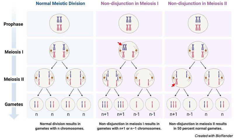
Nondisjunction, or the failure to separate chromosomes between two cells during cell division, causes aneuploidy. Miscarriage is caused by aneuploidy in the germline. A normal woman is a 46 XX, while a typical man is a 46 XY. Trisomy is the most prevalent kind of aneuploidy.
The following are some examples of aneuploidy:
• Loss or acquisition of a portion of a chromosome causes partial aneuploidy or trisomy.
• Monosomy refers to the absence of one of the typical complement’s chromosomes. Monosomy is exemplified by Turner’s syndrome. Females with Turner’s syndrome have only one X chromosome, 45XO. Fertility and reproductive organ growth have both decreased in these people. Patients with Turner syndrome are commonly referred to as mosaics.
• The existence of two copies of a chromosome is known as disomy. It is a common occurrence, but uniparental disomy occurs when both copies of the chromosomes come from the same parent.
• Trisomy refers to the existence of three copies of a chromosome rather than the usual two, implying the presence of an additional chromosome. Edwards syndrome is caused by a trisomy of 18, while Patau syndrome is caused by a trisomy of 13. Trisomy of the Y chromosomes, such as (47, XXX), (47, XXY), and (47, XXY), is also conceivable (47, XYY). The majority of trisomies are not viable and do not survive; just a handful are capable of living. Down’s syndrome is the most prevalent trisomy capable of surviving.
An additional chromosome 21 causes Down syndrome. It can happen once every 750 births. Nearly 75% of cases are discovered before delivery using prenatal screening approaches such as serum screening and US monitoring. Down syndrome patients show signs and symptoms of cognitive impairment.
The Klinefelter’s syndrome, in which males have 47 chromosomes (two X chromosomes and one Y chromosome), is another trisomy that can survive (47 XXY). These people have a normal lifespan, but their fertility is poor and their reproductive sex organs are underdeveloped.
• The existence of four or five copies of a chromosome is referred to as tetrasomy/pentasomy. This is a rarely seen in humans.
Dysfunctional Gamete
We’ve seen how gametes are critical in ensuring the survival of species through sexual reproduction, as well as their importance in fostering biodiversity (especially during the events of gamete formation and fertilization). As a result, if these gametes become dysfunctional, the species’ propagation and species diversity may suffer. Both of these are essential for the species’ survival. We learnt in the previous part how chromosomal abnormalities in humans can result in impaired physiological processes and reproductive capacity in those who are afflicted. Let’s look at how defective gametes affect other species.
Plasmodium Life Cycle
The circulating female and male gametocytes undergo gametogenesis in a mosquito vector during the sexual development of malarial parasite protozoans (Plasmodium spp.).

Observe how gametocytes are divided into two types: macrogametes (female gametes) and microgametes (male gametes) (male gamete). The male gamete fertilises the female gamete, which results in the production of oocysts, which then develop into an ookinete.
However, these males and females are affected by host immunological factors throughout their sexual development, which can make them dysfunctional. Although a defective gamete can engage in fertilisation, fusing a healthy gamete with a dysfunctional gamete leads to the creation of a nonviable zygote that does not survive to the ookinete stage.

Gamete Citations
- Extracellular vesicles: Multi-signal messengers in the gametes/embryo-oviduct cross-talk. Theriogenology . 2020 Jul 1;150:59-69.
- Evolutionary Dynamics of Unreduced Gametes. Trends Genet . 2017 Sep;33(9):583-593.
- Cold case: Small animal gametes cryobanking. Theriogenology . 2020 Jul 1;150:445-451.
- Gametogenesis: A journey from inception to conception. Curr Top Dev Biol . 2019;132:257-310.
Share