Plant Tissue: Definition, Characteristics, and Examples
Plant Tissue
Roots, stems, and leaves are the three primary organ groupings found in plants. These organs, as we know from other areas of biology, are made up of tissues that collaborate to achieve a shared objective (function). Tissues, in turn, are made up of a variety of cells, which are made up of elements and atoms at their most basic level. The many forms of plant tissue, as well as their location and function inside a plant, will be discussed in this section. It’s vital to keep in mind that unique plants may have small changes and alterations to the fundamental tissue types.
The structure and function of plant tissues are used to characterise and classify them. Within a plant, the organs they produce will be arranged into patterns, which will assist in further categorising the plant. The three fundamental tissue patterns seen in roots and stems that serve to distinguish between woody dicot, herbaceous dicot, and monocot plants are an excellent illustration of this.
(1) pith, (2) protoxylem, (3) xylem, (4) phloem, (5) sclerenchyma, (6) cortex, and (7) epidermis are the different plant tissues.
Meristematic Tissues
Meristems or meristematic tissues are tissues in which cells are continually dividing. These areas generate new cells. By comparison, these new cells are compact, six-sided boxlike structures with a lot of tiny vacuoles and a big nucleus. There are no vacuoles in some cases. Vacuoles will develop into a variety of shapes and sizes as the cells age, depending on the cell’s demands. The vacuole has the potential to occupy 95 percent or more of the entire volume of the cell. Apical meristems, lateral meristems, and intercalary meristems are the three types of meristems.
Apical Meristems
At or around the tips of roots and shoots, apical meristems are found. The roots and shoots will grow longer as new cells develop in the meristems. This type of vertical development is referred to as primary growth.
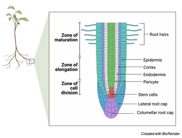
An excellent illustration is the increase in height of a tree. Procambium, protoderm, ground meristems, and procambium, three types of primary meristems, are all developed by each apical meristem. The cells that will become the primary tissues will be produced by these primary meristems.
Lateral Meristems
Plants’ secondary growth is controlled by lateral meristems. Horizontal growth is the most common type of secondary growth. An excellent illustration is the increase in girth of a tree trunk. In the study of plants, there are two types of lateral meristems to be aware of.
The first form of lateral meristem, the vascular cambium, is also known as the cambium. Except at the tips, where the apical meristems are located, the cambium is a thin, branching cylinder that covers the length of the roots and stems of most perennial plants and many herbaceous annuals. The cambium is in charge of producing cells and tissues that increase the plant’s thickness, or girth.
The cork cambium, the second kind of lateral meristem, is a narrow cylinder that runs the length of roots and stems, similar to the vascular cambium. The distinction is that it can only be found in woody plants since it is responsible for producing the outer bark.
Only until the main tissues produced by the apical meristems have begun to mature, will the vascular and cork cambiums, if present, begin to create cells and tissues.
Intercalary Meristems
Because they do not expand in girth, intercalary meristems are seen in grasses and similar plants that do not have a vascular or cork cambium. These plants have apical meristems and the third kind of meristematic tissue at regions of leaf attachment, termed nodes. This meristem is also important for lengthening since it actively produces new cells. The regeneration of cut grass is controlled by the intercalary meristem.
Plant tissues that do not actively generate new cells are known as non-proliferative tissues. Nonmeristematic tissues are the name for these types of tissues. Nonmeristematic tissues are made up of cells produced by meristems and come in a variety of shapes and sizes depending on their function in the plant. Sometimes the tissues are made up entirely of the same cell type, while other times they are mixed. There are simple and complicated tissues to examine, but for the sake of discussion, we’ll start with the basic tissues.
Simple Tissues
(1) parenchyma tissue, (2) collenchyma tissue, and (3) sclerenchyma tissue are the three main kinds of tissue called after the type of cell that makes up its composition.
i. Parenchyma Tissue
Parenchyma tissue is made up of parenchyma cells. Parenchyma cells are the most common cell type in higher plants, and they may be found in nearly every major organ. When these cells are originally created, they are essentially sphere-shaped. These cells, on the other hand, have thin walls that flatten at the points of contact when a large number of them are packed together. They usually have a lot of sides, with the majority having 14 sides. These cells may store a variety of secretions such as starch, lipids, tannins, and crystals. The tissues present in leaves are formed by parenchyma cells with numerous chloroplasts. Chlorenchyma is the name of this kind of tissue.
Photosynthesis is the primary function of this type of tissue, whereas parenchyma tissues lacking chloroplasts are often utilised for food or water storage. Also known as aerenchyma tissue, certain clusters of cells are loosely packed together with connecting air gaps, such as in water lilies. These cells can also grow irregular inner wall expansions, which enhances the plasma membrane’s overall surface area and promotes the transfer of dissolved chemicals between neighbouring cells. If parenchyma cells are mature, they may divide, which is important for healing damage to plant tissues. The majority of the edible parts of fruit are made up of parenchyma cells and tissues.
ii. Collenchyma Tissue
Collenchyma tissue is made up of collenchyma cells. These cells, like parenchyma cells, have living protoplasm and may exist for lengthy periods of time. Their walls are thicker than those of parenchyma cells, which is their major differentiating feature. The walls appear irregular in cross-section. Collenchyma cells are located immediately beneath the epidermis, and they are typically elongated with flexible as well as robust walls. These cells, together with the tissues they produce, offer flexible support for organs like leaves and flower parts as a plant grows. Celery‘strings’ that get trapped in our teeth are a good example of collenchyma plant cells.
iii. Sclerenchyma Tissue
Sclerenchyma tissue is made up of sclerenchyma cells. These cells contain lignin-embedded secondary walls that are thick and strong. Most sclerenchyma cells are dead by the time they reach maturity, but they nevertheless provide structure and support. Sclerenchyma cells come in two varieties:
a) Sclereids are sclerenchyma cells that are dispersed throughout the body at random. They are sometimes clustered together in distinct zones or areas inside other tissues. They’re usually twice as long as they’re wide. The rough feel of some varieties of pears is one example. The grittiness is caused by sclereid cell clusters. Stone cells are another name for scleereids.
b) Fibers can be found in roots, stems, leaves, and fruits in connection with a wide range of tissues. Fiber cells are often longer than they are wide, with a very small hollow in the centre. Textiles, ropes, strings, and canvas items, to mention a few, are now made from fibres from over 40 different plant families.
Secretory Cells and Tissues
Substances that collect within the cell as a result of biological activities can occasionally harm the protoplasm. As a result, it is critical that these components be separated from the protoplasm from whence they arise, or that they be transported outside of the plant body. Although the majority of these chemicals are waste products, some are necessary for plant function. Citrus oils, pine resin, latex, opium, nectar, fragrances, and plant hormones are among examples. Secretory cells are formed from parenchyma cells and can operate as a tissue or on their own. They might have a lot of commercial value.
Complex Tissue
The term “complex tissue” refers to tissues that have more than one cell type. The fundamental tasks of xylem and phloem, the two most essential complex tissues in a plant, are to transport water, ions, and soluble food items throughout the plant. While apical meristems create some complex tissues, the vascular cambium produces the majority of them in woody plants, and is frequently referred to as vascular tissue.
The epidermis and periderm are two more complicated tissues. All plant organs are protected by the epidermis, which is made up largely of parenchyma-like cells. Specialized cells that enable water and gases to flow in and out of the plant, secretory glands, different hairs, cells in which crystals are collected and separated, and other cells that improve absorption in the roots are all found in the epidermis. The outer bark of woody plants is formed by the periderm, which is mainly cork cells. Because of the pockets of parenchyma cells distributed throughout, it is considered a complex tissue.
Xylem
The xylem is an important plant tissue since it is part of the plant’s “plumbing.” Consider the main axis of stems and roots as a network of pipes. It is made up of parenchyma cells, fibres, vessels, tracheids, and ray cells and transports water and dissolved chemicals throughout the body.
The vessels are long tubes made up of individual cells, with open ends on both ends. There may be bars of wall material stretching across the open space on the inside. From end to end, these cells are connected to form lengthy tubes. Vessel members and tracheids die when they reach adulthood. Tracheids have thick secondary cell walls that taper towards the ends. They don’t have the same end holes as the vessels.
The tracheids’ ends overlap, and there are two pits on each side. Water can flow from cell to cell through the pit pairs. While most xylem conduction is up and down, there is some lateral or side-to-side transmission through rays. Rays are long-lived parenchyma cells that emerge from the vascular cambium in horizontal rows. Rays extend out from the centre of the stems and roots of trees and other woody plants, resembling the spokes of a wheel in cross-section.
Phloem
Phloem is an equally essential plant tissue since it is a part of the plant’s “plumbing.” Phloem is primarily responsible for transporting dissolved food molecules throughout the plant. The sieve-tube component and companion cells in this conduction system do not have secondary walls. Both xylem and phloem are produced by the vascular cambium’s parent cells. Fibers, parenchyma, and ray cells are generally included.
Sieve tubes are made up of end-to-end sieve tube components. Unlike vessel members in xylem, end walls do not have apertures. The end walls, on the other hand, are lined with tiny holes through which cytoplasm flows from cell to cell. Sieve plates are the name of these porous connectors.
Sieve-tube members do not have nuclei at maturity, despite the fact that their cytoplasm is actively involved in the conduction of food items. It is the companion cells that are nestled between the sieve-tube members that are responsible for food conduction in some ways. A polymer called callose is found in living sieve-tube members. As long as the cell contents are under pressure, the callose remains in solution.
If an insect injures a cell and the pressure decreases, the callose will form as a healing mechanism. The callose and phloem protein, on the other hand, will be transported to the closest sieve plate, where they will form a clog. This stops additional sieve tube contents from leaking, and the injury isn’t always deadly to the plant’s total turgor pressure.
Epidermis
The epidermis is a fascinating plant tissue that is both complicated and intriguing. The epidermis is the outermost layer of cells on all plant organs, according to official definitions (roots, stems, leaves). Because the epidermis is in close touch with the environment, it is subject to its circumstances and limitations. The epidermis is usually one cell layer thick, although there are certain outliers, such as tropical plants, where the layer can be many cells thick and serve as a sponge.
The cuticle is a waxy protective coating formed by cutin, a fatty substance produced by most epidermal cells. One of the primary determinants of how much water is lost by evaporation is the thickness of the cuticle. The cuticle also provides some resistance to bacteria and other disease organisms at no additional cost. The wax palm, for example, produces enough cuticle to be commercially valuable: carnauba wax.
Polishes, candles, and even phonographic recordings are made from other wax materials. The absorptive surface area of root hairs is increased by epidermal cells. Root hairs are tubular extensions of the main root body that are completely made up of epidermal cells. Leaves are not ignored. They feature a lot of tiny pores called stomata that are bordered by guard cells, which are specialised epidermal cells. Because they have a distinctive form and contain chloroplasts, guard cells are distinct epidermal cells. Other modified epidermal cells, such as glands or hairs, may be used to repel insects or minimise water loss.
Periderm
The epidermis is sloughed off and replaced by a periderm in woody plants as the cork cambium begins to develop new tissues to increase the girth of the stem or root. The periderm is made up of cork cells that are semi-rectangular and boxlike. The outermost layer of bark will be this. When these cells reach maturity, they are no longer alive. The protoplasm, however, secretes a fatty material called suberin into the cell walls before the cells die. Suberin protects the tissues beneath the bark by making the cork cells waterproof. The cork cambium produces pockets of loosely packed cork cells in some areas.
Suberin is not incorporated into the cell walls of these cork cells. Lenticels are loose patches that extend through the periderm surface. Lenticels are responsible for gas exchange between the air and the inside of the stem. Lenticels are found at the bottom of deep cracks in tree bark.

Plant Tissue Citations
- Ice-cap. A high-throughput method for capturing plant tissue samples for genotype analysis. Plant Physiol . 2004 Jul;135(3):1162-9.
- Ups and downs of tissue and planar polarity in plants. Bioessays . 2004 Jul;26(7):719-29.
- The lipids of plant tissue cultures . Adv Lipid Res . 1976;14:171-211.
- Rice virus release from the planthopper salivary gland is independent of plant tissue recognition by the stylet. Pest Manag Sci . 2020 Sep;76(9):3208-3216.
Share