Primary cultures denote those cultures which consist of cells before subculturing.
It represents the mixture of cell types present in a given tissue and hence they are closer to the tissue architecture in vivo.
In primary cultures the tissue pieces can be grown as explants cultures or tissues can be subjected to enzyme treatment and dissociated cells are grown as monolayers.
Distinguishing Features of Organ Cell Culture
Architecture of tissues in vivo is retained.
Their functionality is also retained, since they maintain their differentiated character.
The tissue is cultured at the liquid-gas interface.
Grids, gels and sponge matrix which maintain 3D structure of organs are used as substrates.
Attempts are made to deliberately minimize proliferation and migration of cells.
Advantages of Organ Cell Culture
Organ cultures are used for studies involving cell interactions, functional properties of cells, effects of external agents or effects of products of one organ on another.
Characterization of these cultures can be easily carried out from histological studies.
Biochemical differentiation is also possible.
Disadvantages of Organ Cell Culture
1. Organ cultures cannot be propagated for long periods.
2. Each experiment requires a new culture.
3. Quantitation of results is very difficult.
4. High inter-sample variation is required for reproducibility and homogeneity of results.
Requirements of Organ Cell Culture
Unsterile-fertilized chick eggs incubated for 10 days.
Sterile-surgical instruments, 65 mm pertidishes, PBS-A with antibiotics, 35 mm petridishes, RPMI 1640 with 20% FCS, trypsin 50 ml conical flasks, magnetic bars.
Organ Cell Culture Procedure
1. Wipe precleaned eggs with savlon and iodine.
2. Hold the egg with broad side up and tap the shell gently with scalpel to crack it uniformly all around.
3. With a sterile forceps gently remove the shell pieces and displace the CAM to expose the embryo.
4. Lift up the embryo by inserting one arm of a blunt forceps below the neck of the embryo and transfer it to a petridish with PBS-A.
5. Wash the embryo twice with PBS-A, decapitate and wash again twice.
6. Dissect out all the visceral organs wash the remaining parts of the embryo with PBS-A, mince it with sharp scissors and transfer to a 50 ml conical flask.
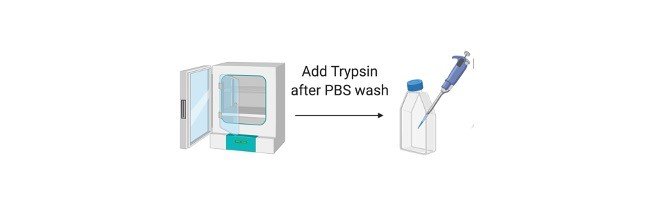
7. Add 10-15 ml trypsin, put a magnetic bar in the flask and keep the flask on the magnetic stirrer with a gentle speed to avoid frothing for 30 mins.
8. Remove the flask from the stirrer, allow the pieces to settle, transfer the supernantant to a centrifuge tube and centrifuge for 10 mins at 1500 rpm.
9. If necessary, repeat the step of trypsinization once or twice more.
10. Pool all the cell suspensions, make a cell count and add about 1 X 10^5 cells/ml in 35 mm petri dishes and incubate in the CO2 incubator.
Observation: Within 24 hrs cells form monolayer and show semi confluency in 4-5 days.