Top 15 Best Statistical Software Free in 2022 (Statistics and Data Analysis)
What Are Statistical Software?
Statistical software, or statistical analysis software, refers to tools that assist in the statistics-based collection and analysis of data to provide science-based insights into patterns and trends. They often use statistical analysis theorems and methodologies, such as regression analysis and time series analysis to perform data science.
List of Top 15 Best Statistical Software Free in 2022 (Statistics and Data Analysis)
(1) SAS® software
Whether you’re a professor, teacher, student or independent learner, you can get easy access to powerful SAS software via the cloud. Setup is easy, too. After you get set up, a broadband internet connection is all you’ll need to run the best analytics software in the world.
PSPP is a stable and reliable application. It can perform descriptive statistics, T-tests, anova, linear and logistic regression, measures of association, cluster analysis, reliability and factor analysis, non-parametric tests and more. Its backend is designed to perform its analyses as fast as possible, regardless of the size of the input data. You can use PSPP with its graphical interface or the more traditional syntax commands.
The Statistical Lab is an explorative and interactive tool designed both to support education in statistics and provide a tool for the simulation and solution of statistical problems. The graphical user interface is designed to make complex statistical relations easy to understand. It connects and displays data frames, frequency tables, random numbers or matrixes in a user-friendly statistical worksheet allowing users to run calculations, conduct analyses and perform multiple simulations and manipulations.
Statistical software for fast and easy interpretation of experimental data in science and R&D in a technical environment. This statistical package helps with analysis and prevents making false assumptions. In short it makes statistics faster and easier, suitable for less experience users but advanced enough for more demanding users.
IBM® SPSS® Statistics is a powerful statistical software platform. It offers a user-friendly interface and a robust set of features that lets your organization quickly extract actionable insights from your data. Advanced statistical procedures help ensure high accuracy and quality decision making. All facets of the analytics lifecycle are included, from data preparation and management to analysis and reporting.
Fast. Accurate. Easy to use. Stata is a complete, integrated software package that provides all your data science needs—data manipulation, visualization, statistics, and automated reporting.
(7) Minitab: Data Analysis, Statistical & Process Improvement Tools
Data is everywhere, but are you truly taking advantage of yours? Minitab Statistical Software can look at current and past data to discover trends, find and predict patterns, uncover hidden relationships between variables, and create stunning visualizations to tackle even the most daunting challenges and opportunities. With powerful statistics, industry-leading data analytics, and dynamic visualizations on your side, the possibilities are endless.
A versatile statistics tool purpose-built for scientists-not statisticians. Get a head start by entering data into tables that are structured for scientific research and guide you to statistical analyses that streamline your research workflow. No coding required.
DataMelt is a free software for numeric computation, mathematics, statistics, symbolic calculations, data analysis and data visualization. This multiplatform program combines the simplicity of scripting languages, such as Python, Ruby, Groovy (and others), with the power of hundreds of Java packages.
Powerful mathematics-oriented syntax with built-in 2D/3D plotting and visualization tools. Free software, runs on GNU/Linux, macOS, BSD, and Microsoft Windows. Drop-in compatible with many Matlab scripts
SOFA is a user-friendly statistics, analysis, & reporting program. It is free, with an emphasis on ease of use, learn as you go, and beautiful output. SOFA lets you display results in an attractive format ready to share. And SOFA will help you learn as you go.
Dataplot® is a free, public-domain, multi-platform (Unix/Linux, MacOS, Windows 7/8/10) software system for scientific visualization, statistical analysis, and non-linear modeling. The target Dataplot user is the researcher and analyst engaged in the characterization, modeling, visualization, analysis, monitoring, and optimization of scientific and engineering processes.
SciPy is a set of open source (BSD licensed) scientific and numerical tools for Python. It currently supports special functions, integration, ordinary differential equation (ODE) solvers, gradient optimization, parallel programming tools, an expression-to-C++ compiler for fast execution, and others. A good rule of thumb is that if it’s covered in a general textbook on numerical computing (for example, the well-known Numerical Recipes series), it’s probably implemented in SciPy.
Zelig is an easy-to-use, free, open source, general purpose statistics program for estimating, interpreting, and presenting results from any statistical method. Zelig turns the power of R, with thousands of open source packages — but with free ranging syntax, diverse examples, and documentation written for different audiences — into the same three commands and consistent documentation for every method.
MacAnova is a free, open source, interactive statistical analysis program for Windows, Macintosh, and Linux written by Gary W. Oehlert and Christopher Bingham, both of the School of Statistics, University of Minnesota. In spite of its name, MacAnova is not just for Macintosh computers and not just for doing Analysis of Variance.
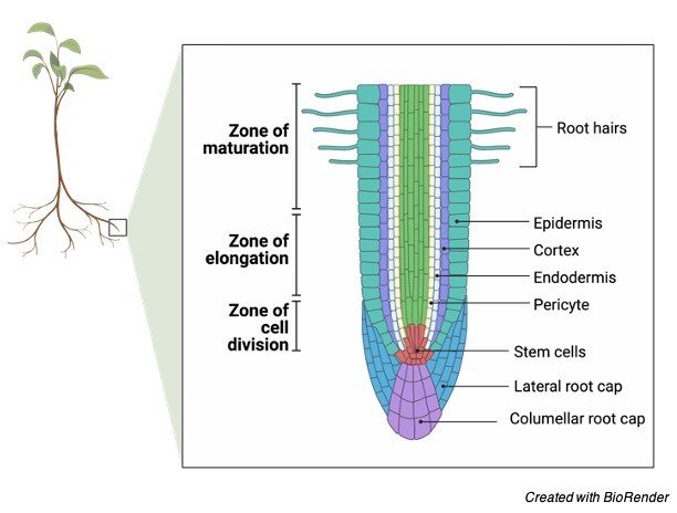
The area of tissue from which new growths are formed in plants is called meristem. The cells at meristem continually proliferate and do not differentiate. The new leaves arise from the meristem tissue, which depends upon the signals received by it. It also gives rise to flowers or roots of the plant. A plant cannot produce new cells without the meristem.
Apical Meristem Function
The apical meristem is responsible for the growth of the plant and its length and height. The location of apical meristem is at the ends of roots, known as root apical meristem, or at the tops of shoots, which are known as shoot apical meristem.
This meristem is responsible for the primary growth of the plant. The vertical growth is promoted by the axillary buds, which are exerted by the presence of an apical bud.
Shoot Apical Meristem Function
The shoot apical meristem is developed to become one of three primary meristems that are the protoderm, ground meristem, and procambium. The shoot apical meristem is mainly composed of undifferentiated cells found above the ground.
The epidermal tissues of the plant are formed by the protoderm while the ground develops into the cortex and pith of the plant. And the xylem and phloem, collectively known as vascular bundles are formed by the procambium.
The leaves of the plant are also included in the shoot that grows from the sides of the apical meristem. A bump or an axillary bud is formed by the beginning growth of the leaf at the node.
The axillary bud remains dormant if the terminal bud is in close proximity to it. However, the increased distance or removal of the terminal bud results in the disappeared or diminished exerting apical dominance.
It allows the growth of leaves at the lateral buds of the apical meristem. The shoot apical meristem converts into the inflorescence meristem when the angiosperm plant becomes ready to bloom and emerge various flower parts such as petals, sepals, stamens, carpels, etc.
Root Apical Meristem
It is found below the ground. The root apical meristem is responsible for the growth and development of a plant’s root. The root apical meristems have the ability to yield two types of tissues at the same time and can produce cells in a bilateral direction.
Among these two types of tissues, one typically comprises the main roots of the plant and the other type consists of a root cap, the main roots are responsible for continuous growth and supply proliferative, undifferentiated cells for continued growth while the root cap protects the apical meristem.
As the root cap grows deeper into the soil, the cells of the root cap are continuously being shed and replaced by new cells, which are provided by the main root. This is typical of the taproot.
The lateral growth of the root is conducted by the lateral root meristem, which helps in efficient water supply and nutrient absorption in the plant. It also helps in nutrient storage and stability for aerial growth.
Basal Meristem Function
Basal meristem is also called intercalary meristem. It is located between mature, differentiated tissues. The intercalary meristem is distinctly different from the apical meristem, which is located relatively near an apical meristem.
The vertical growth of the plant is promoted by the apical meristem and the intercalary meristem works independently of the apical meristem. However, it works in the base of the plant, not at the tip of the plant tip. The grasses and other plants can grow continuously after being cut due to the intercalary meristem.
Lateral Meristem Function
The vertical growth of the plant is controlled by the apical meristem, similarly, the lateral meristem is responsible for the lateral growth of the plant. Lateral growth is also known as secondary growth.
The lateral growth is conducted around an already established stem thus it is known as secondary growth. There are two types of lateral meristem in woody plants, named the vascular cambium and the cork cambium.
The vascular cambium is similar to the procambium of the apical meristem, which is responsible for the development of wood and increased width of the plant. The periderm is developed from the cork cambium, similar to the protoderm.
The primary epidermis growth is produced from the protoderm. And the epidermis is replaced by periderm to produce bark, which acts as a shield and protects the plant from physical damage and water loss by a waxy layer of suberin.
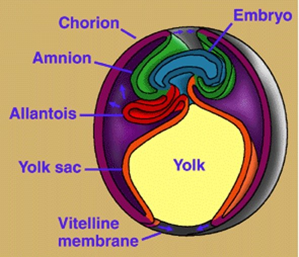
The vertebrates which have a fetal tissue known as the amnion are called amniotes. A membrane that surrounds the fetus and is derived from the fetal tissue is called the amnion. It protects the fetus and can be found within the egg, or can simply enclose the fetus. For instance, lizards and birds, it found within the egg or enclose the fetus within the uterus.
Amniotes Description
Most of the vertebrates, excluding fish and amphibians are amniotes. Amphibians and fishes are known as amniotes, which means “without an amnion”. The species often lay their eggs in water to protect them from any damage.
Image Credit: https://www2.gwu.edu/
Most amniotes are terrestrial animals thus they have to protect their fetus under the weight of gravity. The amnion protects the developing fetus. Whales are the only exceptions of amniotes because they are aquatic animals.
It may be due to the development of amnion in whales before their move to the sea. Some marine turtles also live in the sea but return to land to lay their eggs.
Characteristic of Amniotes
The fetus of amniotes is surrounded by three membranes, which are the amnion, the top chorion layer, and the waste-absorbing allantois. In general, the amniotes share several characteristics such as they all are developed from a common ancestor and the ancestor has developed the amnion character.
Most of the egg-laying species consist of the amnion. For example, the birds and reptiles, and some mammals consist of this protective layer. However, the shell is not present in the human egg but in many ways, the development of the human egg is similar to the chicken eggs.
Animals Considered Amniotes
Sauropsid Amniotes
Amniotes are divided into two divisions, the sauropsid amniotes, and the synapsid amniotes. The reptiles and birds are included in the sauropsid amniotes. Formally, many different groups are included in this group, but they are separated from the synapsids because they share many derived characteristics.
However, both the groups are evolved around the same time from a common ancestor. The ancestor was likely not terrestrial. After the evolution of both sauropsids and synapsids, both had to make some adaptations to sustain themselves in terrestrial environments.
The heart, lungs, and kidneys reflect the differences mainly. The lungs of synapsids differ from the lungs of sauropsids that have foveolar lungs. The small chambers present in foveolar lungs open to a common space.
The heart of sauropsids lacks the ventricle and some sauropsids have almost 4 chambered hearts. The process for waste excretion is also different in the sauropsid amniotes than the synapsids.
Sauropsids excrete uric acid. Much of the water can be absorbed in the cloaca in which the substance precipitates out of the urine. Thus, uric acid become a more water-efficient way of excreting nitrogenous wastes than the synapsid method. The turtles, lizards, crocodiles, and birds, which lay eggs are included in sauropsids.
Synapsid amniotes
The sauropsids are separated from the synapsid amniotes since millions of years ago, but they have slight differences. They excrete all urea as nitrogenous waste. Urea is concentrated in the synapsid kidney and excreted with a small amount of water.
The strategy is much more efficient than the excretion of straight ammonia but it is not water-efficient as uric acid. Ammonia is usually excreted by fish and amphibians. Synapsids consist of 4 chambered hearts, which have a partition between ventricles.
Thus, the efficiency of oxygenating the blood is improved due to separate paths for blood. The structure of the lungs is also different in synapsids than the sauropsids. They consist of an alveolar lung which has many branches of the trachea instead of small pockets from a central chamber.
The branches end at an alveolar sac. Synapsids consist of only three extant groups, all of them are mammals. However, the amniotic sacs are present in all of these amniotes but they reproduce by different methods.
For instance, the monotremes lay eggs in nests and after the eggs hatch, they feed them milk like other mammals. The marsupials are considered as a median between the monotremes and the placental mammals.
In the marsupials, the young are developed in the uterus but born at a very early age. They must climb along with the mother into the marsupial pouch, where the mother feeds them milk for their proper development.
The rest of the synapsid amniotes are represented by the placental mammals. In these animals, a placenta or oxygen and nutrients passing maternal membrane are used to feed the offspring within the womb.
They produce the largest offspring among all amniotes but they have fewer offspring compared to sauropsid amniotes.
Evolution of Amniotes
The evolution of amniotes may occur when first terrestrial animals are venturing onto land. The two main groups of amniotes were divided due to the much different terrestrial environment.
The division may occur around 400 million years ago, in the Devonian period. Later, various considerable differences have evolved between these groups. As discussed above, they differ in their anatomy and physiology.
At the time, they also diversified due to the new terrestrial environment which provided them several new niches for the animals to fill. An organism named parieasaur, a cow-sized organism could have been a common ancestor of modern amniotes.
It is a large reptile-looking organism from the Devonian period that may have primitive hearts, lungs, and kidneys and likely had an amnion. Thus it is likely one of the first amniotes.
The set of glands between the bladder and the penis is called the prostate gland. A component of seminal fluid, called prostate fluid is secreted by this gland, which helps sperm cells swim. Skene’s gland is the female homolog of the prostate gland, but in this article, we only focus on the prostate gland.
Prostate Gland Description
The size of the prostate is about the size of a walnut (1.5 in). It is penetrated by the urethra that directs urine out of the body. An alkaline seminal fluid is expelled into the urethra by the prostate gland during ejaculation.
The alkaline nature of this fluid helps to counter the acidic environment of the vagina and protects sperm in the vaginal tract. The prostate gland also helps in sperm ejection.
Image Credit: https://www.cancer.org/
The function of the prostate gland is simple but when it becomes swollen, infected, or damaged by disease, it can cause pain and discomfort. The gland blocks the urethra and holds urine in the bladder when the gland swells.
The swollen prostate gland causes several problems such as pain during urination and an inability to empty the bladder at old age, which can lead to further complications if not corrected.
Prostate Gland Function
The primary function of the prostate gland is the secretion of prostate fluid. The prostate fluid is a seminal fluid, which ejects during ejaculation. The seminal fluid is ejected into the urethra with the help of prostate muscle.
The weight of the muscle is about twenty grams and surrounds the urethra just below the bladder. The drainage tubes are known as vas deferens shuttle sperms into the site of the prostate gland.
The proteolytic enzymes exit the prostate through ducts. Before ejaculating, the ducts open into the urethra. The prostate squeezes with the help of its surrounding, it closes the opening found between the urethra and bladder.
The fluid is directed into the urethra and pushes the semen out. Enzymes, zinc, and citric acid are present in the seminal fluid. The semen becomes slightly basic due to the fluid produced by seminal vesicles while the prostate fluid is slightly basic.
The basic nature of semen is to protect it and to prolong the lifespan of each sperm. An enzyme, called PSA or prostate-specific antigen is another element of prostate fluid. The function of this enzyme is to liquefy seminal fluid and to allow sperm to swim easily.
The levels of PSA in the patient’s blood can be measured by a blood test. A high level of prostate fluid increases the risk of prostate cancer. However, the swollen prostate gland can cause many difficulties but its site makes it a great location for delivering fluid.
Prostate Gland Location
In males, the prostate gland is the largest accessory gland. The location of the prostate gland is above the urethral sphincter and inferior to the neck of the bladder.
The prostate is located in front of the rectum, which makes it easy for physicians to conduct DRE. Digital rectal exams are conducted to inspect the health of the gland. The prostate is monitored by doctors for any signs of disease, such as swelling.
Prostate Gland Structure
There are four anatomical lobes in the prostate gland. But a histological parsing into zones is a more significant delineation.
• The central zone surrounds the ejaculation dust. This zone is derived from the Wolffian duct located in human embryos.
• The transitional zone is the area near the center, which surrounds the urethra. It is originated from the urogenital sinus.
• The peripheral zone forms the body of the prostate gland. The area originated from the urogenital sinus and is located toward the back.
The gland is well-innervated and notably vascular. The prostate gets the supply of oxygenated blood from the prostatic arteries and the oxygen-depleted blood drains into the prostatic venous plexus and enters the iliac veins of the pelvis.
The prostate gland innervates in a little simpler manner and is innervated by the inferior hypogastric nerve plexus. A bundle of nerves, which innervates the smooth muscle of the prostate is called a plexus.
Prostate Gland Pathology
The urination could be blocked by a swollen prostate by pressing against the urethra. This will irritate the bladder and the surrounding area. In older gentlemen, gland swelling or urinary discomfort occurs more frequently.
Benign prostatic hyperplasia (BPH) or the enlargement of the prostate gland is very common in old men and up to half of the men over sixty suffer from this. Between the ages of seventy and eighty, this statistic jumps to ninety.
Frequent urination and leaking are the symptoms of this affliction, which can be treated by alpha-blockers to relax the muscles around the urethra. 5-alpha reductase inhibitors also help in reducing the levels of DHT testosterone and shrink the prostate.
Another condition where the prostate tissue is inflamed by an infection is called prostatitis, which can be treated by antibiotics. All prostate growth is not benign. According to the National Cancer Institute, over 200,000 men are diagnosed with prostate cancer each year.
The five-year survival rate for prostate patients is only 29 percent, which means that under 30,000 deaths are caused by this disease each year. In America, prostate cancer is the third leading cause of death.
Prostate cancer can be treated by a combination of surgery, radiation, and chemotherapy. Sometimes, it becomes necessary to remove the prostate gland because it can lead to additional tissues.
Aix galericulata or the Mandarin duck is also known as yuan-yang in Chinese. The bird is native to China, Japan, and Siberia and closely related to the North American wood duck. Mandarin duck is a beautiful bird where the male is more colorful than the female and has quite striking plumage like most other duck species.
The male mandarin duck has a red/ orange face with a white crescent above his eyes. It consists red bill and the rest of his plumage is red, orange, brown, purple, and green colored.
The male also consists of a sail of feathers in his back during the breeding season. While female ducks have brown and grey feathers and a light bill, which has a tinge of pink. Female also consist a white eye-ring.
Mandarin Duck Habitat and Nesting
The mandarin ducks live in pools, rivers, lakes, marshes, fast-flowing streams, and swamps surrounded by dense forests. The courtship rituals in mandarin ducks are much elaborated. At a beginning of the mating season, these ducks form pairs and look for a nest.
The female is accompanied by the male on this nest hunt. The nest of the ducks can be up to 32.8 feet above ground in a hole in a tree. Nine and twelve eggs laid by the female at a time. The eggs are incubated for up to four weeks.
The eggs hatch within a few hours of each other. The mother will coax them out of the nest by calling to them from the ground. Before the move to feeding grounds, the chicks will free fall to the ground from the nest.
The chicks are ready to fledge after 45 days after hatching and will fly off to join another flock. Habitat destruction is the main threat to the Mandarin duck populations. The predators include raccoons, minks, eagles, snakes, and otters.
Fun Facts About Mandarin Duck!
The fascinating plumage of Mandarin ducks makes them the most beautiful duck species. In China, Japan, and Korea, it is presented as a symbol of love and fidelity because they often form long bonds with their partner.
Mandarin ducks sometimes form pairs for life and usually, they pair up for several breeding seasons. These ducks also show several interesting biological concepts. Let’s understand and take a closer look at these concepts.
i. ZW Sex-determination System
Sex is determined by the presence of specific chromosomes in humans and other animal species. For instance, humans have an XY system of chromosomes in which the sex is determined by a pair of chromosomes in an individual.
The females have two homologous chromosomes (XX) while the males have two different types of chromosomes (XY). The Mandarin ducks have a ZW sex-determination system like most other birds. This system is different from the XY system in most mammals.
Besides birds, the ZW system is also observed in several species of fish, crustaceans, and some insects such as moths and butterflies. The ZW system of sex- determination is relatively widespread in nature.
During the incubation period, the male duck stays around the female while once the eggs hatch, he leaves the female to rear the chicks and he molts and sheds his colorful plumage. During this time, the male is unable to fly because he also sheds his primary flight feathers.
During this time, the male also resembles a female because his bright plumage is gone and only brown and grey color remains. Thus he blends with his environment and also hides from its predators.
iii. Dabbling Ducks
Dabbling ducks include eight genera and 50-60 species, which also includes Mandarin ducks. However, there are many differences between these ducks and other duck species but they are named after the way they feed.
These ducks usually live in shallow waters such as flooded fields and marshes. Instead of diving, they feed by tipping up in the water and their rear end out of the water with their head down.
Their feeding manner is defined as dabbling in which they move their bill around in the water. They have flat, broadbills and float high on the surface of the water. This way of feeding helps them to feed on small insects living on the surface of the water and to forage through floating algae and plants.
The legs of dabbling ducks are more central than the diving ducks that helping them to walk well on land thus they can also graze. The dabbling ducks are strong fliers and able to take a flight straight from the water, unlikea diving ducks.
Lovebirds are chunky, short-tailed birds belonging to the genus Agapornis. These birds are native to savannas of sub-Saharan Africa and Madagascar and live in forests. There is a total of nine species of lovebirds among which eight species are distributed across Africa.
And one species is called the grey-headed lovebird or Madagascar lovebird found in Madagascar. The species is the only lovebird, which is endemic to this island.
Lovebirds mostly occur in green colors like small parrots but some members also have orange, yellow, grey, black, or red colors on their heads and neck.
They have a prominent ring around their eyes and their beaks are relatively large and sharp. The rosy-faced lovebird is the largest species of lovebird, distributed from Angola to South Africa.
Lovebird Habitat
Lovebirds are social birds and always found in flocks, and forage together sometimes. They obtain their nutrition from seeds, fruits, and berries and are herbivores. Some species of lovebirds are specialists that only feed upon particular plant material whereas some other species are generalists and feed on whatever they can find.
The homes of lovebirds are generally made on tree holes, rocks, and shrubs. In some species, the members build their nests together while in other species they pair off and build a nest away from the flock.
The birds make pairs for their whole life and are monogamous thus they are named “lovebirds”. Courtship rituals are also seen in these birds in which males feed small pieces of food to the female, in some species males dance and sing to impress the female.
Around 4 to 6 eggs are laid by the female during the breeding season. The incubation period is of approximately 20 days after which the eggs hatch and both the parents take care of the chick for one month of age.
Lovebird Predators
Lanner falcons are one of the predators of lovebirds, who often seek refuge in thorny bushes to avoid capture. Besides this, habitat destruction and capture for the pet trade are some other threats to lovebirds.
Their bright colors, small size, and high energy makes them popular pets. If they are kept alone and bond with humans, they are known to be extremely affectionate.
Lovebirds are facing threats due to habitat destruction but besides this six species are listed as least concerned species and three species are listed as vulnerable and near threatened among which the black-cheeked lovebird is listed vulnerable and the Nyasa and Fischer’s lovebirds are listed as near threatened.
Fun Facts About Lovebird
There are several biological adaptations made by the lovebirds, which help them to live in their environment. They are the smallest species of parrots and can perform acrobatics in the air. Let’s understand some biological concepts about lovebirds!
i. Head movements during flying
Lovebirds have great flying skills and they are also able to maneuver quickly in dense areas. Researchers study their swift and agile flying skills by using high-speed cameras to film the birds.
With the help of the captured footage, they discovered that these birds can move their heads very quickly. Lovebirds can move their heads up to 270 degrees at a speed of 2700 degrees per second.
The speed is similar to insects and this swift movement is one of the fastest animal movements. Thus, their ability helps them to see in all the directions while flying in the air, which means that they get more time to see and react to the environment and helps to avoid collisions, enabling them to keep a clear line of sight during twisting and turning.
ii. Beak adaptations
The robust, hooked bill is the characteristic feature of lovebirds, which is also present in lovebirds. The bill looks very similar to the bill of owls and hawks from distance but actually, the bill of lovebird have the upper and lower mandibles sharper, more uniform curve.
The upper mandible of the lovebird’s beak has hooked and fits into the lower mandible. The bills of lovebirds are broad and powerful and thus enable them to crack seeds and tear them into fruits.
The beak has continuous growth because it is made of keratin. These birds nibble their beak on hard objects like branches to ensure that their beak has a perfect length and is sharp.
iii. Monogamy
In some parts of the animal kingdom, monogamy is a fascinating behavior. In some animals, when the offspring had much better chances of survival in presence of both the parents, monogamy was likely evolved.
Lovebirds can often be observed feeding and pruning each other which resembles their strong bond with each other. They form mating pairs for their whole life. Unlike other bird species, who make pairs for only one or more breeding seasons, lovebirds make bonds for up to 15 years or form mating pairs for life.
They are monogamous, which has various advantages. For instance, the birds can share parental responsibilities. After laying eggs, the female sits on her nest to incubate the eggs for approximately three weeks and the male brings her food.
After the eggs hatched, both the parents take care of their brood where usually males gather food while females stay in the nest. Thus in monogamous pair, one parent is always able to protect the chicks from predators.
Another advantage of monogamy is that they won’t have to waste their energy trying to find a mate after bonding. There is not any requirement and advantage to waste energy searching for a new mate at each breeding season if the bond between the birds is right
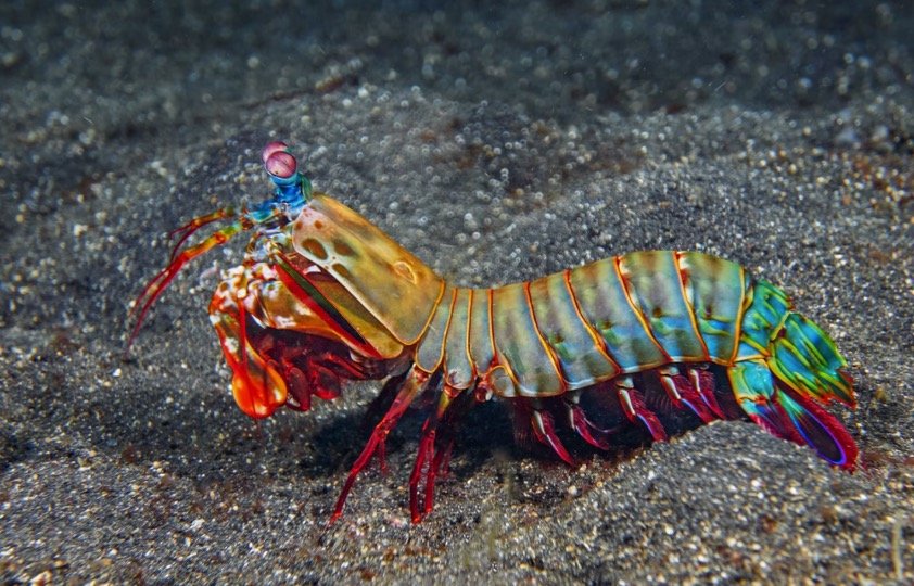
Mantis shrimp are an extremely fascinating group of shrimp species. They are true predators, and take nutrition from tiny organisms or scavenge on dead organisms. The mantis shrimps have modified their forelimbs to shaft or stun their prey. Based on their features, mantis shrimps are divided into subgroups.
The forelimbs are used as clubs by “smashers”. They can create forces unequaled in the animal world due to the massive power behind their modified forelimbs. The club can reach speeds of nearly 50 mph in a fraction of a second and over 1,500 newtons force created by this, which is similar to a 340-pound object falling directly onto the prey.
But, a sonic wave is also created by the acceleration of the club that smashes into the prey after the actual club and can cripple much larger prey. Comparatively, the sharp points on their limbs are used by “Spearers” that help them to impale small fish and other prey.
The speed and precision of the strike of Spearers are amazing. Unlike Smashers, the habitat of Spearers includes soft substrates such as sand. They wait for a fish to swim by and impale it at lightning speed.
The coloration of most mantis shrimp is very beautiful and striking, which helps them in communication and camouflage. The color of some species is very dark that helps them in blending with the seafloor.
The peacock mantis shrimp is very bright colored that helps it to scare off any predator. If the camouflage doesn’t work, many potential predators get injured or scared by their powerful strike.
Fun Facts About Mantis Shrimp
The mantis shrimp is not cool enough, but there are many interesting biological concepts related to these organisms. Let’s take a closer look.
i. Hunting Adaptations
We can see the adaptations made by crustaceans by the two different types of mantis shrimps- Smashers and Spearers. However, both types were evolved from a common ancestor nearly 200 million years ago, but the use of front limbs is quite different in both.
The direct force is used by the Smashers by their mantis-like front legs to strike their prey, on the other hand, Spearers modified their front legs into sharp barbs to impale and capture prey.
The prey is also different in both groups. Spearers usually prey upon soft, fleshy fish, while Smashers eat other crustaceans, snails, and oysters. The shells of these animals can be broken apart from their club-like limbs so they can get the nutrients inside.
Maybe the process of speciation in this group started by the difference in usage and prey type, which may have been the driving factor. Today, mantis shrimp represents 450 different species found in coastal regions all over the world.
ii. Compound Eyes
To become an incredible predator, mantis shrimps made another adaptation besides their weapon-like appendages. Like many arthropods, mantis shrimps consist of compound eyes. The function of these eyes is similar to human eyes, though the construction is very different.
The receptor cells of these eyes are right at the surface, instead of a single lens that funnels light onto a retina. The eyes of mantis shrimp are considered the most complex and functional eyes of any animal.
In humans, different colors and wavelengths of light can be detected by 3 types of cells, mantis shrimp have up to 16 different types of cells in their eyes. Due to this, several wavelengths that are far beyond human perception can be seen by mantis shrimp.
It can also detect both infrared and ultraviolet light, which cannot be seen by humans. It also provides a clue about the bright colors of mantis shrimp.
iii. Coloration as Communication
The coloration of many organisms helps them in communication. For instance, many insects including bees display “warning coloration”, which is recognized as dangerous by other animals. Similarly, peacocks and other animals use colorful coloration to attract mates.
Over time, females select more colorful mates, which leads to the birth of very colorful males and females. Mantis shrimp seem to be using a little of both. They use bright coloration to warn their potential predators.
However, according to some researches, mantis shrimps use bright coloration to attract mates and communicate with other members. Some species signal to their mates and other shrimp invading their space by using fluorescence.
The mantis shrimp can see patterns and colors of light, which is not visible to the human eye due to their amazing compound eyes. However, the exact mechanism and process of communication via colorful signals is not fully understood.
The reason behind this is the need for complex scientific instruments to measure what light is displayed by the mantis shrimp and how they process light signals.
The slowing down of enzyme-catalyzed chemical reactions is called allosteric inhibition. The proper functioning and maintenance of our bodies’ equilibrium are done by these metabolic processes and the process is regulated by allosteric inhibition.
All the important molecules are broken and built up by these metabolic processes. From the digestion of food to the repairing of our muscles, the metabolic processes are fundamental for everything.
Lock and Key: Substrate Binds to Enzyme at the Active Site
A series of chemical reactions consist of metabolic processes, which produce end products. Enzymes are the key drivers of metabolic processes. The reactions are catalyzed by these specific proteins. These enzymes reduce the amount of required energy and speed up important chemical reactions.
First, an enzyme bind to a substrate. A product is then created by this reaction. At the next metabolic step, the product serves as a subsequent substrate for a different enzyme. Finally, until a final product is created at the end, the chain of reactions occurs.
The interesting fact is that an enzyme binds to a specific substrate. Thus the enzyme can be compared to a lock and the substrate is compared with the key. A specific enzyme only binds with a specific substrate. The location, where an enzyme binds to a substrate is called the “active site”.
The wrong key will not fit the specific lock on an enzyme. The enzyme cannot catalyze a reaction if the substrate cannot fit into an active site. Even if the substrate is correct for the specific enzyme, an allosteric inhibitor can prevent the enzyme from having the correct conformation.
Allosteric Inhibition and Enzymatic Activity
Allosteric inhibition is used to control the speed of metabolic reactions. They deactivate the enzyme and thus slow down the enzymatic activity. A molecule that binds to the enzyme at an allosteric site is called an allosteric inhibitor. This site is located at a different location from the active site.
The enzyme changes its 3D shape after binding with the inhibitor. Allosteric inhibition is a form of noncompetitive inhibition. It means that at the active site, the inhibitor does not compete directly with the substrate but indirectly changes the composition of the enzyme.
The enzyme becomes inactive after changing its shape and cannot bind with its corresponding substrate. The formation of subsequent products will be slowed down by this.
The allosteric inhibitor can be compared with a locksmith. The lock (enzyme) is changed by the locksmith (allosteric inhibitor) so that the key (substrate) will no longer be able to open the lock (enzyme).
Allosteric Inhibition Prevents the Over-accumulation of Products
Allosteric inhibitors prevent the body from creating unnecessary products and wasting energy. If a metabolic pathway is compared with an assembly line at a factory, a machine alters the products at each station in the assembly line before passing it to the nest station.
Then the intermediate products are moved from station to station by the assembly line until they get the final product at the end. For example, pants are produced in this factory. At the first station, a pair of pants are cut from raw material by a machine, then the hems of the pants are stitched together by a machine at the second station.
At the third station, the zippers are attached by a machine, and finally, at the fourth station, the products (pants) are tagged and shipped in the shipment pile. The assembly line is smooth and there is no hold-up when the machines are working properly and stations will produce products at similar rates.
However, if the machine at the third station breaks down that attaches the zippers, there is now a hold-up. We must stop the first two stations of the supply to prevent products from piling up.
In this way, the supply and demand for each intermediate product are controlled and it is ensured that they are equal at each station. Similarly, the chain of reactions can be slowed down by allosteric inhibition. In this way, the over-accumulation of unnecessary products is prevented.
Examples of Allosteric inhibition
ATP in cellular respiration is an example of an allosteric inhibitor. This metabolic process is operated as a feedback loop. The speed of upstream reactions is controlled by downstream products. Phosphofructokinase is an enzyme involved in glycolysis. It converts ADP into ATP.
The ATP serves as an allosteric inhibitor when there is too much ATP in the system. The ATP slows down the conversion of ADP by binding with phosphofructokinase. Thus, ATP prevents its unnecessary production by itself because when there are already adequate amounts of ATP, it is no need to produce more ATP.
The antibiotic penicillin, which is an important drug acts as an allosteric inhibitor. Penicillin has saved millions of lives by helping the body to kill harmful bacteria. The enzyme DD-transpeptidase helps the harmful bacteria to create strong cell walls.
Penicillin binds to this enzyme and counteracts this process. Penicillin inhibits the formation of bacterial cell walls by acting as an inhibitor. The surrounding fluids of the bacterial cell can then push itself in through osmosis with a weak wall and the cell burst and die.
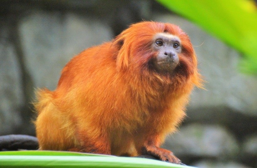
The tamarin is a small monkey that belongs to the genus Saguinus. The genus comprises 13 species that are native to New World. The tamarins are distributed in the jungles of the Amazon basin and other parts of Central and South America and live in the treetops.
Most of the species of tamarin are endangered in their jungle habitat. Appearance The most notorious species of tamarin include the golden lion tamarin (S. Ursula), which has an impressive golden-brown mane of fur similar to those of male lions. The tail is relatively long and its face is small, black.
Other tamarin species are black with some interesting mustache-like features. The weight of 13 species ranges from 7.8-31.7 oz and their length ranges from 5.1- 11.8, not including their weights.
Tamarin Diet and Predators
The range of tamarin occurs in tropical rainforests and lives in trees. They rest on hollow trees at night and during the day, they forage for food by climbing branch to branch. Their long fingers help them in snagging insects, fruits, and small lizards and birds.
Tamarins are small primates and have several predators such as snakes prey on them and their young and wild cats, some species of birds also feed on tamarin. To avoid predation, they move swiftly in the forest canopy at high speed. They hide in tree hollows at night to avoid predation.
Social Structure and Breeding of Tamarin
Tamarins are social animals and live in family groups of up to 40 individuals. However, a typical family group consists of about 10 members. The males are also involved in parental care and also carry their offspring around between feedings.
The gestation period of around 140 days after that the female gives birth to two young, which are usually twins. The new monkeys are raised by the males and other juveniles in the family, they often carry for them throughout the day but when required, return them to their mothers.
New tamarin starts eating solid food at the age of one month. They reach full maturity at the age of two years and become an independent member of the family. The life span of tamarin is about 18 years, although some species live closer to about 10 years.
Conservation Status of Tamarin
The Amazon rainforest basin is the major habitat of tamarin and also one of the most biodiverse areas in the world. Amazon basin is also considered as one of the most threatened areas in the world.
The tamarin and thousands of other species are continuing to strip from their natural habitats due to deforestation and the extension of agricultural land.
The IUCN red list for threatened species listed the golden lion tamarin as endangered species after the improvement from its previous critically endangered status.
Fun Facts About Tamarin
The habitat of tamarin includes one of the highly important and rare parts of the world. The area is relatively small and productive, which provided habitat to many closely related species. The tamarin show many interesting biological concepts to learn about it.
i. Difficult to Categorize
The taxonomy of tamarin is not straightforward. Originally, there are ten species in the genus Saguinus. Further, based on their unique facial hair patterns, they are divided into ‘morphotypes’.
Later, they are classified based on dental measurements and separated into two clades, and eventually, the genus consists of 15 species and no subspecies. A genetic analysis was conducted by the researcher in 2016 on the genus and other primates.
This study suggests that some members of the genus have emerged from other members about 10 million years ago. The members include the saddle-back tamarin but they emerged earlier than other monkeys and marmosets of genera Callithrix, Cebuella, and Mico thus the saddle-back tamarin was moved to genus Leontocebus.
ii. The Golden Touch
A beautiful color morph has evolved by the red-handed tamarin, which is quite interesting. The species is native to wooded areas north of the Amazon River and is well known for its conspicuous golden hair. It contains golden hair on hand which is in contrast to its black fur. The red-handed tamarin also has some gold-colored flecking on the anterior side and rump.
iii. Fierce Competition
Some species of tamarin are relocating from their natural habitat due to being threatened and global climate change, changing weather patterns in many areas. Thus due to fierce competition for remaining habitat, it can add to the demise of other tamarin species and it also causes harsh effects on the ecosystem.
For instance, the red-handed marmoset is expanding into the historical range of the pied tamarin, which is causing inter-specific competition and leading to the replacement of pied tamarin by the red-handed marmoset.
It also highlights the challenges linked with conservation efforts because the introduction of threatened species to a new range to save it, may cause harm to the native species of this range.
A tiger subspecies, known as the Siberian tiger is native to far eastern Russia, China, and North Korea. Genetically, the Caspian tiger native to Central and Western Asia is extinct now and closest to Siberian Tiger.
The body of these tigers comprises notorious transverse strips all over the body with red-rust or rust-yellow colored coarse fur. In comparison with other tiger species, the ground coat of the Siberian tiger is often pale and changes between seasons and populations.
These tigers have strong legs and a long tail with relatively short and extended bodies. The length of the Siberian tiger is up to 60 inches and they weigh up to 675 lbs. historically, larger tigers were targeted by hunters, even females weighing at least 220 lbs with most remaining tigers falling short of this.
Generally, tigers are solitary animals. They are territorial animals and claim their large territories by scent-marking. They hunt any prey that falls within their territory and aggressively defends it. The large deers and boars, young of even larger animals are the prey of Siberian tigers.
On availability, they also eat rabbits and badgers. They track their prey and then camouflage to ambush it. The females give birth to two to six cubs at a time after a 3-month pregnancy. The male tigers are involved a little in the parental care of cubs.
The cubs are unable to hunt for 18 months and remain with their mother for at least two to three years and then find their territory. In some cases, the female offspring shares a part of their territory with their mother when they become adults.
Current and Historic Range of Siberian Tiger
The historical range of the Siberian tiger ranges from the Korean Peninsula, northern China, and Mongolia. In different regions, they are also known are “Amur tiger”, “Manchurian tiger”, “Korean tiger”, and “Ussurian tiger”.
Now the remaining population is found in a mountainous area in southwest Primorye Province of Russia and a part of Siberia, thus they get the name “Siberian Tiger”. Only about 350 tigers remained in wild in 2005 but currently, the population is stable since, even increasing to about 550 individuals in 2014 due to intensive conservation efforts.
Habitat loss and illegal poaching are the major threats to the viability of tigers. Now, Siberian tigers are listed as “endangered” by the IUCN.
Fun Facts About Siberian Tiger
Tigers are the most famous animals in the world. They are well known for their mysterious, elusive, dangerous, and beautiful characters. In the 20th century, 3 species from a total of 9 tiger subspecies went extinct, and even more, are endangered. So this proves that we are failing to conserve them despite the collective awareness.
i. The Great Traveler
According to various genetic studies, the Siberian tigers share common ancestors with Caspian tigers. They traveled from Eastern China via the Gansu-Silk Road corridor, colonized Central Asia, and ultimately reach Siberia.
The Siberian tigers are also known as great travelers and travel for large distances. They even travel up to 1000 km in search of prey or mates, but their range is now limited.
According to some records, the Siberian tigers travel up to 60 km per day. Thus, tiger conservation essentially requires the conservation of large areas or tracts of undisturbed wilderness.
ii. Rare Neighbors
Besides the tiger, another large feline also lives in this region. Generally, leopards live at a higher elevation than tigers but the Amur leopard’s habitat overlap with a Siberian tiger in the Changbai Mountains. The Amur leopards are one of the most evasive cats in the world. The IUCN listed these leopards as critically endangered.
iii. Not a Man-eater
The Siberian tigers look very dangerous and are often seen as a threat but opposite to this, they are very elusive and avoid humans to eat. In all cases, when they are reported becoming aggressive towards humans, it is mainly due to any injury or being ill.
Occasionally, an individual tiger does such behavior due to the dwindling population of prey species. However, there are some terrifying exceptions to this rule. John Vaillant, in this book “The Tiger”, described the story of a hunter named Vladimir Markov, a poacher in Far East Russia.
Before stealing a part of its kill, he shot and wounded a tiger. The injured tiger stalked the hunter and waited at the door for him to come home before it killed him and ate him. This rare case is an apparent act of vengeance in which the tiger waits up to two days for the hunter before killing him.
Although in India, some populations of tigers kill humans relatively regularly, this is thought to be an extraordinarily rare behavior of these tigers. The retaliation killings by people led by this, which further increase the threat to their populations.