• Absorption of Digested Products

    Absorption of Digested Products

    Share

    Similar Post:

    Continue Reading
  • Lactate Dehydrogenase: Definition, Types, & Examples

    Lactate Dehydrogenase Definition

    Lactate Dehydrogenase is an enzyme, converts the end product of glycolysis i.e., pyruvate into lactic acid and is found in all most every organism. This process utilizes one NADH that is the energy transferring molecule. As a result, it generates NAD+, which allows the glycolysis pathway to continue.

    Lactate Dehydrogenase, What is Lactate Dehydrogenase,2

    What is Lactate Dehydrogenase?

    Lactate conversion is essential in hypoxic or anaerobic conditions in the cell when there is no availability of oxygen. For the production of ATP, the cell is dependent on the glycolysis pathway only in less availability of oxygen.

    This pathway requires the continual input of NAD+ to generate ATP. Lactate Dehydrogenase enzyme catalysis fermentation reaction leading to the formation of lactic acid that is responsible for the burning or muscle cramps in muscle cells during intensive exercising. The most important product of this pathway is electron carriers NAD+.

    Lactate Dehydrogenase, What is Lactate Dehydrogenase, 1

    Lactate Dehydrogenase enzyme is found in all cells of the human body where it works to maintain homeostasis when hypoxic conditions occur. In the process of exercising the oxygen level within the muscles is depleted. Without the presence of oxygen that functions as the final electron acceptor in ETC, ATP synthesis drops.

    For the muscle cells to work ATP is required for this they become dependent on glycolysis in absence of oxygen. But glycolysis pathway requires the supply of electron carriers to function and produce ATP.

    Lactose dehydrogenase enzyme catalysis is the removal of electrons from NADH to produce NAD+ that is employed in the glycolysis pathway. ATP production is less in this pathway when compared to ETC, but it helps the cells to work and generate energy even in absence of oxygen.

    Lactate Dehydrogenase Function

    i. Producing Energy Without Oxygen

    While the process of oxidative phosphorylation in the mitochondria produces the most energy, some energy is produced by the breakdown of glucose into pyruvate. This process, glycolysis, requires NAD+ but produces ATP. The cell can use this small amount of ATP to keep the cell operating until oxygen returns.

    Instead of using the pyruvate in the Krebs cycle, the pyruvate is converted to lactic acid via lactate dehydrogenase. This process regenerates NAD+, which is needed to continue glycolysis. Continuing glycolysis produces a small amount of ATP, which allows the cell to survive.

    ii. Converting Lactic Acid into Energy

    When the level of oxygen is back to normal, the enzymatic function of this enzyme is reversed. That will lead to the production of pyruvate that can then move to mitochondria and be broken down releasing a large amount of ATP.

    Even simple exercises may cause a lack of oxygen in some tissues, but the presence of lactate dehydrogenase ensures the supply of ATP. But this causes increased concentration of lactic acid in muscles that can cause the burn that some experience. When oxygen again rushes to these tissues, this sensation will subside.

    Lactate Dehydrogenase Test

    As lactate dehydrogenase only acts in absence of oxygen it can be employed to analyze tissue damage and its extent. For example, people who recently had a heart attack will have high quantities of this enzyme in their blood that has been released from damaged tissues like heart tissue in this case.

    By testing the enzyme form, they can determine the type of enzyme as different tissues produce a different type of this enzyme. If the enzyme is LDH-1, it indicates that the enzyme is from heart tissue. Depending on its level in spinal and blood fluid, the intensity of damage can be ascertained and the period of the trauma can also be determined.

    It can also serve as a diagnostic tool for other kinds of diseases like monitoring internal tissue damage, certain cancer treatment evaluation, and monitor conditions. It can be utilized with other kinds of tests to analyze the damage better. The most significant indicator is the level of LDH in blood and its type.

    Lactate Dehydrogenase Level in Human Population

    Newborn babies have a high level of LDH in their blood, which measures up to 450 units per liter. This level decreases as children age, in the case of infants it is 250 U/L, while in the case of children it declines to 170 U/L.

    In a normal healthy adult, the level of this enzyme is 200 U/L. A level higher than this indicates tissue damage. The spinal fluid has a level of 40-70 U/L of LDH, a rise beyond this level indicates bacterial infections.

    The treatment of cancers can be monitored by the amount of LDH in the blood. This can help the doctors understand if therapies like chemotherapy are working on the correct tissue.

    This test can also be employed for anemia, meningitis, encephalitis, pre-eclampsia, HIV, identify hypothyroidism and liver disease. The presence of isoforms of LDH makes it an excellent diagnostic measure as the tissue damaged can be identified.

    Lactate Dehydrogenase Isoforms

    This enzyme comprises 4 hetero-subunits that work as a unit. These 4 subunits are encoded by different genes and have different structures. Five different isoforms can be observed in the case of humans that are primarily located in different tissues.

    Due to these isoforms, it is easy to identify the tissue that produced this enzyme. For example, LDH-1 isoform is found in tissues of the brain, blood cells, and the heart. While LDH-3 form is seen in the case of lungs.

    Commercial Uses of Lactate Dehydrogenase

    It is also a commercial enzyme that is utilized to create many dairy products like kefir, yogurt, and cottage cheese. It is the enzyme responsible for making milk sour. It is employed by wineries and brewers to give a characteristic sour taste to their wine or beer. This sour taste is attributed to the production of lactose.

    Lactate Dehydrogenase Citations
    • Elevated lactate dehydrogenase (LDH) can be a marker of immune suppression in cancer: Interplay between hematologic and solid neoplastic clones and their microenvironments. Cancer Biomark . 2017 Jul 4;19(4):353-363.
    • Lactate Dehydrogenase Inhibition: Biochemical Relevance and Therapeutical Potential. Curr Med Chem . 2019;26(18):3242-3252.
    • Lactate dehydrogenase A: A key player in carcinogenesis and potential target in cancer therapy. Cancer Med . 2018 Dec;7(12):6124-6136.
    • Images are created with BioRender.com

    Share

    Similar Post:

    Continue Reading
  • Amino Acid Chart: Structure, Types, & Examples

    Amino Acid Chart

    Amino acid, any of a group of organic molecules that consist of a basic amino group (―NH2), an acidic carboxyl group (―COOH), and an organic R group (or side chain) that is unique to each amino acid.

    Amino Acid Chart, Amino Acid, Essential Amino Acid,2

    Amino acids play central roles both as building blocks of proteins and as intermediates in metabolism. The 20 amino acids that are found within proteins convey a vast array of chemical versatility. The precise amino acid content, and the sequence of those amino acids, of a specific protein, is determined by the sequence of the bases in the gene that encodes that protein.

    Amino Acid Chart

    Amino Acid Chart, Amino Acid, Essential Amino Acid,1

    Amino Acid Types

    i. Essential Amino Acid

    Amino acids are of 3 types: non-essential, essential, and conditional. Essential Amino Acid comprise 9 amino acids that form a group, these amino acids cannot be synthesized de novo inside the body so they are important to ingest in the diet.

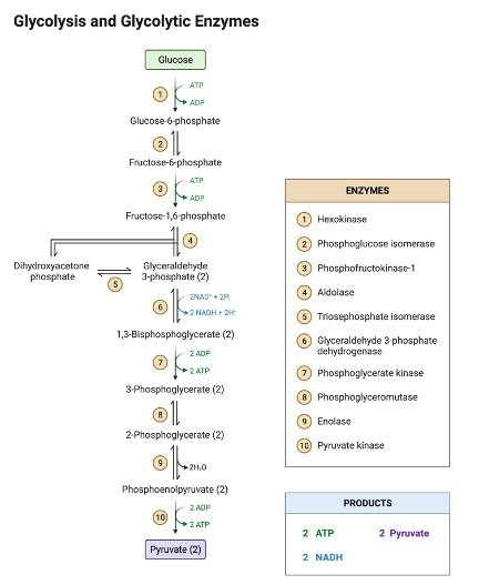
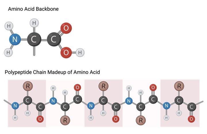
    Amino acids form the monomers that makeup protein polymers with the help of peptide bonds. Depending on the type and number of amino acids that make a protein they can vary in structure and function.

    Histidine: This amino acid is essential during the development and growth of children, but it is not required in adults unless kidneys are impaired. It functions in growth, maintaining the nervous system, and forms the metabolite of histamine neurotransmitters.

    One of its most significant functions is to regulate the concentration of heavy metals like molybdenum, copper, iron, manganese, and zinc and to metabolize them. If there are high level of trace metals in the system, but histidine amount is low then the depletion of histidine stores occur resulting in mineral-enzyme deficiencies.

    Isoleucine: They are often taken as supplements to increase endurance in athletes. It together with valine and leucine comprise 70% of all proteins found in the human body.

    They function in hemoglobin synthesis, tissue repair, and regulating energy levels and glucose levels. As they are relatively safe to consume in large quantities they are a popular ingredient of supplements in sports.

    Leucine: Leucine is one of the important members of the BCAA essential amino acid group. They have a role in fat catabolism without any decline in muscle mass as a result, they are incorporated in weight loss supplements.

    Weight loss can be increased by combining intake of leucine with vigorous exercise. Vegans usually have low leucine levels as it is found in dairy and meat products.

    Lysine: These amino acids aids in calcium absorption and are important for nervous system function and to maintain healthy muscle movement. It is also involved carnitine and collagen synthesis. Legumes are rich source of lysine for vegans.

    Deficiency of this amino acid causes dizziness, retarded growth, nausea, fatigue, and infertility. It can be administered to neurological patients to lower seizure events, but lysine-0restricted diets are needed for pyridoxine-dependent epilepsy.

    Methionine: It is found abundantly in meat, whole-grain foods, and dairy products and hence not required as a supplement. If methionine metabolism is impaired then it affects fatty acids and lipid biosynthesis that can lead to atherosclerosis.

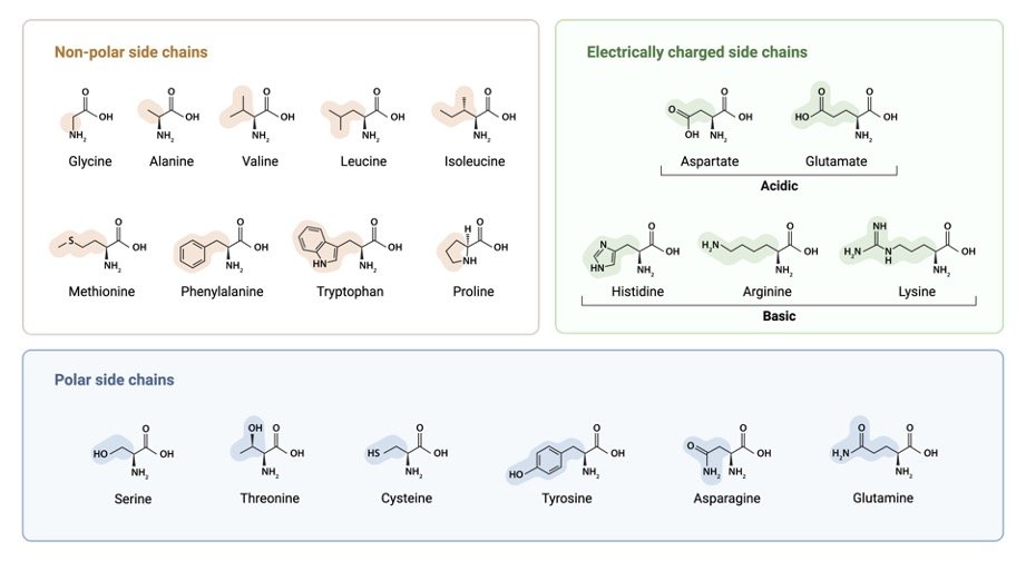
    It contains elemental sulfur in its structure. The other amino acid that contains sulfur is cysteine. Sulfur is crucial in the formation of anti-oxidants.

    They are taken in form of supplements suffering from liver ailments and people that suffer from dominance of hormone Low methionine supplements also improve cancer outcome and cell longevity according to researchers.

    Phenylalanine: It is involved in the biosynthesis of adrenaline, tyrosine, and noradrenaline the latter increases memory and mental alertness, decreases appetite, and boosts mood.

    Phenylketonuria refers to the disorder where phenylalanine hydrolase is lacking due to which the metabolism of phenylalanine is impaired and it accumulates in the body. This causes mental retardation is no intervention is done to treat this condition.

    Threonine: This amino acid along with methionine and aspartic acid promotes the metabolism of fat in the liver that helps prevent steatosis. They are also important for maintaining the integrity of the nervous system.

    They are often taken as supplements by people suffering from Lou Gehrig’s disease and multiple sclerosis. It is involved in the synthesis of elastin, glycine, collagen, serine, and muscle production. Research is focusing on employing this amino acid in colitis therapy.

    Tryptophan: It is one of the common health store supplements and ingredients that boost mood and energy levels. It is the precursor of melatonin, serotonin, structural proteins, and enzymes so it has become a popular ingredient in the health industry to treat conditions like migraine.

    The emergence of various research studies focusing on blood-brain barrier and gut synthesized serotonin, highlights the significance of this amino acid. It has clinical significance and is administered to reduce ADHD symptoms, alleviate anxiety, treat menopausal depression and restless leg syndrome.

    Valine: This amino acid is a part of BCAAs besides isoleucine and leucine that exhibit a different structural form than the rest of amino acids and are often utilized in dietary supplements. It is abundantly found in green leafy vegetables and kidney beans.

    This amino acid is involved in many physiological functions like it can enhance sleep quality and calm the nervous system in times of stress and improve cognitive processes.

    This is also involved in the recovery of tissues, growth, and repair and is endorsed as a supplement by athletes for building endurance. They also form a component of weight loss supplements as they decrease appetite.

    ii. Non-Essential Amino Acids

    They comprise 12 amino acids that can be synthesized in the body itself, but certain people may take further provide themselves with their supplements or high protein diets.

    These non-essential amino acids include arginine, aspartate, alanine, cysteine, serine, tyrosine, taurine, glycine, glutamate, proline, glutamine, and asparagine. The deficiency of these can be inborn and can form dysfunctional proteins.

    For instance, if there is a low synthesis of glycine amidinotransferase and arginine it can cause muscular impairments and mental retardation.

    Another example is the lack of glutathione synthetase that can lead to progressive neurologic disorders, oxidative stress, hemolytic anemia, and metabolic acidosis.

    iii. Conditional Amino Acids

    6 amino acids are conditional that are tyrosine, proline, glutamine, glycine, cysteine, and arginine. These amino acids are synthesized by the human body but under some circumstances their production is limited. Such conditions denote natural temporary physiological conditions like stress conditions or in babies born preterm cannot synthesize arginine.

    Amino Acid Chart Citations
    • A common periodic table of codons and amino acids. Biochem Biophys Res Commun . 2003 Jun 27;306(2):408-15.
    • Amino acid kinetics and the response to nutrition in patients with cancer. Int J Radiat Biol . 2019 Apr;95(4):480-492.
    • Amino Acid Transport Across the Mammalian Intestine. Compr Physiol . 2018 Dec 13;9(1):343-373.

    Share

    Similar Post:

    Continue Reading
  • Solvent: Definition, Types, & Examples

    Solvent Definition

    Solvent is defined as any molecule that can dissolve other molecules (solutes). Solvent molecules pull apart the solute molecules such that they are uniformly distributed in the solvent.

    The solvent can be gas, liquid, or solid. The homogenous solution formed as a result of the dissolution of solutes cannot be physically separated but a chemical process ned to be employed for their separation.

    Types of Solvent

    There are 2 types of molecules non-polar or polar. Polar molecules are charged and carry different electrical charges on different sides, while nonpolar molecules do not have a static charge. Both these kinds do function as solvents.

    i. Polar Solvent

    The negative and positive ends of the atom interact both with each other as well as the solute in a polar solvent. Solute dissolves as the electric charges of solvent pull on different atoms of the solute.

    For example, Salt an ionic compound is dissolved in polar solvent as the oppositely charged molecules are pulled apart. The positive ions of solute are pulled by the negative side of the polar solvent, while the positive ions are pulled by the positive side of the solvent. This leads to uniform distribution of solute in the solvent.

    ii. Nonpolar Solvent

    Their mechanism of dissolution is also similar to polar solvents. They often form spontaneous dipoles that have opposite charges linking bonds. This causes the adjacent solvent molecules to form dipoles.

    These momentary interactions are responsible for the dissolution of nonpolar solutes. But when we compare polar and nonpolar solvents, the interaction in polar compounds is much stronger. It is also the reason why the polar and nonpolar solvents do not mix.

    Solvent Examples
    i. Water

    It is the universal biological solvent that is very important. Cells of all living entities are dependent on water. The unique structure of water aids in its properties as a solvent.

    Oxygen atoms in water being more electronegative pull the electrons towards them that renders them electrically negative. The hydrogen atoms of water only get a small share of shared electrons that gives them a positive charge. This results in making water a strong dipole molecule that can help in the dissolution of a wide variety of solutes.

    The molecules and ions in the cell can be easily dissolved in water as it is a polar solvent. Some non-polar substances are also present in a cell that cluster together and shield themselves away from water.

    The bilipid membrane is also formed as a result of this property of water. In the membrane, the phospholipids are present such that the hydrophilic head that projects outside while the non-polar tail is shielded from the aqueous environment in the bi-lipid membrane that comprises 2 leaflets.

    When the phospholipid layers are brought together, the differential properties of the head and tail of phospholipid lead to the formation of a water barrier. Special proteins like carriers and channels are required for the passage of molecules inside and out of cells.

    These solute molecules are to be transported along the concentration gradient with the aid of solvent. If the solvent gets saturated then no more solutes can be dissolved. The ocean can be considered a big solution of various chemicals and salts. When raindrops fall on the soil, they dissolve some solutes.

    These are then carried through water and flow downstream into the river and then ultimately into the ocean. Many aquatic organisms rely on these solutes as they act as important salts or nutrients.

    ii. Solvents in Everyday Cooking

    Deglazing is a common procedure of cooking where the sticky bottom part of the pan is dissolved into a solvent. Both kinds of solvents can be employed to remove the caramelized part utilizing heat.

    Oil, a non-polar substance is often used to fry a variety of food items as they create a hot solution. This can infusion of solutes in oil to the fried food. Water can also be employed for deglazing for creating soup, gravy, or other sauces.

    Solvent Citations
    • Solvent-terminated dispersive liquid-liquid microextraction: a tutorial. Anal Chim Acta . 2018 Aug 3;1016:1-11.
    • Solvent production from xylose. Appl Microbiol Biotechnol . 2018 Oct;102(20):8707-8715.
    • Inverse Solvent Isotope Effects in Enzyme-Catalyzed Reactions. Molecules . 2020 Apr 21;25(8):1933.

    Share

    Similar Post:

    Continue Reading
  • Glutamic Acid: Definition, Types, & Examples

    Glutamic Acid Definition

    This non-essential amino acid has a molecular formula C5H9NO4. When the carboxyl group of this amino acid loses a proton under physiological conditions it gets a negative charge and now it forms glutamate amino acid.

    Glutamate is found abundantly in the brain and it also acts as a neurotransmitter. Glutamic acid comprises the building block of many proteins.

    What is Glutamic Acid?

    Glutamic acid is represented by symbols Glu or E. Like with all amino acids it also has an amino-terminal end, side-chain, and carboxyl-terminal end. The side chain in the case of this amino acid has a carboxylic acid group. The GAA and GAG codons code for the polypeptide chain.

    Glutamic Acid, What is Glutamic Acid,2

    Glutamic acid like other amino acids except glycine has both L and D-form stereoisomers. L-form is normally seen in the case of the living organism. The D-stereoisomer form of glutamic acid in cells of the liver and in the cell wall of bacteria.

    The blood-brain barrier cannot be crossed by glutamate amino acid as a result it needs to be synthesized in the brain itself from α-ketoglutarate or glutamine and it cannot be obtained by the diet.

    Glutamic Acid vs Glutamate

    Glutamate can be formed from glutamic acid when it loses a proton. Glutamic acid has the formula CH2CH2COOH, while glutamate has the formula CH2CH2COOH. Glutamate is basically the anion of glutamic acid.

    This aliphatic amino acid has a pKa value of 4.1so in an environment of pH more than 4.1, it will exist in the polar negatively charged form. Hence, glutamic acid is mostly observed as glutamate in physiological conditions in the human body.

    Glutamic Acid vs Glutamine

    Both these amino acids are often confused as they have similar names. Glutamic acid has an amine group instead of a carboxy group as in Glutamine that is represented as Gln or Q.

    Food that is high in glutamic acid like yeast, shellfish, and soy sauce give an umami flavoring that has a pleasant savory taste. Glutamate is often employed as a flavor enhancer in form of MSG.

    Glutamic Acid Function

    Glutamic acid comprises various proteins and it or specifically glutamate also functions as a primary excitatory neurotransmitter in the CNS. As a result, it is highly abundant in nervous tissue and in the brain.

    Glutamic Acid, What is Glutamic Acid,1

    Neurotransmitters are signaling molecules or chemicals in the nervous tissue. Acetylcholine, dopamine, and adrenaline are all examples of neurotransmitters. Glutamate receptors are located on the plasma membrane of astrocytes and neurons.

    These include kainite, AMPA, metabotropic receptors, and NMDA. Glutamate bind to the receptors to excite them as it is an excitatory neurotransmitter that will increase the likelihood of an action potential or nerve impulses that function in signaling.

    Glutamate like other neurotransmitters is stored as vesicles at chemical synapses. Glutamate is released via exocytosis when a nerve impulse travels to the synapse. This released neurotransmitter will bind glutamate receptors in the adjacent cell.

    Out of the 4 glutamate receptors AMPA, kainite, and NMDA are ionotropic that on activation lead to the opening of ion channels. In the case of metabotropic receptors, a second messenger is involved that functions in signaling.

    Glutamate as a neurotransmitter is involved in synaptic plasticity that is important for the memory and learning process.

    This enzyme aids in the conversion of glutamate to gamma-aminobutyric acid (GABA). GABA is an inhibitory neurotransmitter that forms from glutamate and binds to GABA receptors.

    This binding is inhibitory and decreases the likelihood of the formation of the action potential. This binding can have a sedative effect, for instance, alcohol and drugs like benzodiazepines activate GABA receptors.

    Clinical Relevance of Glutamic Acid

    Impaired glutamate signaling can result in dysfunctions and resulting disorders in the brain.

    Neurodegenerative Disease: Several neurodegenerative disorders can be traced to the chronic excitation by glutamate in neurons. For example, Alzheimer’s and Huntington’s disease, and other motor neuron disorders. The mechanism behind this is glutamate excitotoxicity that can impair nerve cells and even kill them.

    Psychiatric Disorders: In the case of psychiatric disorders like bipolar disorder and schizophrenia excessive glutamate activity has been observed. Pharmacological drugs like ketamine that specifically target the glutamate system are being considered for treatment in these cases.

    Substance Abuse: It also takes part in reward pathways and is thus involved in addiction development and maintenance. Cognitive processes like sensitization, habit learning, reinforcement, craving and conditioning, relapse, and craving.

    Glutamic Acid Citations
    • Gamma-fluorinated analogues of glutamic acid and glutamine. Amino Acids . 2003 Apr;24(3):245-61.
    • Glutamic acid, twenty years later. J Nutr . 2000 Apr;130(4S Suppl):901S-9S.
    • Microbial production of poly-γ-glutamic acid. World J Microbiol Biotechnol . 2017 Sep 5;33(9):173.
    • Images are created with BioRender.com

    Share

    Similar Post:

    Continue Reading
  • Protostome: Definition, Types, & Examples

    Protostome Definition

    These organisms form a clade that exhibits protostomy during early development. They along with Xenacoelomorpha and Deuterostomes comprise the group Bilateria. The organisms constituting this group are bilaterally symmetrical and triploblastic.

    What is Protostome?

    In embryo development, two gametes—sperm and an egg—fuse to form a zygote. After a zygote forms by syngamy of the 2 gametes, it is divided into 2 poles by an axis. The bottom pole forms the vegetal pole, while the to pole forms the animal pole.

    The zygote divides repeatedly by cleavage to form a mass of blastomere cells called a morula. The pattern of cleavage is what distinguishes protostome and deuterostome.

    Protostomes exhibit spiral cleavage wherein the cleavage occurs long angle resulting in spirally arranged blastomeres. The arrangement of cells changes such that blastoderm, a layer of cells envelope blastocoel that is a yolk or fluid-filled cavity and this forms a blastula.

    Gastrulation starts at this stage leading to the formation of 3 germ layers. These 3 germ embryonic cells in a triploblastic organism give rise to all the organs and tissues. The development of blastopore starts the process of gastrulation.

    The blastopore cells start migrating inwards to create an inner tube that will later develop into an endoderm that gives rise to the GI tract and digestive system. The outer layer ectoderm forms the nervous system and the epidermis.

    The layer between these 2 layers is known as mesoderm and it gives rise to muscles and connective tissue. Another significant distinguishing feature that sets apart protosomes from deuterostomes is the purpose of the blastopore. In the case of protosomes, the blastopore will develop into the mouth while in deuterostome it develops into the anus.

    In protostome the cavity opposite to the blastopore develops into the anus. The name Protostome is derived from the Greek words proto and stoma that means that the mouth develops first.

    Types of Protostomes

    Two taxonomic groups come under Protostomes:

    Lophotrocozoa: This group includes organisms that grow by extending the skeleton size as in mollusks that add calcium carbonate to the edge of their shells to grow larger.

    Brachiopods, mollusks, annelid worms, rotifers, bryozoa, and Platyhelminthes comprise this group. Examples of this group include squid, leeches, earthworms, and bivalve mollusks.

    Ecdysozoa: This group comprises the nematodes, arthropods, and tardigrades. They possess a 3-layered cuticle, that has a soft interior and an exoskeleton which is the hard exterior.

    They periodically shed and re-grow their exoskeleton to grow via the process of ecdysis. They unlike other protostomes do not undergo spiral cleavage during embryonic development. Examples of ecdysozoans include crustaceans, insects, roundworms, spiders, etc.

    Protostome Body Plans

    These triploblastic and bilaterally symmetric organisms can have 3 different body plans:

    i. Worm-like: These types of protostomes are characterized by a cavity inside their body that is well developed and is known as coelom that helps in the circulation of body fluids and functions like a hydrostatic skeleton that employs fluid pressure for movements.

    Based on their feeding system they can be further classified:

    a. Spoon worms or Echiura utilize a structure known as proboscis to acquire and move food. They are present in front of the mouth region and trap food and then using cilia move it to the mouth region.

    b. Penis worms or Pripulida are characterized by the presence of a toothed throat that can be extended inside out to capture the prey and once the prey is captured they retract its back for consumption.

    c. Ribbon worms or Nemertea employ proboscis that is present inside the body above the gut. The proboscis has a barbed tip and it is extended to capture prey by entangling it or by using venom.

    ii. Arthropod Body Plans: Arthropods have segmented bodies that have 3 parts that include head, thorax, and abdomen. They comprise crustaceans, all insects, myriapods, and arachnids. They also have jointed appendages and are the most abundant taxonomic group.

    They are named after the presence of jointed limbs and have a chitinous exoskeleton that also contains sclerotin proteins that harden them. The muscles are attached directly to the exoskeleton for movement. The coelom is reduced and is involved in providing space for organs and the circulation of fluids.

    iii. Mollusk Body Plan: These marine groups comprises Chitons, Bivalves, Gastropods, and Cephalopods. Their body plan has 3 major parts:

    a. The foot: this is responsible for movement and is constitutes a large muscle at the base of the animal.

    b. The visceral mass: it contains external gills and internal organs. Thye help foot in movement.

    c. The mantle: forms the layer that envelops visceral mass and secretes calcium carbonate to form shells. 

    Protostome Citations
    • Evolutionary convergence of higher brain centers spanning the protostome-deuterostome boundary. Brain Behav Evol . 2008;72(2):106-22.
    • Body-plan evolution in the Bilateria: early antero-posterior patterning and the deuterostome-protostome dichotomy. Curr Opin Genet Dev . 2000 Aug;10(4):434-42.
    • Calcium signaling and endoplasmic reticulum dynamics during fertilization in marine protostome worms belonging to the phylum Nemertea. Biochem Biophys Res Commun . 2014 Aug 1;450(3):1182-7.

    Share

    Similar Post:

    Continue Reading
  • Isomer: Definition, Types, & Examples

    Isomer Definition

    Isomer comprise 2 molecules that differ in terms of their structure but are represented by the same molecular formula. The number of atoms would be the same for these isomers but their arrangement would differ due to which they would exhibit different physical properties even though they have the same molecular formula.

    The functional groups associated with each molecule may differ which alters their properties. The molecules can be converted into their respective isomers by the process of isomerization. This may be spontaneous or not.

    Types of Isomer

    Isomers are classified into 2 types stereoisomers and structural isomers.

    i. Structural Isomers

    These molecules differ in respect to the functional groups and the specific attachment and arrangement of atoms. All the different isomers will have different IUPAC names and as they comprise different functional groups they may be classified into different groups.

    They are of different types including position isomers that differ in accordance to the position of the functional group, chain isomers that differ in regard to their branching patterns, skeletal isomers that have different carbon chains, and functional groups isomers.

    Tautomer is a kind of structural isomer that undergoes spontaneous interconversions between the different isomers. It will exhibit different properties based on the isomer conformation it exists in. Some of these spontaneous conversions are so rapid that it is difficult to isolate the different forms.

    ii. Stereoisomers

    These isomers will have identical bond structures but the geometric positions of atoms and functional groups will differ. Different types of stereoisomers include diastereomers, conformational isomers, and enantiomers.

    Enantiomers are non-superimposable mirror images that comprise chiral centers. Diastereomers may or not consist of chiral centers and are not mirror images of each other. Conformational isomers showcase different rotations around single bonds.

    Isomer Examples

    Methoxyethane and Propanol: The C3H8O chemical structure has many isomers like methoxyethane isomers and propanol isomers. The propan-2-ol and propan-1-ol comprise the propanol isomers that differ in regard to the position of an oxygen atom that is either present on the central carbon atom or the terminal carbon respectively.

    In the methoxyethane isomer, the oxygen atom is placed on the center instead of being linked to a single carbon atom, and due to which it is ether.

    Methylacetylene and Allene: These comprise C3H4 isomers that differ in the kind of bonding shown by them. Allene shows 2 double bonds between the carbons, whereas methylacetylene has only one single bond and one triple bond.

    Fulminate and Cyanate: They constitute isomers of CNO. In the case of fulminate, the N atom is linked to both the O and C atoms. While in cyanate, both N and O are linked to the central C atom.

    Glucose and Fructose: They are the most common examples and are C6H12O6 isomers. They differ in regard to the placement of a double-bonded O atom. The O is positioned on the first C in glucose, while in fructose it will be located on the second C in fructose.

    Importance of Isomer

    Isomers are significant in the case of pharmaceuticals, as generally one isomer of a compound will exhibit the desired reaction or effect. For example, ibuprofen will target the binding site and bind to it generating relief from pain in only one isomer form.

    Another example is cisplatin that comprises an anticancer drug, while transplatin that is its isomer shows no anti-cancer traits. One isoform of thalidomide was used to treat morning sickness in pregnant women in the 1950s, but its stereoisomer acted as a teratogen causing birth defects in children.

    In the human body, these isomers are critical for enzyme function. The structure of an enzyme including its functional group, orientation, and bond length is vital to its functioning as they bind very specifically to the active site of an enzyme.

    The isomers of enzymes that vary in their structure won’t be able to bind to the active site. For example, the isomer of the enzyme triose phosphate isomerase is not able to catalyze the interconversion of dihydroxyacetone and (R)-glyceraldehyde phosphate.

    Isomer Citations
    • Single-isomer drugs: true therapeutic advances. Clin Pharmacokinet . 2004;43(5):279-85.
    • Pentanol isomer synthesis in engineered microorganisms. Appl Microbiol Biotechnol . 2010 Jan;85(4):893-9.
    • Single isomer cyclodextrins as chiral selectors in capillary electrophoresis. J Chromatogr A . 2020 Sep 13;1627:461375. 

    Share

    Similar Post:

    Continue Reading
  • Barr Body: Definition, Types, & Examples

    Barr Body Definition

    Barr Body is also referred to as sex chromatin and represents the inactivated X chromosome present in the somatic cells of females. Females have 2 X chromosomes in their sex chromosome constitution, while males have one Y and X chromosome each.

    What is Barr Body?

    In the somatic cells of females, one X-chromosome is inactivated randomly by the lyonization process resulting in the formation of a Barr body. This occurs for dosage compensation as females have 2 X chromosomes which would otherwise produce double the amount of gene products compared with males.

    In all organisms where the sex determination is by XY chromosomal method, this process of X-chromosome inactivation can be observed. This process helps in balancing the X chromosome expression in males and females.

    Barr Body, What is Barr Body, 1

    The Barr body occurs as a result of heterochromatinization, while the active X chromosome is observed in a euchromatin state. The inactivated DNA is condensed and packed to repress the genes such that their transcription is hindered. While in the active X chromosome it is not packaged to freely allow molecules involved in transcription to interact with it.

    Which X Chromosome becomes the Barr Body?

    The selection of the X chromosome that has to be inactivated is done by a random process in the embryonic stage of any particular cell and it stays in this state for the rest of its life. The cells resulting from the division of these cells will all have the same X-chromosome inactivated. The number of Barr bodies is given as one less than the number of X-chromosomes present.

    For instance in Turner’s syndrome, no inactivation of the X chromosome would occur even though the individual is female and in Klinefelter’s syndrome, one of the X chromosomes would be inactivated and would be present as a Barr body.

    Mechanism of X-inactivation and Barr Body

    Two genes Tsix (Xist reversed) and Xist (X-inactive specific transcript) are found in the X-inactivation center (XIC) that is located near the centromere on the X chromosomes. Xist is responsible for the inactivation of a gene and Tisx represses Xist. The chromosome that produces more expression of the Tsix gene will have less probability of being inactivated.

    Initially, both X chromosomes in the female somatic cell express equal levels of Xist RNA, but once the process of inactivation starts the chromosome to be inactivated will express more Xist RNA that will then coat the whole chromosome and silence it. The presence of Tsix in other chromosomes represses the expression of the Xist gene that maintains it in the active form.

    Not all the genes of the inactivated X chromosome are silenced. The Xist gene along with some other genes escapes heterochromatinization. So, a female that has Turner’s syndrome will still express fewer genes than a normal female.

    Genetic Disorder of Barr Body

    In the case of individuals with Turner’s syndrome that only has one X-chromosome, the individual may survive even though they don’t have a Barr body and express lesser genes. They won’t be able to develop like a normal average. Chromosomal abnormalities where there are more than 2 X chromosomes in an individual can lead to developmental dysfunctions.

    In normal females, random inactivation can cause the maternal chromosomes to be silenced half the time, and the paternal one in the other half. But if one of them is repressed more than the other then depending on their ratio this may result in some problems.

    Barr Body Citations
    • Significance of the Barr body in human female tumors. Cancer Genet Cytogenet . 1981 Nov;4(3):269-74.
    • The disappearing Barr body in breast and ovarian cancers. Nat Rev Cancer . 2007 Aug;7(8):628-33.
    • Deciphering the Role of the Barr Body in Malignancy: An insight into head and neck cancer. Sultan Qaboos Univ Med J . 2017 Nov;17(4):e389-e397.
    • Images are created with BioRender.com

    Share

    Similar Post:

    Continue Reading
  • Neurogenesis: Definition, Types, & Examples

    Neurogenesis Definition

    Neurogenesis refers to the generation of new neurons. These neurons arise from stem cells or progenitor cells during embryonic and early development, it can continue into adulthood in organisms like fishes, amphibia, and insects. The adult mammalian nervous tissue was thought to be incapable of this process, but recent research findings prove this as not true.

    Is Neurogenesis True?

    Even in adults many tissues could regenerate or replicate themselves, in event of growth or repair, but it was thought that this was not the case for neurons. One of the reasons behind this was the complexity of the structure of neurons; they have cell bodies from which one long axon emerges and there are also dendrites that are highly branched that rise from these cell bodies.

    This nerve cell is part of a networking functioning unit that makes its replication a difficult process. Another reason is that damage to the brain or spinal cord causes irreversible damage hinting that there is a lack of neuronal regeneration. Death of neurons with aging showed symptoms of cognitive decline as these cells are not being replaced.

    Lab Studies of Neurogenesis

    Research lab studies on organisms like shrews and rats demonstrate the significance of neurogenesis. New labeling and imaging techniques aided in proving the fact that new neurons were born also in the adult brain. Environmental processes played a key role to alter neuronal degeneration with age.

    Exercises and cognitive processes strengthen and enhance neurogenesis while stress impacted this process negatively. It was discovered in 2005, that older mice who were still active on wheels showed a higher rate of neurogenesis than sedentary mice.

    Neurogenesis in Adults

    Research on adult hippocampal neurogenesis has revealed significant data on this process. Neuronal regeneration occurs in 2 regions of the adult mammalian brain; one of them is the hippocampus.

    This center of memory is found deep in the brain and has a tiny sea horse shape and helps in the processing of short-term memory into long-term memory. It also helps with spatial learning and navigational ability.

    The other region is the subventricular zone where neurogenesis can be seen. This region comprises stem cells and progenitor cells. More research is needed to understand what happens to the new neurons formed here.

    Some of them migrate to the olfactory bulb, but there is major purpose is still to be discovered. The presence of these progenitor cell-enriched sites may make it easier for neuron generation.

    Neurogenesis and Hippocampus

    The hippocampus region is susceptible in the early stages of Alzheimer’s disease, which may damage the area and also affect the disease’s progression. Hippocampus is critical for navigation and memory and possible damage may impact their functioning as in Alzheimer’s patients that get lost easily and find it hard to remember things. Besides neurodegenerative diseases, hypoxic conditions may also damage this region.

    Such conditions of low oxygen availability can occur during a heart attack, carbon monoxide poisoning, sleep apnea, and other dangerous situations like drowning. Impairment of this region can also cause anterograde amnesia, where the person struggles with making new memories.

    How to Enhance Neurogenesis?

    Recent research has indicated that neurogenesis does occur in the adult brain. The factors that can potentially increase this process are being investigated. Exercise, sex, intermittent fasting, and learning can increase the neurogenesis rate.

    Foods like blueberries and dark chocolate that are rich in flavonoids, omega-3-fatty acids found in fishes like salmon, and resveratrol found in red wine can also enhance this process. Some factors can also decrease neurogenesis like sleep deprivation, stress, alcohol, and diet high in saturated fat.

    Thus up to a limit changes in behavior and diet can enhance the rate of neurogenesis. Further research needs to be done to come up with a therapeutic technique for reversing hippocampal damage and damage caused by aging.

    Neurogenesis Citations
    • Adult Hippocampal Neurogenesis: A Coming-of-Age Story. J Neurosci . 2018 Dec 5;38(49):10401-10410.
    • Nutritional Factors Affecting Adult Neurogenesis and Cognitive Function. Adv Nutr . 2017 Nov 15;8(6):804-811.
    • Adult neurogenesis: beyond learning and memory. Annu Rev Psychol . 2015 Jan 3;66:53-81.

    Share

    Similar Post:

    Continue Reading
  • Phylum Chordata: Definition, Characteristics, & Examples

    Phylum Chordata Definition

    Chordate belong to the phylum Chordata that comes under the Deuterostomes kingdom that is characterized by the development of the anus that precedes the development of the mouth in the embryonic stage.

    Chordata phylum comprises a large variety of organisms including invertebrates that lack a backbone and vertebrates that possess a backbone. This phylum includes 3 subphyla that are Urochordata, Cephalochordate, and Vertebrata. They generally have a bilaterally symmetrical shape though exceptions are there.

    Phylum Chordata Characteristics

    The following characteristics are exhibited by all the organisms that come under this phylum at some stage of their lives or during embryonic development.

    Pharyngeal Slit: This can also be seen in invertebrates where they function in filter-feeding. Whereas in vertebrate fishes they develop into gill arches the function that supports gills. In other vertebrates like humans, these gill slits can only be seen in the embryonic stage and disappear later in development. This tissue may develop into eustachian tubes or jaws in terrestrial vertebrates.

    Dorsal Hollow Nerve Cord: This dorsal hollow tube comprises nerve fibers that will develop into the CNS that consists of the brain and spinal cord. The vertebral column shields this nerve cord. The vertebral column consists of bones that form our backbone. Whereas in non-chordates the nerve cords are located either laterally or ventrally and are solid.

    Notochord: This skeletal flexible rod is made of cartilage and functions to support the structural frame of the organism. It is present between the nerve cord and the digestive tube. The phylum derives its name from this rod. In vertebrates, it is the precursor of the spine or backbone.

    Post-Anal Tail: This posterior elongation provides balance, helps the aquatic organisms in propelling themselves in water, and is utilized by certain terrestrial vertebrates in signaling or in finding a mate. This tail disintegrates as it shrinks and can be seen as a tailbone during embryonic development in apes and humans.

    Chordata Subphylum

    Vertebrata: This subphylum is also known as Craniata as the comprising organisms have a cranium case that protects their head and is known as a skull. They are also distinguished by the presence of a closed circulatory system, unique motor and sensory cranial nerves, internal organs, and a highly evolved brain. This subphylum includes amphibians, fish, mammals, reptiles, and birds. Their evolution can be studied using paleontological pieces of evidence like fossils as due to the presence of cartilage and bone, their remains were preserved.

    Cephalochordata: Also called lancelets this subphylum comprises marine filter-feeding organisms. These have segmented small elongated and soft bodies due to which they did not fossilize well. They bury themselves in substrates and are seen in soft bottoms with exposed head regions consisting of tentacles that help in capturing the prey. Though they resemble fish, they do not have a brain, limbs, scales, and backbone like them. They have a greater number of pharyngeal slits than the fish.

    Urochordata: This sub-phylum is also referred to as Tunicates and comprises sea salps and squirts. They are also soft-bodied organisms and thus their fossils are rare. These marine organisms are barrel-shaped filter feeders that are not segmented. The tail can be seen in the larval stage when it is free-living. But in adults, they lose the tail and attach to a substrate. In adults, the only chordate characteristic visible is the pharyngeal slits.

    Phylum Chordata Examples

    Lampreys: Belonging to subphylum vertebrates this chordate is a filter feeder during its larval stage. The adult of this jawless fish is a parasite that latches on to other fish with its oral disk that comprises teeth. They are not obligate parasites and the adults may live off the reserve they built in the larval stage. The adult exhibits external gills, nerve cord, cartilaginous skeleton, and a notochord. In the larval stage, they utilize a mucus-secreting organ that helps to trap the food in their pharynx and also show a post-anal tail.

    Sea Squirts: This barrel-shaped organism belongs to the subphylum Tunicates. The larval stage resembles a tadpole and has a dorsal nerve, post-anal tail, notochord, and pharyngeal slits. In their adult stage, they are primarily filter feeders that have 2 siphons that are openings that are employed to draw water during filter feeding. They acquire their food phytoplankton by this method and drain the excess water through the other siphon.

    Phylum Chordata Citations
    • The notochord: structure and functions. Cell Mol Life Sci . 2015 Aug;72(16):2989-3008.
    • Evolution of the Chordate Telencephalon. Curr Biol . 2019 Jul 8;29(13):R647-R662.
    • Onto-phylogenetic aspect of myotomal myogenesis in Chordata. Folia Biol (Krakow) . 2004;52(1-2):1-12.

    Share

    Similar Post:

    Continue Reading