• Cetrimide Agar Test: Principle, Procedure, Results, and...

    Cetrimide Agar Test

    Many of the biochemical tests are identified and performed in the laboratories and in experiments to differentiate a characteristic of a microbe and they are differentiated from other organisms.

    One such biochemical test is cetrimide agar test. Cetrimide agar test is performed in order to identify the Pseudomonas aeruginosa and it also helps in differentiating it from other organisms.

    Cetrimide Agar

    Cetrimide agar is one of the selective and differential mediums which is used for isolating the Pseudomonas aeruginosa from other clinical and non-clinical specimens.

    Cetrimide is considered as one of the selective agents which inhibits the detergent activity of most of the bacteria.

    Cetrimide agar is also known as Pseudomonas cetrimide Agar or Pseudosol agar.

    What is Cetrimide Agar Test?

    Cetrimide agar test works on the principle based on the ability of the organisms to grow in the presence of cetrimide.

    Cetrimide is one of the toxic quarterly ammonium detergents. This ammonium detergent is toxic to most of the bacteria except few organisms such as Pseudomonas aeruginosa.

    P. aeruginosa plays active role in causing infections in the urinary tract and other infections like bacteremia, which occurs especially in humans.

    This Pseudomonas aeruginosa is usually identified on their basic characteristics such as production of pyocyanin, and its blue colour its ability to soluble in water, presence of a phenazine Pigment, morphology of their colony and its characteristic grape like odour.

    Pseudomonas aeruginosa is the only species of Pseudomonas or its gram-negative rod which has the capability to excrete pyocyanin, and this capability is usually detected by performing this cetrimide agar test.

    Cetrimide Agar Test Objective

     Aim of this test is to identify the species, Pseudomonas aeruginosa and to differentiate it from other micro-organisms.

     To detect the ability of various organisms to grown on the cetrimide agar.

    Cetrimide Agar Test Principle

    As mentioned above, cetrimide is a quaternary ammonium salt, and it also acts as a cationic detergent in most of the bacterial cells.

    This cationic detergent is considered as one of the toxic substances. Cetrimide reduces the surface tension of the medium at the time of point of contact which results in complex precipitants which denatured the effect of proteins that were present on the bacterial membranes.

    Where as the inhibition of growth in various micro-organisms is observed in this cetrimide agar medium including Pseudomonas species except P. aeruginosa.

    When cetrimide is Brought in contact with the bacteria there will be release of nitrogen and phosphorus in the bacterial cell.

    In such case, the organisms like P. Aeruginosa and other species of pseudomonas cannot withstand this germicidal activity.

    This media also contains gelatine peptone which provides necessary nutrients for Pseudomonas aeruginosa.

    Sodium chloride helps in maintaining the osmotic equilibrium in the cetrimide medium and it also prevents the change of pH during the growth in the medium.

    Micro Organisms Tested

    Cetrimide agar is usually used for testing the isolated colonies of non-glucose fermentation, gram negative rods which are suggestive for Pseudomonas aeruginosa.

    Cetrimide Agar Test Reagent

    Cetrimide agar can be purchase commercially in the format of dehydrated powder or it can be prepared in the laboratory using the necessary chemicals and materials available.

    Supplies:

     Sterile inoculating loops

     Sterilized sticks

     Uv light

    Cetrimide Agar Test Procedure
    Media Preparation

    About 46.7grams of dehydrated powder is added in a beaker containing 1000 millilitres of distilled water. Instead of dehydrated powder, media prepared in lab can also be used.

    The prepared medium is now heated till it boils, such that the medium dissolves completely.

    The boiled medium is then transferred into the tubes and it is sterilized in an autoclave at a temperature of 121ºC or at 15 lbs pressure for about 15 minutes.

    After autoclaving the tubes are taken out and placed in a slanted position and it is allowed to cool till the temperature reaches about 40 to 45ºC.

    The slanted position is maintained until it develops a butt in a depth of 1.5 to 2.0 cm.

    Cetrimide Agar Test

    A well isolated colony is collected from an 18 to 24-hour culture using sterile inoculating loops or needles.

    The cetrimide agar tubes are then inoculated by streaking the surface of the slants along the medium, in the direction of froth and back and it is also ensured for proper inoculation with the inoculating stick.

    The caps of the tubes should not be closed tightly so that the medium gets an adequate aeration.

    Further the tubes are incubated aerobically at a temperature of about 35 to 37ºC for about 7 days.

    The test tubes are checked daily, and they must be kept at least for 7 days without discarding in order to get the proper and appropriate results.

    Cetrimide Agar Test Result

    Positive Result: A positive result is usually determined by observing the growth in the medium, and sometimes along with a growth there will also be a change in colour from yellow green to dark blue green. Positive results are usually detected in P. aeruginosa.

    Negative Result: Negative result is usually determined when there is no change in colour or no growth is seen. Negative results are usually detected in Escherichia coli.

    Cetrimide Agar Test Citations

    Share

    Similar Post:

    Continue Reading
  • Catalase Test: Principle, Procedure, Results, and Uses

    Catalase Test

    Many of the biochemical tests are identified and performed in the laboratories and in experiments to differentiate a characteristic of a microbe and they are differentiated from other organisms. One such biochemical test is catalase test.

    Catalase test is mostly used to detect the production of catalase enzymes in the aerobic organisms.

    What is Catalase Test?

    Catalase enzyme is one of the common enzymes which is found in all living beings which survive in oxygen and it also catalyses the decomposition of hydrogen peroxide, releasing water and oxygen.

    Catalase is considered as one of the important enzymes in the pathogen organisms as it helps in protecting the organism from oxidative damages from the reactive oxygen species.

    Catalase enzyme also results in neutralising the bactericidal effects of the hydrogen peroxide, and its concentration in bacteria has been correlated depending upon the pathogenicity of the organism.

    Catalase test is being used from olden days in order to differentiate the positive catalase organisms from the negative organisms like streptococci.

    Catalase positive organisms includes Staphylococci. This test is very much useful in identifying the characterises of most of the bacteria.

    In aerobic conditions only 3% H2O2 is used, whereas in case of anaerobic conditions 15% of H2O2 is used.

    Catalase Test Purpose

     The main aim of this test is to detect the ability of the organism to produce the enzyme catalase.

     To differentiate the catalase positive organisms from the catalase negative organisms.

    Catalase Test Principle

    The metabolic activity of the aerobic and the facultative anaerobic micro-organisms has the capability to produce toxic by-products like Superoxide radical and hydrogen peroxide.

    Where these products are toxic to the organisms and they may also result in lysis of the cell if the cell is not broken.

    Considering the pathogenic organisms, different mechanisms are used in order to break the products into non-toxic substances.

    The bacteria which is capable of synthesising the enzyme catalase, hydrolyses the hydrogen water into gaseous oxygen and water that results in the liberation of gas bubbles.

    Thus, the production of this enzyme, catalase protects the organism from suffering those lethal effects of the hydrogen peroxide which is accumulated at the end of the aerobic metabolism.

    The presence of this enzyme can be detected by adding hydrogen peroxide in the bacterial inoculum, which thus results in the liberation of rapid air bubbles.

    If the enzyme is absent then there will be no liberation of such air bubbles.

    Micro Organisms Tested

    The organisms that can be used for catalyse test includes the colonies of young bacteria, which are left in a culture of 18 hour or less than that, and mostly agar media is preferred here.

    In case of anaerobes the colonies should be exposed to the air for about 30 minutes prior to the test.

    Catalase Test Reagents

    • Hydrogen peroxide Reagent

    • But the reagent of hydrogen peroxide various for each organism, where as it is 30% for Neisseria, 15% for anaerobes, 3% for other bacteria. 

    • Glass slide

    • Sterile glass sticks

    • Sterilised Platinum loops or wires

    Catalase Test Procedure

    The procedure for catalase test includes more than one method. The methods used are slide or drop catalase test, tube method, heat-stable catalase test, Semiquantative catalase test, Capillary tube method and cover -slip method.

    Here the Heat-stable catalase test is mostly used for differentiating the species of Mycobacterium, the semi-quantative catalase method is mostly used for identifying the species, Mycobacterium tuberculosis.

    From all these methods, the test which are most commonly performed in clinical bacteriology includes slide or drop catalase method, as this method requires only a small number of organism and it also works relatively as an uncomplicated technique.

    1. Slide Method

    To perform this slide method, first the microscopic slide is placed inside a petri dish. But it is also not necessary that the slide must be placed inside a petri dish as it is used only to limit the catalase aerosols, that carries viable bacterial cells.

    Then a small amount of organism is collected from a well isolated colony from an 18 to 24-hour culture using the inoculating loops and it is placed on the microscopic slide.

    No agar should be picked up along with the colony when the culture is taken from the blood agar.

    About one drop of 3% of hydrogen peroxide is added upon the organism that is placed on a microscopic slide, using a Pasteur pipette or a dropper.

    Then we can observe the formation bubbles which is formed against a dark background.

    2. Tube Method

    In tube method about 4 to 5 drops of 3% of hydrogen peroxide is added into a test tube.

    Then the small amount of organism is picked up using an inoculation loop from an 18 to 24-hour colony and it is placed into the test tube.

    Then the test tube is placed against the dark background and we can observe the bubbles that are formed immediately.

    Catalase Test Quality Control

    The organisms listed below are tested foe positive and negative results.

     If the result is positive then it denotes the presence of Staphylococcus aureus.

     If the results are negative then it denotes the presence of Streptococcus pyogenes.

    Catalase Test Result

     The positive test is usually determined by appearance of immediate bubbles

     Whereas if only one or two bubbles are appeared, then it indicates the weak reaction

     The negative test is indicated by no bubbles are only few bubbles.

    Catalase Test Citations

    Share

    Similar Post:

    Continue Reading
  • Citrate Utilization Test: Results, Purpose, and Principle

    What is Citrate Utilization Test?

    Many biochemical tests are studied and experimented in the laboratories. These tests help us to find the nature of the micro-organisms and their characteristics against certain enzymes and indicators used. One such test is citrate Utilization test.

    Citrate tests are usually performed to determine the ability of the bacteria to utilize sodium citrate as the source for carbon and inorganic ammonium hydrogen phosphate as the source for nitrogen.

    This citrate utilization test is possible only for the organisms which are capable of fermenting the citrate. This processes usually takes place with the help of an enzyme citrase.

    Characteristic of Citrate Utilization Test

    Citrate utilization test is often performed along with IMViC test. IMViC test is an abbreviated form of Indole, Methyl Red, Vogues-Proskauer and citrate test.

    This test helps in differentiating the organisms on the basis of their ability to use citrate as a source of energy. This citrate is test mostly performed with other IMViC tests which helps us to differentiate Gram-negative bacilli of the Enterobacteriaceae family from other species.

    It is considered as one of the important tests as it has a capability to detect the species-level identification in the family of Enterobacteriaceae.

    However, it also has exception with the few species of the Enterobacteriaceae family. Some species such as Salmonella, Edwardsiella, Serratia Providencia, Citrobacter, Enterobacter, etc. gives a positive reaction in a citrate utilization test, and other species like Escherichia, Morganella, Shigella and Yersinia gives a negative reaction during this test. Proteus is considered as citrate variable.

    Citrate utilization test is most commonly called as Simmon’s citrate test; as it helps in utilizing Simmon’s citrate agar that contains a major source of energy in the form of carbon.

    This medium also comprises of ammonium hydrogen phosphate as the sole source of nitrogen.

    The organisms which gives positive result during this test has the capability to ferment citrate during the presence of an enzyme citrate.

    Citrate Utilization Test Principle

    Citrate agar is used to test the ability of an organism whether the particular has citrate as the source of energy.

    The agar medium contains citrate as the source of carbon and the other inorganic ammonium salts as the source of nitrogen.

    Thus, the growth of organism in the medium is usually indicates by the amount of citrate that is utilized, as citrate is one of the intermediate compounds in the process of Krebs’s cycle.

    The enzyme citrase breaks the citrate into oxoacetate and acetate.

    Oxoacetate is again broken down into pyruvate and carbon dioxide. Hence the carbon dioxide released, induces the metabolism of ammonium salts, and results in the formation of ammonia or sodium carbonate, which increases the alkalinity in the medium.

    On adding an indicator bromothymol blue, there will be a shift in pH and the color of the medium changes from green to blue, when the pH is above 7.6.

    Thus, the growth of the organism is detected by change in colour of the medium, as a result Citrate metabolism determines a positive citrate test.

    Citrate Utilization Test Requirements

    Media: For citrate utilization test, simmon’s citrate is generally used as the medium to test the ability of the organism to utilize citrate as a sole source of energy.

    Simmon’s citrate agar is now a days sold commercially in the form of dehydrated powder by commercial vendors.

    At the same time, it can also be prepared in the laboratory using the necessary ingredients.

    The ingredients and their composition are listed below;

    IngredientsGram/liter
    Ammonium dihydrogen phosphate1.0
    Magnesium sulphate0.2
    Dipotassium phosphate1.0
    Sodium citrate2.0
    Sodium chloride5.0
    Bromothymol blue0.08
    Bacteriological agar15.00

    Supplies:

     Inoculating loops or needles

     Incubator

    Citrate Utilization Test Procedure

    About 24.28 grams of dehydrated powder or the media prepared in laboratory are added in a beaker containing 1000 milliliters of pure distilled water.

    Then the solution is heated until it boils, so that the medium dissolves completely.

    Further the dissolved medium is shifted into the tubes and the tubes are sterilized in an autoclaved at a temperature of 121ºC for about 15 minutes.

    After completion of the autoclaving process, the tubes are taken out and they are cooled at temperature of about 40 to 45ºC in a slanted position and this position is maintained till obtaining the butts in a depth of about 1.5 to 2.0cm

    Utilization Test

    An isolated colony is taken from an 18 to 24-hour culture using a sterile inoculating loop.

    Then the citrate agar tubes are inoculated by streaking the surface of the slant, in the back and froth direction using the inoculating sticks.

    The caps of the tubes are usually left loosened in order to ensure adequate aeration and they are incubated aerobically at a temperature of 35 to 37ºC for about 4 days.

    This test is examined each day.

    It must be kept for a minimum to 4 days to finalize the results.

    The change in color is observed if present.

    Citrate Utilization Test Quality Control

    Aseptic simmon citrate agar is maintained in a forest green color which is slightly opaque at the gel slants.

    The agar is inspected for the evidence of freezing, any contamination, dehydration, cracks or formation of bubbles prior to use.

    If any tubes are seen as blue perform usage then it must be discarded.

    Positive control of the tests includes Klebsiella pneumoniae, and the negative control include Escherichia coli.

    Citrate Utilization Test Results

    Positive Result: Positive test is usually determined by growth in the medium along with the color change. Here the color changes from green to deep blue along the direction of the slant.

    Negative Results: The test is concluded as negative if there is no growth or change in color in the medium, such that the color remains the same, green.

    Citrate Utilization Test Citations

    Share

    Similar Post:

    Continue Reading
  • Decarboxylase Test: Purpose, Indicator, and Results

    What is Decarboxylase Test?

    Decarboxylase test is one of the biochemical tests. This test is commonly used to differentiate the members of the family Enterobacteriaceae on the basis of their ability to produce the enzyme decarboxylase.

    However, the metabolism of amino acids differs depending on their characteristic as aerobic or anaerobic facultative anaerobes and as well as in gram-negative organisms.

    Metabolism of amino acids occurs either by decarboxylation, deamination or hydrolysis depending on the metabolization of amino acid in an organism.

    Decarboxylation often occurs in the presence of an enzyme deaminase and catalyzation occurs by breaking the bonds and it also binds to the rest of the amino acid in the amino group.

    Three different decarboxylase enzymes are produced by the organisms, which catalyze the metabolism of amino acids, ornithine, arginine decarboxylase, decarboxylase, and lysine decarboxylase.

    The production of these enzymes is taken as an important parameter for differentiating the bacteria present in the family of Enterobacteriaceae.

    Apart from differentiating and identifying the Enterobacteriaceae, Ornithine decarboxylase teat is considered as one of the paramount importance especially in separating the members of the Klebsiella-Enterobacter Serratia group and also in identifying the species Proteus.

    Decarboxylase Test Objective

     To detect the ability of an organism to produce an enzyme known as decarboxylase.

     To differentiate the members of the family Enterobacteriaceae on the basis of their ability to produce an enzyme decarboxylase.

    Decarboxylase Test Principle

     The medias of Arginine, lysine and orthenine decarboxylase are used to detect the ability of an organism to decarboxylate or hydrolyze the amino acid and forms an amine which produces an alkaline ph.

     The basal medium is formed by using Moeller’s formula.

     The medium contains meat peptones and beef extracts which supplies nitrogenous nutrients to support the growth of bacteria.

     The media is composed of glucose which is a fermentable carbohydrate.

     The enzyme pyridoxal acts as a cofactor and enhances the decarboxylase activity. Bromocresol purple and cresol red is used as a pH indicator.

     Amino acids namely arginine, Lysine and ornithine are added once in the basal medium and it also helps in detecting the production of enzyme which decarboxylate or hydrolyze these substrates.

     After fermenting the glucose in the medium, acids are produced. These acids have the capability to lower the pH in the medium which results in change in color from purple to yellow.

     If the organism produces an enzyme decarboxylase, it results in decarboxylation or hydrolysis of amino acids, due to the response in the acid pH.

     Decarboxylation thus results in alkaline end products known as amines, which causes the medium to get back its original color, purple.

     In case if the organism does not ferment glucose, then the medium does not change into yellow, but in these cases too, the tests are performed by including a control without amino acids for comparison.

    Decarboxylase Test Requirements

    Media: Decarboxylase test medium Base is generally used for testing the amino acid decarboxylase activity. Where as the other medias like Motility-indole-ornithine medium and Lysine iron agar can also be used.

    Reagents:

     Mineral oil

     Vaspar

     Liquid paraffin

     Petroleum jelly, which is maintained at 56ºC in the liquid form.

    Supplies:

     Sterile sticks

     Inoculating loops

     Incubator

    Decarboxylase Test Procedure
    1. Preparation of Media

     Initially 9.02 grams of dehydrated powder is filled into a beaker containing 1000 milli liters of pure distilled or deionized water.

     The prepared solution is then heated till it boils, so that the medium dissolves completely.

     The media is initially divided into four equal parts. When one part is tube without adding any amino acid and the tube is labelled as control.

     Then the remaining three parts are dispensed into each of the tubes and L-lysine hydrochloride, L-arginine hydrochloride, and L-ornithine hydrochloride are separately added to bring a final concentration of about 0.5%.

     About 3 to 4 ml of the media is dispensed in a screw capped tubes and they are sterilized by autoclaving them at a 10 lbs. pressure in 115ºC for about 20 minutes.

     In order to avoid the false alkalinization on the surface of the medium, liquid paraffin is added to height of about 5mm before sterilizing.

    2. Decarboxylase Test

     For Glucose-fermenting organisms

     A drop of 18-24hour brain heart infusion broth culture is add in each of the three decarboxylase broths.

     Here the control tube is not used for glucose-fermenting organisms.

     4mm layer of sterile mineral oil is added to each of the tubes and the tubes are incubated for 4 days at a temperature of 35 to 37ºC in an ambient air.

    Glucose-Nonfermenting Organisms

     A suspension is prepared using brain heart infusion broth and it is prepared from an overnight culture on a sheep blood agar.

     Each of the four-decarboxylase broth is inoculated with four drops of the prepared suspension.

     Then a sterile oil is added to a height of about 4mm.

     The tubes are then incubated for 4 days at a temperature of about 35 to 37ºC in an ambient air.

     The tubes are then observed for color change for 24, 48, 72 and 96 hours.

    Decarboxylase Test Results

     A positive test is found out by change in color of the medium from turbid purple to faded out yellow purple color.

     A negative test is detected by a bright yellow color or no change in color.

     The control tube must retain, its original color or sometimes it turns yellow. If turbidity or alkalinity of the purple color is seen in the control tube, then the test is considered as invalid one.

    Decarboxylase Test Citations

    Share

    Similar Post:

    Continue Reading
  • Methyl Red Test: Results, Procedure, and Principle

    Methyl Red Test

    Methyl red test helps us to detect the production of sufficient acid during the process of fermentation of glucose and maintenance of conditions such as pH.

    The pH of an old culture is sustained below the value of 4.5 which is shown by the change in color of the indicator which is added at the end of the period on incubation.

    MR-VP broth was developed by Clark and Lubs which allowed both MR and VP tests to be performed in the same medium of inoculation by aliquoting the different portions of the tube.

    What is Methyl Red Test?

    Methyl red test is commonly known as MR test and it is used to detect the ability of the organism to produce and maintain the stable products of acids by fermenting the glucose.

    Whereas methyl red test along with VP test is always performed together; as they are physiologically related to each other and they are performed in MRVP broth.

    All of the members of the family Enterobacteriaceae has the capability to convert the substance of glucose into pyruvic acid by using Embden-Meyerh-of pathway, whereas the bacteria can metabolize the pyruvic acid further.

    This test is one of the commonly performed biochemical test, which is used to detect the ability of the microorganism.

    Methyl Red Test Principle

    Generally, some bacteria have the ability to use glucose and convert it into a stable lactic acid, acetic acid or formic acid end product.

    The bacteria first metabolize through mixed acid pathway to produce stable acid.

    The acid production varies from species to species depending on the specific enzymatic pathways which is seen in the bacteria.

    This acid thus produced decreases the ph. value about 4.5 or in some cases even less than that, which is indicated by change in color from yellow to red when methyl red indicator is added.

    During methyl red test, the bacteria which tests the growth of bacteria in a broth medium containing glucose.

    When the bacteria have the ability to use the glucose by producing stable acid, the color changes from yellow to red on adding the methyl indicator in the broth culture.

    Where as the mixed acid pathway gives 4 mol of acidic products which mainly involves lactic and acetic acid, 1 mol of neutral fermentation product, mostly ethanol is used here, 1 mol of carbon dioxide and 1 mol of H2 per mol of glucose is fermented.

    When a large quantity of acids is producing, they cause a significant decrease in pH in the culture medium.

    Methyl Red Test Reagents

    MRVP Broth:

     Usually, the ph. in the MRVP broth is maintained at 6.9.

     1 litre of deionized water

     7.0 grams of Buffered peptone

     5 grams of Glucose

     5 grams of Dipotassium phosphate

    Preparation of Methyl Red Solution

     Initially 0.1 gram of methyl red is dissolved in 300ml of 95% of ethyl alcohol.

     Then sufficient distilled water is added to make about 5oo ml of methyl red solution.

     Then the prepared solution is stored at a temperature of 4 to 8 ºC in a brown bottle.

     This solution which has been prepared can be stored up to one year.

    Methyl Red Test Procedure

     Before inoculating the medium, it is allowed to equilibrate at a room temperature.

     Then the organisms are taken from the 18 to 24-hour pure culture, and it is lightly inoculated in the medium.

     Then the medium is incubated aerobically at a temperature of 37ºcelsius for about 24 hours.

     After incubating for 24 hours, 1 ml of aliquot broth is added into a clean test tube.

     Again, the broths are incubated for additional 24 hours.

     Two to three drops of methyl indicator are added to the aliquot.

     Then the change in color is observed immediately.

    Methyl Red Test Results

     In positive reaction, the color changes to distinct red

     Some of the species like, Escherichia coli, Yersinia sps gives positive results in Methyl red test

     Whereas, in negative reaction, the color remains yellow and there will be no change in color

     Organisms such as Enterobacter aerogenes, Klebsiella pneumonia, etc. gives negative results in Methyl red test.

    Methyl Red Test Uses

    Basically, methyl test is paired up with Voges Proskauer test and it is used in differentiating the members of the family Enterobacteriaceae. But in recent times they are used to characterize the other groups of bacteria including those of Actinobacteria.

    Methyl Red Test Limitation

     Usually, biochemical tests are recommended to perform on pure cultures for the complete identification.

     Methyl red test can be performed only if the medium is incubated for at least a minimum of 48 hours. If the tests are let to run very early them it results only in false-positive results.

     It is also important to take a note that only light inoculums are used in methyl red tests because when heavy inoculums are used the growth of bacteria will be inhibits and it results in invalid results

     Incubation can be followed for a maximum of five days, which is necessary for this test.

    Methyl Red Test Citations

    Share

    Similar Post:

    Continue Reading
  • Novobiocin Susceptibility Test: Purpose, Procedure, Risks, &...

    Novobiocin Susceptibility Test

    Novobiocin Susceptibility test is usually done to differentiate the coagulase-negative Staphylococci and to identify and isolate as Staphylococcus saprophyticus, which is usually novobiocin resistant.

    It is generally performed on urine isolates, especially the urine from women of the reproductive ages, as they contain coagulase negative.

    Novobiocin is an aminocoumarin antibiotic, and it is produced from actinomycete Streptomyces nivens, and has its own antibacterial property.

    Kloos and Schleifer in the year 1975, detected the simplified scheme for differentiating coagulase-negative Staphylococcus spp.

    That includes novobiocin disk test. Staphylococcus saprophyticus, is one of the gram-positive coagulase negative species of Staphylococci.

    It is an uropathogenic bacterium which acutes acute infection in the urinary tract.

    This infection is uncomplicated, it is often found in young and middle-aged female patients.

    Unlike other Coagulative Negative Staphylococcus, Staphylococcus saprophyticus is rarely resistant to the antibiotics and it is active against gram-positive organisms.

    S. saprophyticus is one of the promising resistant of the antibiotic novobiocin.

    Hence screening for coagulase-negative staphylococci from the urine cultures for novobiocin resistance is reliable presumptive for identifying saprophyticus.

    Novobiocin Susceptibility Test Objective

    Novobiocin susceptibility test helps to determine the susceptibility pattern of the bacterium with the antibiotic novobiocin.

    Novobiocin Susceptibility Test Principle

    As mentioned above, novobiocin is an antibiotic which interferes with the unpacking and repacking of DNA during replication of DNA and the bacterial cell cycle.

    Novobiocin binds with the DNA gyrase and restricts the activity of adenosine triphosphate.

    Susceptibility of the novobiocin is determined by placing a novobiocin antibiotic in a paper disk which is kept on a agar plate and then it is seeded with the organism under investigation.

    As organism multiplies during the period of incubation and it produces lawn of confluency growth, cells are exposed to antibiotic diffusing into the agar from the paper disk.

    If the bacterial species are susceptible to antibiotic novobiocin, then there will be a formation of transparent zone of inhibition around the area of the disk, which represents the area where the antibiotic concentration prevented the bacterial growth.

    If there is no zone of inhibition then, it is said that the organism is resistance to the specific antibiotic used. Usually in the laboratories, Mueller-Hington agar plate is used.

    The agar plate is seeded with the test organism to produce a confluent growth on the surface of the agar.

    After seeding the test organism is completed, a novobiocin antibiotic disc is applied on the surface of the agar, and incubation is done and further the sensitivity of the organism to the antibiotic determined by performing a Kirby-Bauer method.

    Novobiocin Susceptibility Test Plate

    Novobiocin disks are prepared by impregnating 5µg of novobiocin into a high quality of about 6mm diameter filter paper disks or it can also be purchased, as it is also commercially available.

    Novobiocin Susceptibility Test Procedure

     First the test isolated is kept in the pure culture for about 18 to 72 hours. Suspension is prepared of the test isolate in the tryptic soy broth which is equal to a Mc Farmland 0.5 standard or equivalent.

     Sterile swab is immersed in the suspension and it is rotated against the sides of the tube above the fluid level to remove the excess inoculum.

     Then the expressed swab is collected and inoculated into a blood agar or into the Muller Hinton plate by streaking the swab on the entire agar plate.

     This process is repeated in both the plates.

     After streaking the agar surface is allowed to dry for at least 15 to 20 minutes before applying it on the novobiocin disk.

     Using a sterile swab, lawn of growth is prepared on the entire surface of the plate in three directions and also along the entire edge of the plate.

     Then the forceps are dipped in an alcohol and flamed.

     Then the aseptically flames are used for applying the novobiocin disc to the surface of the inoculated plate.

     The discs are genteelly pressed down using a sterile forceps to ensure whether they adhere to the agar surface.

     The plates are aerobically incubated for about 18 to 24 hours at a temperature of about 35 to 37ºC.

     The diameter of the zone of inhibition is measure using a metric ruler or sliding capillaries.

    Novobiocin Susceptibility Test Results

    Positive Results: In case, if the results are positive, then the zone of inhibition will be greater than 16mm in diameter and it also indicates that the specific organism is sensitive to the antibiotic.

    Negative Results: If the results are negative, then there will be a zone of inhibition which is less than or equal to 16mm, which indicates the novobiocin resistance.

    Novobiocin Susceptibility Test Significance

     The CoNS is subdivided into two based on their novobiocin pattern of susceptibility. Here the CoNS group demonstrates the novobiocin susceptibility including epidermis, S. Captitits, S. haemolyticus, S. hominis, S. lugdenensis, S. warneri, S. saccharolyticus and other species.

     The novobiocin resistant group consists of species such as S. saprophyticus, S. xylosus, and kloosii.

     This test is basically used for differentiating Staphylococcus saprophyticus from other Coagulase negative staphylococci, especially in the clinical specimens.

    Novobiocin Susceptibility Test Citations

    Share

    Similar Post:

    Continue Reading
  • Lipase Test: Purpose, Procedure, Risks, and Results

    What is Lipid and Lipase Test?

    Lipid is the common term in biology which is used to denote the type of fats present in our body.

    Generally, fats are formed by linking the ester bonds between three molecules of the fatty acids along with one molecule of glycerol.

    Where simple fats are known as triglycerides or triacylglycerols, as they are composed of glycerol with three long fatty acids chain.

    The enzyme which breaks simple fats into smaller components of the fatty acids is known as Lipase.

    As in human, some microorganisms also contains lipase and they are classified depending on their ability to synthesize and secrete lipase.

    Different types of simple fats are used for determining the type of microorganism to be tested.

    Lipase is one of the enzymes presents in our pancreas, and it is synthesized in pancreases and released in to our digestive tract, only when we swallow food materials.

    Lipase thus helps in breaking down the fats present in our food materials.

    Specific levels are maintained in producing and utilizing the lipase such as maintaining the normal digestion and the cellular functions.

    But abnormally levels of the enzyme present in the blood indicates some of the health issues.

    However, Serum lipase test helps in measuring the amount of enzyme lipase present in our body.

    Lipase test helps in detecting the health issues such as acute pancreatitis, chronic pancreatitis, celiac disease or pancreatic cancer.

    Thus, increase level of lipase is noted in our body when we have any of these issues.

    Lipase Test Objective

     The aim is to determine the ability of the micro-organism to produces the lipase.

     This test also helps to determine the bacteria on the basis of the activity of lipase.

     Here the variety of lipid substrates, including corn oil, olive oil and the soyabean oil are used commonly to identify the differential characteristics, between the members of the family Enterobacteriaceae, Neisseria, staphylococcus and Clostridium.

     Several varieties of fungal species can also be detected by lipolytic ability.

    Lipase Test Principle

    Generally, fats are formed by the linkage of esters between three molecules of fatty acids along with one molecule of glycerol. Simple fats are also known as triglycerides or triglycerols.

    Lipases breaks the simple fats into its smaller components as fatty acids and glycerol.

    Many types of bacteria are classified based on their ability to produce lipases.

    For determining and identifying the type of organism and the variety of simple fats used and are being tested.

    However, the smallest and simplest test components of triglycerides is tributyrin, which is considered as a common constituent in the lipase testing media.

    But tributyrin is too large to enter the cell, so lipase is released to break it into smaller components, which becomes easy for the cells to uptake.

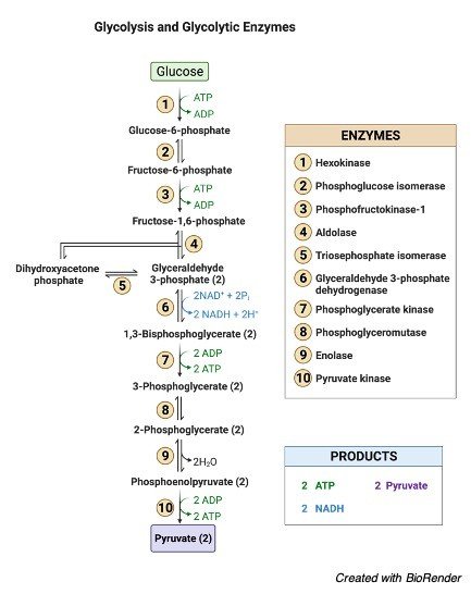
    After hydrolysis, the glycerol is converted into dihydroxyacetone phosphate, an intermediate form of glycolysis.

    The fatty acids are catabolized by the process β-oxidation; which converts it into a variety of end products which can be used by the cell for the producing energy.

    Tributyrin oil is one of the types of lipid known as triglyceride. Other lipase tests are used as a various source of fat involving soybean oil, corn oil, olive oil, peanut oil, egg yolk.

    Tributyrin agar is a differential medium, which tests the ability of an organism to produces an exo-enzyme, known as lipase, that hydrolyses tributyrin oil.

    Tributyrin oil is prepared in the form of an emulsion, so that the agar appears opaque. When the plate is inoculated with a lipase-positive organism, clear zones will appear around the growth as the evidence of lipase activity. If no clear zones appear, the organisms are lipase negative.

    Lipase Test Media

    Tributyrin agar: Peptic digest of the animal tissue at 5.0 gram/litre, Yeast Extract of 3.0 gram/litre, Agar 15.0 gram/litre, pH is maintained at 7.5.

    Lipase Test Procedure

     First the tributyrin agar medium is inoculated in a single line streaking of the organism.

     Inoculated medium is incubated in a gas-Pak jar immediately after streaking anaerobically at a temperature of 35 to 37ºC for about 24 to 48 hours.

     Then the clear zone can be observed around the area of bacterial growth.

    Lipase Test Results

     In case of positive result, a clear zone is observed around the area of bacterial growth.

     If the test is negative, then there will be not be any clear zone around the area of bacterial growth.

    Lipase Test Importance

     The lipase test, is generally used for detecting the enumerate lipolytic bacteria, specifically in diary products which contains high amount of fats.

     It also helps in detecting the variety of other lipid substrates such as olive oil, corn oil, and soya bean oil and also helps in selecting the characteristics of other members such as Enterobacteriaceae, clostridium, staphylo coccus and Neisseria.

     Clinically the blood test for lipase helps in detecting most of the acute pancreatic diseases and other characteristics symptoms related to pancreas.

     Lipase test is often performed either amylase test which helps in detecting the pancreatic diseases.

    Lipase Test Citations

    Share

    Similar Post:

    Continue Reading
  • Optochin Susceptibility Test: Principle, Steps, and Importance

    Optochin Susceptibility Test

    Optochin susceptibility test is usually done for identifying the species of Streptococcus pneumoniae.

    Differentiating Streptococcus pneumoniae from the other species of Streptococci variants depends on demonstrating optochin susceptibility, bile solubility, reaction with a specific DNA probe, or detection of species-specific capsular polysaccharides.

    Most of the clinical microbiology laboratories depends on the optochin susceptibility test.

    Initially Optochin susceptibility was first described for differentiating pneumococci, from other α-hemolytic streptococci in the year 1915, but this test is being virtually unused in the laboratories in the mid-1950s.

    Optochin is a chemical named as ethylhydrocupreine hydrochloride which is completely soluble in water.

    Optochin is used to identify Streptococcus pneumoniae, and alpha hemolytic Streptococcus which is mostly susceptible to the chemical, Optochin.

    Where as the species of alpha-hemolytic Streptococcal species are resistant to optochin.

    This test helps in determining whether bacterium is either sensitive or resistant to the chemical.

    This test is also widely used in the form of filter paper discs or impregnated with ethylhydrocupreine hydrochloride, and they are applied directly on the inoculated plates before incubating.

    Optochin test is one of the tests which can be performed in shorter time than the other tests like bile solubility test.

    Features of Optochin Susceptibility Test

    Streptococcus pneumonia is most commonly found in the respiratory tract of the humans, as other pathogens like Streptococci, and it has a hemolytic pattern which helps in distinguishing it from the other alpha-Streptococci and the Lacto cocci.

    Optochin is a chemical that is used in the presumptive identification of alpha-hemolytic Streptococcus pneumoniae, which is sensitive to optochin.

    Where as the other species like alpha-hemolytic Streptococcus species are resistant to optochin.

    Optochin is completely soluble in water.

    This test is widely used in the form of filter paper disks.

    Bowen and Jeffries in the year 1955 impregnated the disks with the reagent ethylhydrocupreine hydrochloride that are applied directly to inoculated plates before incubation to demonstrate the susceptibility of pneumococcus for identification.

    Optochin Susceptibility Test Principle

    Streptococcus pneumoniae is generally found in the respiratory tract and it also has its hemolytic pattern which is identical to other alpha-hemolytic Lacto bacilli and streptococci.

    Streptococcus pneumoniae is sensitive to the chemical optochin, which is not able to form colonies in its presence and as and its change in surface tension and causes the cell membrane of Streptococcus pneumonia to lyse.

    Optochin is a chemical which is quinine derivative and it has a capacity to soluble in water where all the alpha-hemolytic streptococcal species are resistant to this chemical.

    Optochin susceptibility test is usually employing in identifying the streptococcus which is sensitive to the chemical optochin.

    For determining the susceptibility of the organism, Streptococcus pneumonia, filter paper disks are impregnated with the chemical optochin and it is used in a disk diffusion test.

    This test is usually performed on blood agar, which is a zone for creating inhibition of the lysis of the cell membranes in the Streptococcus pneumonia cells surrounding the disk which helps in determining the positive test.

    This test is very easy to perform and it also economic and has a sensitivity of more than 95%.

    Optochin Susceptibility Test Requirements

    • 5% of sheep blood agar.

    • Optochin disks- each disk should be impregnated with 5µg of optochin

    • Incubator

    • Sterile forceps

    • Sterile inoculating loops

    Optochin Susceptibility Test Procedure

     With a help of an inoculating loop, 2 to 3 streaks are drawn on a well isolated colonies of a pure culture medium, which is tested on 5% of blood agar.

     Optochin disks are placed on the inoculated surface of agar with the help of sterile forceps.

     Ensure whether the disks are adhered firmly to the surface of the agar by using a sterile forceps or loops.

     Then the plates are placed in an incubator at temperature of about 35 to 37ºC.

     Observe the growth on the surface of the blood agar plates and finally the inhibition of the diameter zone is measured including the diameter of the disk plate.

    Optochin Susceptibility Test Results

    Positive Results:

     If the zone of inhibition is detected as 14mm or greater than that and a diameter of 6mm disk, it results in the positive condition.

     If the zone of inhibition is formed lesser than 14mm the strain is identified as pneumococcus only if it is soluble in bile.

    Negative Results:

     If there is no detection of any formation of zones, the test is negative and it indicates the absence of Streptococcus pneumoniae.

    Optochin Susceptibility Test Limitations

     Usually, Streptococcus pneumoniae are isolated and they must be incubated in a carbon-dioxide enriched environment, as some isolates will grow poorly or it does not grow at all.

     Optochin susceptibility is one of the presumptive tests which is recommended as one of the important biochemical tests which is used to perform the complete identification process.

     Any zone of inhibition which is less than 14mm is not enough for performing the test of pneumococci, as the strain is important for identifying the pneumococcus with confirmation by a positive solubility test of bile or serology is performed.

    Optochin Susceptibility Test Citations

    Share

    Similar Post:

    Continue Reading
  • Deoxyribonuclease Test (DNase Test): Principle, Steps, and...

    Deoxyribonuclease Test

    Deoxy ribonucleic acid is one of the large molecule sized polymer, which is being composed of multiple nucleotides in the form of monomer and it is large in size and it is not able to enter cell membrane of the bacteria directly.

    Thus, microorganisms like bacteria produces an enzyme known as Deoxyribonuclease, which helps in breaking down the DNA into smaller monomers, so that the cell membrane can easily absorb or intake it.

    The nucleotides thus taken are used for making nucleic acid in the bacteria, and it also acts as a source of nitrogen, carbon and phosphorus.

    Some microorganism even produces extracellular DNase which breaks down the larger mass of DNA into smaller monomers, so that it is easy for the taking up the vital nutrients that are necessary for the organism through cell membrane by means of protein transports that are present on the membrane of the cell.

    But the degradation of DNA is a virulent factor, which causes serious infections in the hosts cell and it results in various drastically symptoms.

    The ability to produce DNase can be used in differentiating a different pathogenic organism. Thus, Deoxyribonuclease is an enzyme which helps in catalyzing the hydrolytic cleavage of phosphodiester bonds, which forms the back bone of the DNA, which results in degradation of the DNA.

    Deoxyribonuclease is also considered as one of the types of nuclease, a generic form for enzymes that are capable of hydrolyzing phosphodiester bonds.

    DNA ribonuclease test is often called as DNA hydrolysis test or DNase test, which helps in determining the ability of the organism to hydrolyze DNA and to utilize it as a source of energy and carbon for the growth.

    What is Deoxyribonuclease Test (DNase Test)?

    Deoxyribonuclease test is one of the biochemical tests which is being performed to differentiate the organisms on the basis of their ability to produce the enzyme Deoxyribonuclease.

    This test is used presumptively to differentiate one of the species of bacteria known as Staphylococcus aureus, which also plays an important role in producing deoxyribonuclease, the enzyme from the other species of staphylococci.

    As Staphylococcus aureus produces heat stable enzyme, a thermo nuclease.

    To detect this enzyme, initially the organisms are destroyed using heat and then the free DNase reacts in the medium.

    This test also gives positive results for Vibrio, Helicobacter, Moraxella, Aeromonas, Serratia.

    Deoxyribonuclease Test Principle

    This test is used to determine the ability of a particular organism to produce the enzyme DNase.

    DNase are the extracellular endonucleases, which cleaves the DNA and there by releases free nucleotides and the phosphates.

    To detect these enzymes, DNase agar is used to detect the hydrolysis of the DNA, without the use of any indicators.

    When without using any of the indicator, DNA agar helps us to observe the hydrolysis of DNA by clearing the agar after adding HCL.

    This acid helps in precipitating the unhydrolyzed DNA and makes the medium opaque.

    Hence, DNase producing the colonies produces, colonies which hydrolyses the DNA and results in clear zone around the growth of bacteria.

    When DNase is left to react with methyl green it produces, mint green color by combining with DNA.

    On hydrolyzing DNA, complex is released and the methyl green changes into a colorless compound at changes its structure when a pH of about 7.5.

    Hence it results in the formation of clear halo like appearance around the areas where the organisms producing DNase are grown.

    If toluidine blue O is added to the DNase agar.

    It results in the formation of complex in the DNA and changes its structure on hydrolyzing the DNA resulting in bright pink color.

    Deoxyribonuclease Test Materials

    Media: DNase agar or DNase agar with methyl indicator is generally used.

    Reagents: Hydrolytic acid is used only when DNase agar is used without using an indicator.

    Supplies used: Inoculating loop, Bunsen burner.

    Deoxyribonuclease Test Procedure

    Initially the Surface of the agar plates are dried before using, Each plate is divided into sections by drawing the line below the glass plate.

    Then agar medium is inoculated by either of the following methods.

    Spot Inoculation Method

    o Touch the colony of the organism under test using a loop and inoculate it on the small area of the test agar plate, in the center of the marked area in any one of the sections, to form a thick plaque of growth about 5-10mm in diameter after incubating.

    o The incubation is done at a temperature of about 37ºC for 18 to 24 hours.

    o Band or Streak line inoculation.

    o Here heavy inoculum is used and a line of about 3 to 4 com long is drawn from rim to the center of the DNase agar plate.

    o Plate is then incubated at a temperature of 37ºC for about 18 to 24 hours.

    DNase Agar Test Without Indicator

    o The plate is being flood with 1N of hydrochloric acid.

    o After flooding, the plate is left to stand for few minutes until the reagent is being absorbed into the plate.

    o Excess hydrochloric acid is decanted and the plate is further examined in a dark background.

    Deoxyribonuclease Test Result

    1. Positive Deoxyribonuclease Test: When DNA is hydrolyzed, methyl green is released which turns the medium colorless around the test organism.

    2. Negative Deoxyribonuclease Test: In case of negative result, there will be no degradation of DNA, and the medium remain green without any change or absence of color.

    Organisms Tested for Deoxyribonuclease Test

    Positive organisms in DNase test:

    o Serratia Marcescens

    o Staphylococcus aureus

    o M. Catarrhalis

    o Campylobacter jejuni

    DNase test negative organisms:

    o Staphylococcus epidermidis

    o Neisseria gonorrhoeae

    Deoxyribonuclease Test Citations

    Share

    Similar Post:

    Continue Reading
  • Glycolysis vs Fermentation: Definition, Function, and Steps

    Glycolysis

    Glycolysis: (anaerobic, occurs in the cytoplasm) Can be performed in all cells.

    Phosphorylated molecules cannot diffuse through the membrane.

    The process of ATP production in glycolysis is called Substrate-level phosphorylation.

    Glycolysis - Definition and Examples - research tweet

    Net Glycolysis Equation

    Glucose + 2ATP + 2NAD+ + 4ADP + 2Pi = 2pyruvate + 2ADP + 2 NADH + 2H+ + 4ATP + 2H2O

    Reduced Glycolysis Equation

    Glucose + 2NAD+ + 2ADP + 2Pi = 2pyruvate + 2NADH + 2H+ + 2ATP + 2 H2O

    "In Glycolysis, 2 net ATP (4 total made, but 2 needed to complete this stage)"

    2 NADH produced (making 4 ATP in ETC for eukaryotes and 6 ATP for prokaryotes) via reduction of NAD+

    2 pyruvate (3 carbons) produced Pyruvate can then be converted into various products:

    1. It can be oxidized to give the acetyl group of acetyl- coenzyme A, which is then oxidized completely to give CO2.

    2. It can be reduced to lactate (a type of fermentation).

    3. It can be converted to ethanol and CO2 (another type of fermentation)

    ADP & AMP is a activator for glycolysis

    ATP and Citrate are inhibitors for glycolysis In order for glycolysis to continue operating the produced NADH must be oxidized back into NAD+

    Fructose can enter glycolysis as fructose 6-phosphate!!

    Fermentation

    Fermentation is anaerobic, occurs in the cytoplasm.

    It includes the process of glycolysis

    Animal tissues reduce pyruvate to lactate under anaerobic conditions, at the same time oxidizing NADH back to NAD+, which is needed for continued glycolysis.

    Reaction is catalyzed by lactate dehydrogenase. Lactate can be recycled; it is carried in blood to the liver where it is converted back to glucose (in a process requiring the input of energy).

    The acidification of muscle explain why continued exercise can cross cramping, aka Acidosis.

    Yeast produce ethanol and CO2

    0 ATP is produced.

    Low [NAD+]/High [NADH] is a driver for fermentation

    Pyruvate Decarboxylation

    Aerobic, occurs in the cytoplasm for prokaryotes, mitochondrial matrix for eukaryotes).

    Both pyruvate and NADH move through the mitochondrial membrane through a large membrane protein called porin.

    The inner mitochondrial membrane, however, is less permeable.

    Although pyruvate moves into the matrix via facilitated diffusion, each NADH (depending upon the mechanism used for transport) may or may not require the hydrolysis of ATP (this is why the NADH in glycolysis only produce 4 ATP instead of 6).

    Pyruvate decarboxylation is the biochemical reaction that uses pyruvate to form acetyl-CoA, releasing NADH, a reducing equivalent, and carbon dioxide

    Total: 

    2 CO2

    0 ATP produced

    2 NADH produced, for the decarboxylation of both pyruvate (making 6 ATP in ETC)

    Glycolysis vs Fermentation Citations

    Share

    Similar Post:

    Continue Reading