Hyaline Cartilage: Definition, Function, and Examples
What is Hyaline Cartilage?
Hyaline cartilage tissue is a kind of cartilage tissue that is also known as hyaline connective tissue or hyaline tissue. It’s the most prevalent kind of cartilage, with a lustrous, smooth look.
Cartilage is a tough and pliable connective tissue that protects bone ends, discs, and joints from wear and strain. At the embryonic stage, cartilage acts as the early skeletal structure in various animals, including humans.
The majority of it is replaced by bone as the animal grows. The cartilaginous skeleton of an adult cartilaginous fish (Chondrichthyes) is preserved. When compared to bone tissues, cartilage tissues are more flexible and elastic.
Around the bones of free-moving joints, hyaline cartilage is present. Articular cartilage is what this is called. The tissue present in the walls of the respiratory tract is another example of hyaline cartilage. The bronchi, nose, trachea rings, and rib tips all fall under this category.
Hyaline Cartilage Etymology
Hyaline cartilage is a kind of cartilage that has a lustrous, white, semi-transparent look with a little blueish tinge, according to biology. The name hyaline comes from the Greek word hyalos, which means “glassy,” suggesting the material’s gleaming, smooth look. The larynx, trachea, and bronchi are all places where it may be discovered.
Hyaline Cartilage Structure
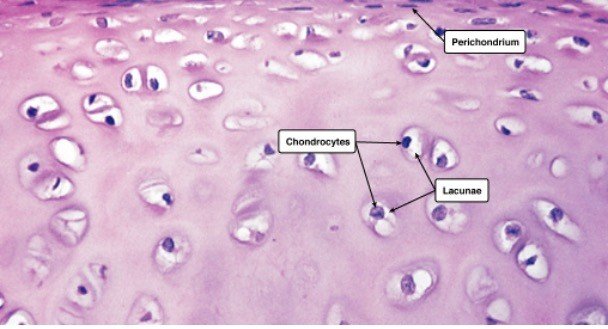
Chondroblasts (or perichondrial cells) generate the extracellular matrix (or ground material), chondrocytes reside in gaps called lacunae, and collagen fibres make up cartilage.
Hyaline Cartilage Location
Mesenchymal cells, which are stem cells present in the bone marrow, give rise to it. Because it lacks blood vessels and nerves, hyaline cartilage has a relatively basic structure. It gets its nutrition from surrounding tissues via diffusion.
Hyaline cartilage has a gleaming, semi-transparent white look with a blueish tint. The name hyaline comes from the Greek word hyalos, which means “glassy,” suggesting the material’s gleaming, smooth look. Surprisingly, as the tissue matures, this look fades. Hyaline cartilage forms the initial skeleton in an embryo, which then changes as the embryo grows. Endochondral ossification is the process that causes this.
Fine type II collagen fibres, chondrocytes (matrix-producing cells), and the extracellular matrix make up hyaline cartilage (or ground substance). Collagen fibres of type II are thinner than collagen fibres of type I. Collagen types I, IV, V, VI, IX, and XI are also found in trace amounts and assist in holding the fibres together.
Glycosaminoglycans (GAGs), proteoglycans, and glycoproteins are abundant in the extracellular matrix, commonly known as the ground material. The extracellular matrix (ECM) covers the gaps between cells and fibres.
Long polysaccharides consisting of amino sugars that attract sodium and potassium ions are known as GAGs. These ions carry water with them. As a result, the amount of water in the extracellular matrix may be controlled.
Sulfated GAGs include chondroitin sulphate and keratan sulphate, whereas non-sulfated GAGs include hyaluronic acid. All of these substances can be present in cartilage’s extracellular fluid.
Proteoglycans and glycoproteins are a combination of amino acids and carbohydrates. They form a gel-like fluid that helps to absorb compression and tension by binding extracellular molecules and components together.
In hyaline cartilage, chondrocytes are the sole cartilage cells. These cells begin as chondroblasts (or perichondrial cells), which generate a cartilaginous matrix before becoming trapped inside it in tiny areas known as lacunae.
Chondrocytes are responsible for the development, repair, and maintenance of the extracellular matrix. Due to their restricted replication ability, chondrocytes have a limited healing capability. They seldom have cell-to-cell communication and are just concerned with preserving their local environment.
The perichondrium covers the hyaline cartilage in most cases. The perichondrium covers the articular cartilage on the ends of growing bones but not in adults. There are two layers to the perichondrium: an exterior layer and an interior layer.
The outer layer is fibrous cartilage that generates collagen fibres, whereas the inner layer contributes to cartilage development by producing chondroblasts or chondrocytes.
Hyaline Cartilage Examples
Hyaline cartilage is a kind of articular cartilage. It differs from normal hyaline cartilage in that the chondrocytes at the surface are flattened. It is 2 to 4 mm thick in people. There are no blood vessels, nerves, or lymphatics in it. It has a thick ECM but sparse chondrocytes.
The chondrocytes take on a more normal shape as they go deeper into the tissue. The cells are located in columns with a calcified matrix in the cartilage’s deep layers. Collagen strands create arches, which provide a robust structural arrangement that can bear pressure.
Type II collagen makes up articular cartilage, although it also contains tiny quantities of type VI, IX, X, and XI collagen.
Different zones make up the articular cartilage. The superficial zone is the first, followed by the intermediate transitional zone, the deep zone, and the calcified zone. There are three areas within each zone.
The pericellular region, territorial region, and interterritorial region are the three.
The superficial zone accounts for around 10% to 20% of the cartilage’s overall thickness. Here you’ll find collagen fibres II and IX. It has a significant number of chondrocytes with a flatter look.
The synovial fluid is in direct touch with the superficial zone, which shields the deeper layers from force and stress.
The intermediate zone runs parallel to the superficial zone and serves as a link between the two levels. This zone accounts for about 40-60% of the overall cartilage thickness.
It is made up of more dense collagen fibres and proteoglycans. The chondrocytes in this sample are spherical and in tiny numbers.
The purpose of the intermediate zone is to defend against compacting pressures. Following the intermediate zone, the deep zone gives the best resistance to compacting pressures. It has the largest proportion of proteoglycans and the least amount of water.
Collagen fibres are organised into columns and chondrocytes are positioned at right angles to the surface. It accounts for around 30% of the overall volume of articular cartilage.
Finally, the cartilage is attached to the bone via the calcified zone. It accomplishes this by attaching the deep zone collagen fibres to the subchondral bone.
Hyaline Cartilage Histology
Hyaline cartilage connective tissue is made up of cells and fibres inside an extracellular matrix, as previously stated. Hyaline cartilage histology explains the appearance of hyaline cartilage under a microscope.
The shape of the chondrocytes might be spherical or angular. The cells in mature cartilage are found in isogenous clusters, each produced from a single progenitor cell. The matrix appears to be optically homogeneous and basophilic.
The explanation for this is that the collagen fibres are obscured by the large quantity of sulfated GAGs in the matrix. Because type II collagen fibres are so tiny, the extracellular matrix appears gleaming and smooth.
Within the extracellular matrix, there is no homogeneous distribution. As a result, the three fundamental zones are visible.
1. The capsular matrix, which is made up of a narrow zone around each lacuna. The greatest concentration of sulfated GAGs may be found here.
2. The capsular matrix is surrounded by a territorial matrix.
3. The interterritorial matrix, which is less basophilic due to a larger amount of collagen and a lower quantity of sulfated GAGs.
Under the microscope, hyaline cartilage may be examined using the hematoxylin and eosin (H & E) staining method as well as the Van Geison staining method. Picric acid and acid fuchsin are used in the Van Geison stain, which turns collagen red.
The staining grows lighter as it gets closer to the territorial matrix’s lacunae. The colour intensities are inverted in the H & E stained sections, although they have higher definition than the Van Geison stain.
The territorial matrix is black in hue, whereas the interterritorial matrix is much lighter. In the H & E technique, groups of chondrocytes may be detected surrounded by these darker regions. Because these chondrocytes come from the same progenitor, they form an isogenous group. Except for articular cartilage, the perichondrium surrounds the cartilage.
Hyaline Cartilage Function
Hyaline cartilage has a small number of fibres and offers a smooth surface for movement as well as a cushion to absorb stress at the point where the bones meet.
The major function of articular cartilage is to create a smooth surface that can resist friction and pressure caused by weight-bearing activities. It supports the softer tissues of the trachea and helps them to retain an open posture.
Hyaline cartilage’s primary purpose is to mechanically support the respiratory system, developing bones, and articular surfaces. The quality of our hyaline cartilage might deteriorate as we get older.
The number of chondrocytes in the surface layer of articular cartilage decreases as people become older, whereas the number of chondrocytes in the deeper layers rises. Additionally, as one gets older, the amount of proteoglycans in the extracellular matrix decreases.
Keratin sulphate levels are also up, whereas chondroitin sulphate levels are down. Hyaluronic acid volume increases as well. Due to its role as a shock absorber and frequent usage in daily activities, hyaline cartilage is prone to wear and strain. All of these characteristics can make hyaline cartilage more vulnerable to injury and illness than other cartilage forms.
Due to a lack of blood flow to the chondrocytes, cartilage tissues are prone to recovering slowly following an injury. This indicates that the matrix is taking a long time to develop. Furthermore, chondrocytes become trapped in lacunae and are unable to move to a damaged region. Scar tissue develops from damaged tissue.
Chondroitin sulphate, an anti-inflammatory mediator that decreases pain, plays a vital role in the extracellular matrix. According to research, its presence slows cartilage breakdown, preventing diseases like osteoarthritis.
Osteoarthritis develops when cartilage wears away, enabling the bones to rub against each other, producing sclerosis (hardening) of the subchondral bone (bone immediately under the cartilage) and inflammation of the synovial membrane, resulting in pain.
Hyaline Cartilage in Other Animals
The skeletons of animals in the Chondrichthyes class are entirely made of cartilage. Sharks and rays are excellent examples. Because cartilage is less thick than bone but yet offers strength, these creatures can move swiftly through the water without expending excessive effort.
Horseshoe crabs, snails, and cephalopods are instances of invertebrates possessing cartilage (predatory mollusks, e.g., octopus and squid). The branchial cartilage of the arthropod Atlantic horseshoe crab (Limulus polyphemus) is abundant in vacuolated chondrocytes, unlike that of any other arthropod.
Another kind of cartilage discovered in this species is endosternite cartilage. It has a higher fibrous content than vertebrate hyaline cartilage. It’s located near the ventral nerve cords and cartilage tissue of the gills.
The cranial cartilage of the octopus (a cephalopod) mimics hyaline cartilage and is one of the only hard sections of the octopus’s body. Cells move from the exterior to the core of the cartilage, causing it to expand. The cartilage of the common cuttlefish (Sepia officianalis) is fibrillar collagen. This cartilage has a development pattern similar to that of vertebrate cartilage.
The odontophore is a cartilage-formed feeding device in gastropods (snails, slugs, or whelks) that offers feeding support. The odontophore is a myoglobin-rich cartilage with a little portion of extracellular matrix and collagen around it.
Finally, cartilage supports the tentacles of feather duster worms (Sabellid polychaetes).
Types of Cartilage
There are three kinds of cartilage in the human body. The most prevalent, but also the weakest, kind of cartilage is hyaline cartilage. Fibrocartilage and elastic cartilage are the other two kinds of cartilage. The descriptions of each cartilage type may be found below.
i. Elastic Cartilage
Consider the distinctions between hyaline and elastic cartilage. Elastic cartilage (also known as yellow fibrocartilage) is a kind of cartilage that gives the body strength and elasticity.
Where can you find elastic cartilage?
The pinna, epiglottis, and laryngeal cartilage, as well as the auditory tube/eustachian tube, are all places where it can be found. Elastic cartilage provides greater elasticity while providing support. A thick network of elastin fibres can be found within it. It doesn’t offer any protection against mechanical stress or compression.
ii. Fibrocartilage
Fibrocartilage connective tissue is a fibrous tissue that is thick, flexible, and supports cartilage.
Where can you find fibrocartilage?
The intervertebral discs of the spine, the jaw, the knee, and the wrist are all places where fibrocartilage can be found. Large bundles of type I collagen may be seen in this fibrocartilage tissue. It is the most durable cartilage.
Fibrocartilage provides resistance to weight-bearing and pressure stresses. Fibrocartilage may be classified into four distinct categories.
1. Intra-articular fibrocartilage is the first group. This works as a cushion between joints that are subjected to a lot of stress and movement. The menisci of the knee are one example.
2. Connecting fibrocartilage, which is present in joints with restricted mobility, such as the intervertebral discs, is the second category.
3. Stratiform fibrocartilage is a kind of fibrocartilage that coats the bone grooves where tendons and muscles are present.
4. Finally, certain articular cavity borders are surrounded by circumferential fibrocartilage, which protects their edges. One acetabular labrum is an example (lining the hip socket).
Elastic cartilage, hyaline cartilage, and fibrocartilage are the three kinds of cartilage. Cartilage is a connective tissue defined by an extracellular matrix rich in chondroitin sulphate and chondrocytes as the cellular component. The most prevalent kind of cartilage is hyaline cartilage.
Hyaline Cartilage Summary
Overall, cartilage is a crucial structural component of the body that may be found in vertebrates and some invertebrates. It is a strong but soft tissue that supports, stretches, and strengthens the body.
Hyaline cartilage between joints demonstrates the importance of cartilage. The cartilage thins as we age, causing inflammation and bone friction.
Researchers in this field are working on studies that will help us better understand the mechanisms that contribute to these diseases and discover strategies to combat/treat/prevent them.

Hyaline Cartilage Citations
Share