Preparation of Cortical Neuronal Cell Culture
In the development and maintenance of the nervous system there is a complex interdependency between neurons and glial cells.
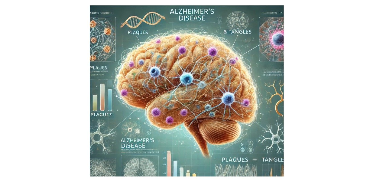
This relationship is vital for their individual differentiation, development, and functionality but also seems to play an important role in progressive neurodegeneration and in the modulation of neurotoxic effects.
Co-cultures of different cells of the nervous system (i.e., neurons, astrocytes, and microglia) represent the easiest approach to:
1) study intercommunication between the different populations of the nervous system,
2) evaluate its relevance in several physiological responses and/or propagation of the damage,
3) study the molecular mechanisms involved. Other possible approaches are to use aggregate cultures of neural and glial cells and the more complex organotypic slices of hippocampus.
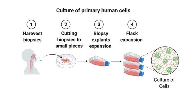
Created with BioRender
Requirements for Cortical Neuronal Cell Culture
Sterile:
1. DMEM-F12 medium
2. B-27 Supplement
3. L-Glutamine
4. HBSS
5. FBS
6. Horse Serum
7. Trypsin (0.1%w/v in sterile calcium free PBS)
8. Cytosine Arabinoside (1mM stock in sterile PBS)
9. Antibiotic mixture
10. Sterile 24 well plates
11. Dissection instruments like scissors, fine forceps, needle, blade
12. Cotton
13. 15ml tubes
14. 0.5ml microfuge tubes
15. Tryphan blue (0.5% w/v in sterile Calcium and magnesium free HBSS)
16. 1ml tips, 200μl tips
17.
Non sterile:
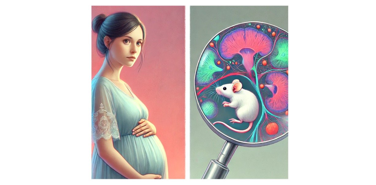
17. Time mated 18 day pregnant female Swiss albino mice
18. Diethyl Ether (for anaesthetizing the animal)
19. 70% alcohol
20. Bench top refrigerated centrifuge
21. Water bath at 37 C
Cortical Neuronal Cell Culture Procedure
1. Neuroglial cultures are obtained from 16 to 18 days old embryonic SA mouse.
2. Anaesthetize the pregnant mouse with ether and remove the fetuses from the uterine horns. Place them in a sterile 60-mm petri dish with ice-cold HBSS.
3. Remove the fetuses from the amniotic sac and remove the brain of the fetuses.
4. Remove the cerebral cortices from the brain and place in ice cold HBSS with Ca and Mg
5. Mince the Cerebral cortices and enzymatically dissociate with 0.5% trypsin in HBSS without Ca++ and Mg++ for 20 min. at 37 ̊C on water bath in 15 ml centrifuge tube.
6. After the enzymatic dissociation mechanically dissociate the cells by means of 1ml pipette. Resuspend the dissociated cells into 2ml of DMEM-F12 medium with 10% Horse serum and 10% FBS
7. Count the number of viable cells using hemocytometer and tryphan blue exclusion method. The number of viable cells should not be less than 80-90%.
8. Plate the cells on to the 6/24 well plate at a density 1×10 – 3×10 cells per well in a culture medium containing DMEM-F12 supplemented with 10% Horse serum
9. After 48 hrs of the plating add Ara C (Cytosine arabinocyde 10μM final concentration). Addition of Ara C will help neurons to out compete in growth from glial cells
10. 24 hrs later remove the existing medium and add fresh DMEM-F12 medium with 10% Horse Serum and then every 3-4 day.
11. All the experiments can be carried out on the confluent cells (After 7-10 days.)
Cortical Neuronal Cell Culture Citations:
Share


