Ribosomes: Function, Definition, Structure, and Formation

  • Reading time:7 mins read

What are Ribosomes?

A ribosome is a complex structure found inside the living cells that produce proteins from amino acids during the interaction called protein union or interpretation.

The interaction of protein amalgamation is an essential capacity, which is performed by all living cells.

Ribosomes are specific cell organelles and found in both prokaryotic and eukaryotic cells.

Each living cell requires ribosomes for the creation of proteins. This cell organelle additionally works by restricting to a courier ribonucleic corrosive (mRNA) and interpreting the data conveyed by the nucleotide arrangement of the mRNA.

The exchange RNAs (tRNAs) involving amino acids, go into the ribosome at the acceptor site. When it gets tie up, it adds amino acid to the developing protein chain on tRNA.

Features of Ribosomes

A ribosome is a complex of RNA and protein and is, in this manner, known as a ribonucleoprotein.

It is made out of two subunits – more modest and bigger. The more modest subunit, where the mRNA ties and is decoded and in the bigger subunit, the amino acids get added. Both of the subunits contain both protein and ribonucleic corrosive parts.

Ribosomes- Function, Definition, Structure, and Formation - research tweet 1

The two subunits are joined to one another by associations between the rRNAs in one subunit and proteins in the other subunit.

Ribosomes are situated inside the cytosol found in the plant cell and creature cell. The ribosome structure incorporates the accompanying: It is situated in two spaces of cytoplasm. Dissipated in the cytoplasm.

Around 62% of ribosomes are included RNA, while the rest is proteins. The construction of free and bound ribosomes is comparative and is related with protein combination.

Ribosomes Function

It gathers amino acids to shape proteins that are fundamental for complete cellular capacities. The DNA produces mRNA by the cycle of DNA record. The mRNA is combined in the core and moved to the cytoplasm for the interaction of protein union.

Ribosomes- Function, Definition, Structure, and Formation - research tweet
Ribosomes during protein synthesis

The ribosomal subunits in the cytoplasm are bound around mRNA polymers. The tRNA then, at that point combines proteins. The proteins blended in the cytoplasm are used in the actual cytoplasm, the proteins orchestrated by bound ribosomes are moved external the cell.

Structure of Ribosome

• Ribosomes are non-layer bound cell organelles made of RNA and proteins.

• Ribosomes are mind boggling atomic machine that make proteins from amino acids in the measure called protein union or interpretation.

• Every cell needs ribosomes to produce proteins. Ribosomes are found in both prokaryotic also, eukaryotic cells.

• Ribosomal construction and capacity are strikingly comparative in all organic entities and organelles.

• In eukaryotic life forms, ribosomes are found in cytoplasm, mitochondria, and chloroplast.

• The cytoplasmic ribosomes are discovered free in the cytoplasm and appended to the endoplasmic reticulum. The endoplasmic reticulum related with ribosomes are called rough endoplasmic reticulum.

• The ribosome found in mitochondria and chloroplast are fundamentally like the prokaryotic ribosomes.

• All prokaryotic ribosomes are discovered free in the cytoplasm of the cell.

• Each E. coli cell contains at least 15,000 ribosomes, which involve almost a quarter of the dry load of the cell. Mammalian cells contain around 10 million ribosomes.

• The ribosomes are made of two subunits, the enormous and the little subunit which involves ribosomal RNA (rRNA) and proteins.

• In prokaryotes, ribosomes are made up from three distinct rRNA molecules, while in eukaryotes ribosomes are made up from four unique rRNA molecules.

• rRNA furnishes the ribosome with its essential structure and capacity and proteins in ribosomes help fill in primary holes and upgrade the amalgamation of proteins.

• The mass units of ribosomes are their Svedberg (S) values, which depend on how quickly the subunits settle to the lower part of test tubes under the centripetal power of a rotator.

• The ribosomes of eukaryotic cells typically have Svedberg upsides of 80S and prokaryotes have 70S ribosomes.

Prokaryotic Ribosome Assembly

Prokaryotes contain 70S ribosomes whilst eukaryotes contain 80S ribosomes. In any case, the construction what’s more, capacity of ribosomes is comparative in the two prokaryotes and eukaryotes.

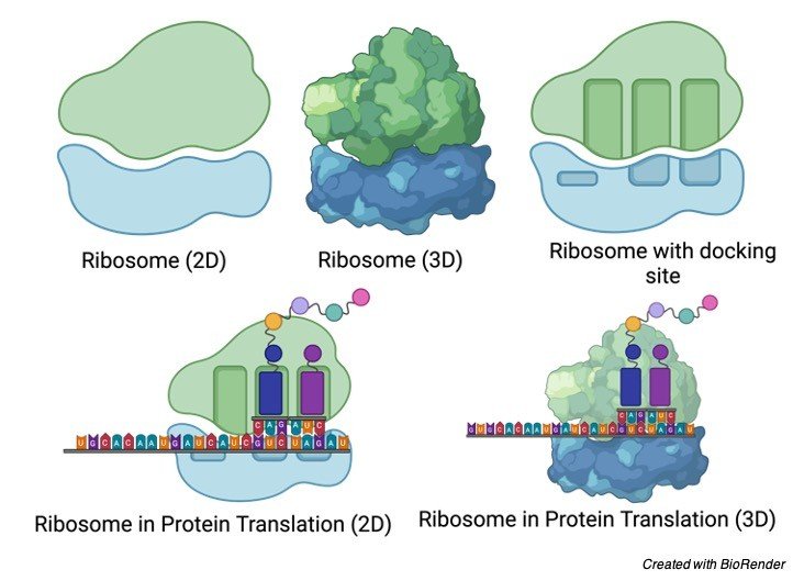
Bacterial ribosomes contain about 65% rRNA and 35% proteins. The measurement of ribosomes is around 18 nm and are made out of two inconsistent subunits with sedimentation coefficients of 30S and 50S and a joined sedimentation coefficient of 70S.

In the last part of the 1960s Masayasu Nomura and partners showed that both ribosomal subunits can be separated into their RNA and protein parts, then, at that point reconstituted in vitro.

The rRNA and proteins precipitously reassemble to frame 30S or 50S subunits almost indistinguishable in construction and action to local subunits.

The primary high-goal designs of bacterial ribosomal subunits were clarified by Thomas Steitz, Ada Yonath, Venki Ramakrishnan, Harry Noller and others. They clarified that the ribosomal subunits are tremendous RNA molecules and the customary spotlight on the protein segments of ribosomes was moved.

In the 50S subunit, the 5S and 23S rRNAs structure the primary center. The proteins are auxiliary components in the complex, finishing the surface.

There is no protein inside 18 Å of the dynamic site for peptide bond development. The high goal structure affirms, the ribosome is a ribozyme notwithstanding the knowledge that the itemized designs of the ribosome and its subunits give into the component of protein amalgamation, they have invigorated another glance at the advancement of life.

The bacterial ribosome is unpredictable, with a consolidated atomic load of approx. 2.7 million.

The two sporadically molded ribosomal subunits fit together to shape a separated through which the mRNA passes as the ribosome moves along it during interpretation.

The 57 proteins in bacterial ribosomes change massively in size and construction. Atomic loads range from around 6,000 to 75,000.

Most of the proteins have globular spaces organized on the ribosome surface. Some additionally have snakelike expansions that jut into the rRNA center of the ribosome, settling its structure.

Eukaryotic Ribosome Assembly

The ribosomes of eukaryotic cells (other than mitochondrial and chloroplast ribosomes) are bigger and more unpredictable than bacterial ribosomes, with a width of around 23 nm and a sedimentation coefficient of about 80S.

They additionally have two subunits, which fluctuate in size among species however on normal are 60S and 40S. Eukaryotic ribosomes contain in excess of 80 unique proteins.

The ribosomes of mitochondria and chloroplasts are to some degree more modest and more straightforward than bacterial ribosomes.

Ribosomes Citations

Share

Similar Post:

Leave a Reply