Author: Admin
-

Non-coding DNA: Sequence, Definition, and Examples
Continue ReadingWhat is Non-Coding DNA?
The non-coding DNA portions are sections of DNA that don’t contain a quality and don’t code for a protein. These locales some of the time alluded to as “Junk DNA” are scattered all through the genome.
The noncoding locales get interpreted however are neither deciphered nor straightforwardly associated with the interaction of interpretation and henceforth no practical protein is created.
Be that as it may, a portion of these locales are thought to have known natural capacity. Certain locales additionally produce records that are included straightforwardly in RNA handling and interpretation, as opposed to being communicated into courier RNAs that encode proteins.
Its incorporates move RNAs (tRNA), ribosomal RNAs (rRNA), little atomic RNAs (snRNA), little nucleolar RNAs (snoRNA), and so on.
The number and variety of these non-coding districts remain basically obscure, even after the finishing of numerous genome arrangements.
A few inquiries like the quantity of non-coding qualities in a genome, their significance, their capacity inside a cell and regardless of whether these huge arrangements of qualities have gone undetected due to their failure to be converted into proteins have consistently stayed a secret.
"Non-coding DNA some of the time alluded to as "Junk DNA" are scattered all through the genome"
To resolve such inquiries, improvement of new precise quality disclosure approaches explicitly focused on non-coding areas is of most extreme need.
The possibility that these classes of qualities have stayed undetected is invigorating, if not distrustful. Non-coding areas are practically missing in bacterial genomes however makes up as much as 90% or a greater amount of the genome in higher organic entities.
Modern comparative genomics studies suggest that genetic variations between any two allied species are more because of the modifications in the non-coding regions slightly than in the protein-coding genes.
Transcription – the process which results in the formation of RNA molecules is the primary way in which genetic information affects a cell’s function.
Studies that employ metabolic labeling of newly synthesized RNA indicated that a vast proportion of nuclear DNA was actually transcribed, and the bulk of this heterogeneous RNA (hnRNA) never access the cytoplasm and hence did not get decoded into proteins.
Novel genomics and RNomic research have cross verified these findings.
Types of Non-coding DNA
I. Non-coding DNA Cis-and Trans-regulatory Components
Cis-regulatory components are arrangements that control the transcription of a close by gene. Numerous such components are engaged with the advancement and control of improvement.
Cis components might be situated in 5′ or 3′ untranslated districts or inside introns. Trans-regulatory components control the transcription of a far-off gene. Advertisers work with the transcription of a specific gene and are commonly upstream of the coding area.
Enhancer groupings may likewise apply exceptionally far off consequences for the transcription levels of genes.
II. Non-coding DNA: Introns
Introns are non-coding segments of a gene, transcribed into the forerunner mRNA succession, in any case eliminated by RNA grafting during the preparing to develop courier RNA.
Numerous introns give off an impression of being versatile genetic elements.
Investigations of gathering I introns from Tetrahymena protozoans demonstrate that a few introns seem, by all accounts, to be narrow minded genetic components, unbiased to the host since they eliminate themselves from flanking exons during RNA preparing and don’t create an articulation inclination between alleles with and without the intron.
Some introns seem to have critical natural capacity, conceivably through ribozyme usefulness that may direct tRNA and rRNA movement just as protein-coding gene articulation, obvious in has that have gotten reliant upon such introns throughout significant stretches of time;
For instance, the trnL-intron is found in all green plants and seems to have been upward acquired for a few billions of years, including in excess of a billion years inside chloroplasts and an extra 2–3 billion years earlier in the cyanobacterial progenitors of chloroplasts.
III. Non-coding DNA: Pseudogenes
Pseudogenes are DNA arrangements, identified with known genes, that have lost their protein-coding capacity or are generally presently not communicated in the cell.
Pseudogenes emerge from retrotransposition or genomic duplication of practical genes and become “genomic fossils” that are nonfunctional because of transformations that forestall the transcription of the gene, for example, inside the gene advertiser area, or lethally adjust the translation of the gene, like untimely stop codons or frameshifts.
Pseudogenes coming about because of the retro transposition of a RNA middle of the road are known as prepared pseudogenes; pseudogenes that emerge from the genomic stays of copied genes or deposits of inactivated genes are non-processed pseudogenes.
Transpositions of once useful mitochondrial genes from the cytoplasm to the core, otherwise called NUMTs, likewise qualify as one sort of normal pseudogene. Numts happen in numerous eukaryotic taxa.
While Dollo’s Law proposes that the deficiency of capacity in pseudogenes is conceivable lasting, quieted genes may really hold work for a few million years and can be “reactivated” into protein-coding sequences and a generous number of pseudogenes are effectively transcribed.
Because pseudogenes are attempted to change without developmental imperative, they can fill in as a valuable model of the sort and frequencies of different unconstrained genetic mutations.
To do again -repeat, transposons, and viral components: Transposons and retrotransposons are versatile genetic components.
Retrotransposon rehashed arrangements, which incorporate since a long time ago scattered atomic components (LINEs) and short mixed atomic components (SINEs), represent an enormous extent of the genomic groupings in numerous species.
Alu successions, named a short sprinkled atomic component, are the most plentiful versatile components in the human genome.
A few models have been found of SINEs applying transcriptional control of some protein-encoding genes.
Endogenous retrovirus groupings are the result of converse transcription of retrovirus genomes into the genomes of germ cells.
Transformation inside these retro-transcribed arrangements can inactivate the viral genome.
More than 8% of the human genome is comprised of (for the most part rotted) endogenous retrovirus groupings, as a component of the more than 42% portion that is conspicuously determined of retrotransposons, while another 3% can be distinguished to be the remaining parts of DNA transposons.
A significant part of the excess portion of the genome that is as of now without a disclosed beginning is required to have discovered its starting point in transposable components that were dynamic such a long time ago (> 200 million years) that arbitrary changes have delivered them unrecognizable.
Genome size variety in somewhere around two sorts of plants is for the most part the aftereffect of retrotransposon sequences.
IV. Non-coding DNA: Telomeres
Telomeres are locales of tedious DNA toward the finish of a chromosome, which give insurance from chromosomal weakening during DNA replication.
Late examinations have shown that telomeres capacity to help in its own dependability.
Telomeric rehash containing RNA (TERRA) are transcripts gotten from telomeres.
TERRA has been displayed to keep up with telomerase movement and stretch the closures of chromosomes.
Non-coding DNA Citations
- Functional variation and evolution of non-coding DNA. Curr Opin Genet Dev . 2006 Dec;16(6):559-64.
- A new method for species identification via protein-coding and non-coding DNA barcodes by combining machine learning with bioinformatic methods. PLoS One . 2012;7(2):e30986.
- Detecting the borders between coding and non-coding DNA regions in prokaryotes based on recursive segmentation and nucleotide doublets statistics. BMC Genomics . 2012;13 Suppl 8(Suppl 8):S19.
- The Genomic Code: A Pervasive Encoding/Molding of Chromatin Structures and a Solution of the “Non-Coding DNA” Mystery. Bioessays . 2019 Dec;41(12):e1900106.
- The mammalian transcriptome and the function of non-coding DNA sequences. Genome Biol . 2004;5(4):105.
- Rate variation in the evolution of non-coding DNA associated with social evolution in bees. Philos Trans R Soc Lond B Biol Sci . 2019 Jul 22;374(1777):20180247.
- Genome defense against exogenous nucleic acids in eukaryotes by non-coding DNA occurs through CRISPR-like mechanisms in the cytosol and the bodyguard protection in the nucleus. Mutat Res Rev Mutat Res . Jan-Mar 2016;767:31-41.
- Searching for functional genetic variants in non-coding DNA. Clin Exp Pharmacol Physiol . 2008 Apr;35(4):372-5.
- Genetic variation: Linear INSIGHTs into non-coding DNA. Nat Rev Genet . 2017 May;18(5):270-271.
- Protection of the genome and central protein-coding sequences by non-coding DNA against DNA damage from radiation. Mutat Res Rev Mutat Res . Apr-Jun 2015;764:108-17.
- The protective function of non-coding DNA in DNA damage accumulation with age and its roles in age-related diseases. Biogerontology . 2019 Dec;20(6):741-761.
Share
Similar Post:
-

Origin of Replication: Definition, Structure, Diagram, and...
Continue ReadingOrigin of Replication
The origin of replication is nothing but a sequence of DNA, at which the replication is initiated on a chromosome, plasmid or a virus. For the small DNAs, which includes bacterial plasmids and other small viruses, where a single origin is sufficient.
Where as the organisms containing larger DNAs have many origins for replicating. The DNA replication is initiated in all of these.
In other cases, if the replication needs to be proceeded from a single origin, it takes a long time to replicate the entire mass of the DNA.
It is said that during each of the process of cell division, usually 30,000 to 50,000 activated origins of replication of DNA are present in humans and it does not remain clear till how these sites are selected and these sites are recognized by the replication factors.
Replication of DNA in the multicellular organisms should accommodate the variations in growth conditions and damage in the DNA.
It also should adapt the changes that is being occurring in the organization of the chromatin which is being associated with the process of cell differentiation and in development of the cell.
However, the selection of origin of replication in the metazoans was being occurred with variety of choices in identifying the cell and also in checking the conditions of the growth.
This also suggests that during the evolution processes, the replication uses its origin to become more controlled by the mechanism of epigenetic factors and mechanisms which affects the chromosomal dynamics and their expression than that of the DNA synthesis.
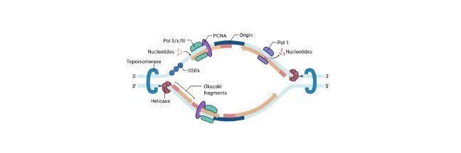
What is Origin of Replication?
The origin of replication, which is also known as replication origin is a particular sequence in a genome where the process of replication is initiated.
Propagation of a genetic molecules between the generations requires accurate and a timely duplication in the DNA by the process of semiconservative replication which occurs prior to the cell division to ensure that each daughter cells receives the full complement of the chromosomes.
This replication process occurs both in prokaryotes and eukaryotes in their genetic material either DNA or RNA, where viruses contain mostly RNA; such as double stranded RNA viruses.
Production of double strands which have been initiated at a discrete site, which are termed as replication origin which further proceeds in a bidirectional manner till all the genomic DNA replicates.
In spites the fundamental nature of this activities, organisms has evolved as divergent variants which control the onset of replication.
Though the replication process is specific in structure and in organizing the recognition sites varies from species to species, where only some common characters are shared.
Characteristics of Origin of Replication
An important need for the replication of DNA is it should occur in extremely high fidelity and should have exact efficiency in one cell per cell cycle which prevents the accumulation of the genetic alterations along potentially deleterious consequences for the survival of cell and in organismal viability.
Incomplete, erroneous or irregular replication of DNA gives rise to the mutations in the chromosomal polyploidy or aneuploidy of the chromosomes along with the variations in the copy number of the gene.
These leads to many diseases and disorders in the cell which also leads to tumor and cancer formations.
In order to ensure complete and accurate duplication in the genome the flow should be maintained correctly to inherit the accurate information to the newly produced cells.
All of these replications should be regulated in an accurate manner along with the cell cycle but it is also important for co ordinating the transcription and DNA repair.
In addition to this, it also originally sequences the commonly having high AT-content across all the cells , as it has number of repeats of adenine and thymine it is easy to separate their base stacking interactions which are not strong as those of guanine and cytosine.
Mechanism of Origin of Replication
DNA replication is divided into different stages. During the initiation the replication machineries which are commonly termed as replisomes are let to assemble on the DNA in a bidirectional fashion.
This assembling of loci constitutes the initiation sites for the process of DNA replication which is also termed as replication origins or origin of replication.
In elongation phases, these replisomes travel in the opposite directions with the replication forks, which unwinds the DNA helix and produces complementary daughter strands of DNA with the help of two parental strands which act as templates, Once the replication is completed, specific termination leads to the disassembly of the replisomes.
As longer the time takes for the duplication of the entire genome before the process of replication, we might assume that the particular location of sites of replication; where the replication starts does not taken into concern.
Although it has been shown that many of the organisms use the genomes regions which they have preferred for the process of replication as their initiation sites.
It is important to regulate the origin of replication because this step likely arises as there is a need to co ordinate the process of replication with the other processes which act on the shared template on the chromatin so that it helps to avoid the DNA strand breaks and the damage that occurs in the DNA.
Origin of Replication Citations
- Mapping the Single Origin of Replication in the Naegleria gruberi Extrachromosomal DNA Element. Protist . 2019 Apr;170(2):141-152.
- Chromosomal origin of replication coordinates logically distinct types of bacterial genetic regulation. NPJ Syst Biol Appl . 2020 Feb 17;6(1):5.
- Do Archaea Need an Origin of Replication? Trends Microbiol . 2018 Mar;26(3):172-174.
- Origin of DNA replication. PLoS Genet . 2019 Sep 12;15(9):e1008320.
Share
Similar Post:
-

Bacteriophage: Definition, Structure, Diagram, and Function
Continue ReadingWhat is Bacteriophage?
Bacteriophage is nothing but an organism which eats a bacterium. As the name suggests that these are viruses which replicate and infect within the cells of the bacteria.
These are commonly called as phages and are found everywhere which is said to be omnipresent.
Bacteriophages contains both DNA and RNA as a nucleic acid in their genomes, which is found encapsulated in a protein coat which also infects other organisms like archaea.
The activity performed by bacteriophages to kill the bacteria is known as bactericidal property.
Which is first observed by Ernest Hanbury Hankins in the Ganges river water, which has the capability to kill cholera bacteria.
There are exists many types of viruses which kills certain bacterias respectively. These viruses act in the same way that how the antibodies affect the cell wall of the bacteria.
They also have the potential to act against antibody resistant pathogenic bacteria.
Features of Bacteriophage
A bacteriophage is made up of a protein coat which is known as capsid, it also encapsulates the genome and also consists a head like structure which is polyhedral in shape. Bacteriophages may be enveloped or non-enveloped.
They also have different shapes. The shapes of bacteriophages include rod-shaped, filamentous, isometric, etc.
The capsid of the bacteriophage is made up many numbers; of capsomeres. The size and shape of the capsomeres varies accordingly depending upon the number of species.
The genome is made up of DNA or RNA, they may be ss or ds DNA or RNA. It may also be linear or colinear.
The genomes which codes for the proteins ranges from four to hundred. Whereas the MS2 bacteriophage genome codes for the four proteins.
However, the largest genome which is present in a bacteriophage is of 735kbp. The tail of the bacteriophage may be long or short, and either be contractile or non-contractile.
The tail contains fibers which helps in anchoring the virus to the bacterial cell wall.
Classification of Bacteriophage
Bacteriophages are classified based on the content of nucleic acid present in there and also based on their morphological characteristics.
There are almost 19 families of bacteriophages, two of these two families belong to the RNA bacteriophages.
Even several types of bacteriophages exist in this environment, but only one type can infect that one specific type of bacteria.
Characteristic of Bacteriophage
They are generally classified into a number of virus families such as Inoviridae, Microviridin Rudiviridae and Tectiviridae.
Like many other viruses’ bacteriophages are also simple organisms which consists a core of genetic material which is surrounded by a protein capsid.
The genetic material present in the bacteriophages may be either RNA or DNA. After infecting a cell, bacteriophages completely take control of the host cells and stops it from producing the components of bacteria and it also forces it to produces viral components which affects the normal activities of the cell.
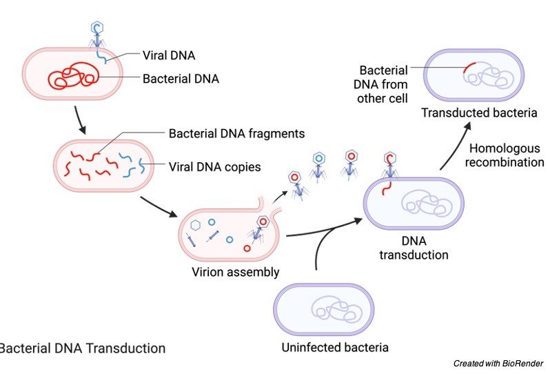
This also eventually induces out the lysis of the host bacterial cell. They are involved in the process of transduction, where the bacteriophages occasionally remove the portion the DNA in the host cell and further transfers it into genome of the host cell.
Structure of Bacteriophage
Generally, a typical bacteriophage is comprising of a head, which is polyhedral in shape, a short collar and a helical shaped tail.
The head of the bacteria phage is polyhedral in shape and consists of about 2000 capsomeres long with the genetic material.

The genetic material is composed of either double stranded DNA or single stranded RNA with is present being enclosed within the head.
The tail of the bacteriophage looks like a hollow tube on its interior side and it is surrounded by a contractile sheath which has about 24 annual rings.
The distal end of the tail is composed of basal plate which has the tail fibers at each of its corner.
Life Cycle of Bacteriophage
When the bacteriophage enters the host cell it inserts its genetic material into the host cell and it undergoes two stages of life cycle namely Lytic cycle and Lysogenic cycle.
Lytic cycle is also known as Virulent cycle, whereas the Lysogenic cycle is known as Temperate cycle.
Lytic Cycle
While undergoing the lytic cycle, bacteriophages tend to infect the host cell and kills it, so that it releases the progeny viruses.
They are several steps involved in the process of lytic cycle as adsorption, penetration, synthesis of phage components, maturation and assembly and release.
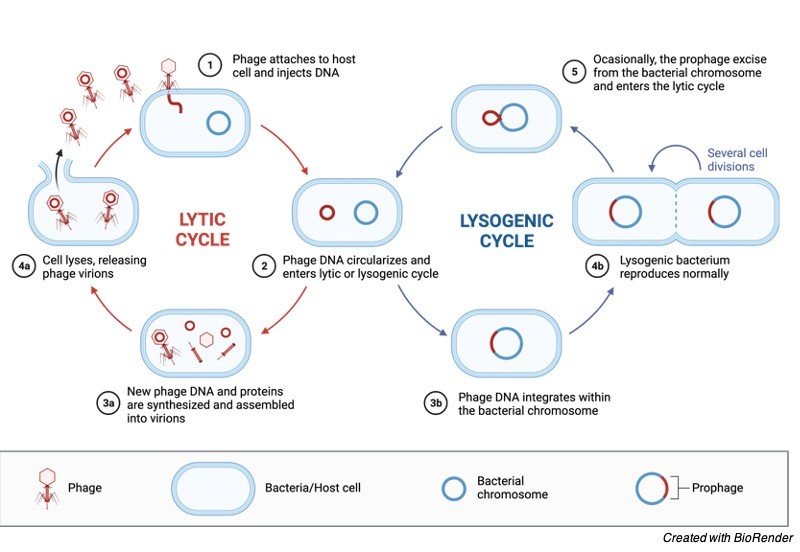
I. Adsorption
This is the first step involved in the process of lytic cycle. Here the bacteriophage attaches itself to the surface of bacteria which is a host cell. It attaches the tip of the tail fibers which attaches specifically to the specific receptor sites on the surface of the host cell.
II. Synthesis of Phage Components
After the release of genetic material from bacteriophage into the host cell many components of new virus particles are released.
The sub units of the bacteriophage which includes the head, tail and late protein appears in the host cell. Whereas the early proteins and specific enzymes helps in carrying out the synthesis.
Components of GH phage are present in the nucleus and cytoplasm of the host cell.
III. Maturation and Assembly
When these cells mature, the head and tail protein of the phage DNA is surrounded by protein coat assemble. Apart from this to these virions are formed by addition of tail structures.
IV. Release
The lysis of the bacterial cell occurs by releasing the progeny during the process of replication, Phage enzymes weakens the cell of the bacteria.
Lysogenic Cycle
It is considered as the other pathway for reproduction of the virus in the host cell. During this phase, the integration of the phage nucleic acid into the host cell genome or either the formation of circular replicon in the host cells cytoplasm take place.
The host cell continuous to live and reproduces normally during this cycle. The genetic material of the phage is called prophage viruses which does not lyse so it is known as lysogenic bacteria.
During the multiplication of lysogenic bacteria, the prophage may lose due to excision.
Bacteriophage Citations
- Bacteriophages and their genomes. Curr Opin Virol . 2011 Oct;1(4):298-303.
- Bacteriophage’s Dualism in Therapy. Trends Microbiol . 2019 Jul;27(7):566-567.
- Bacteriophage electron microscopy. Adv Virus Res . 2012;82:1-32.
- Bacteriophage replication modules. FEMS Microbiol Rev . 2006 May;30(3):321-81.
- Bacteriophage assembly. Viruses . 2011 Mar;3(3):172-203.
Share
Similar Post:
-

Chromosome: Function, Definition, Types, and Structure
Continue ReadingWhat is Chromosome?
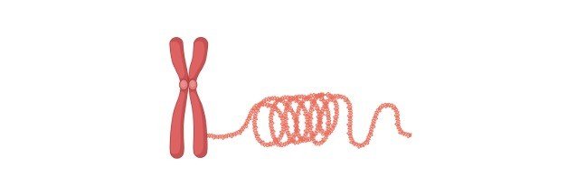
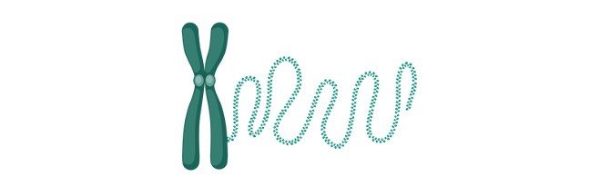
Chromosomes are string like designs present in the core. They are significant in light of the fact that they contain the essential genetic material DNA.
Chromosomes were first found by Strasburger in 1815 and the term ‘chromosome’ was first utilized by Waldeyer in 1888.
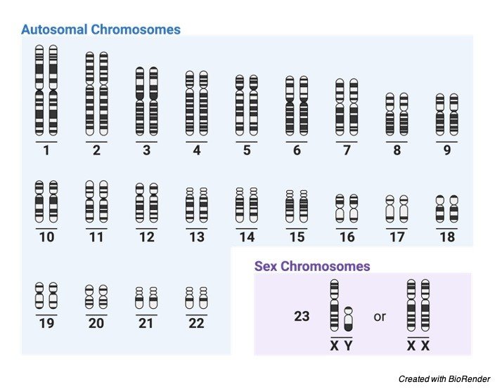
Individuals have 46 chromosomes in their body. These are organized into 23 sets. They assume a fundamental part in cell division, heredity, variety, transformation, fix and regeneration.
In Eukaryotic cells, genetic material is available in the core in chromosomes, which is comprised of exceptionally coordinated DNA molecules with histone proteins supporting its construction.
A chromosome is the construction lodging DNA in a cell. Chromosomes are fundamentally very modern, containing components vital for cycles like replication and isolation.
Every species has a trademark set of chromosomes regarding number and association.
For instance, people have 23 sets of chromosomes- – 22 sets of numbered chromosomes called autosomes, 1 through 22, and one sets of sex chromosomes, X and Y. Each parent contributes one chromosome of each pair to a posterity.
Meaning of Chromosome
“A Chromosome resembles a string and is snaked material, made of proteins. Chromosomes are available in the core of the multitude of cells and contain the essential genetic material DNA, which passes starting with one generation then onto the next”.
Structure of Chromosome
A chromosome has generally 7 sections; Centromere or essential tightening or kinetochore, chromatids, chromatin, optional narrowing, telomere, chromomere, chromonema, and network.
I. Centromere or Kinetochore
It is the essential choking at the middle to which the chromatids or shaft filaments are appended. Its capacity is to empower development of the chromosome during the anaphase phase of cell division.
II. Chromatid
During cell division, a chromosome is isolated into 2 indistinguishable half strands joined by a centromere. A chromatid is every 50% of the chromosome joined.
Every chromatid contains DNA and isolates at Anaphase to shape a different chromosome. The two chromatids are joined to one another by the centromere.
III. Chromatin
It is a complex of DNA and proteins that structures chromosomes inside the core of eukaryotic cells. Atomic DNA is profoundly dense and folded over atomic proteins to fit inside the core.
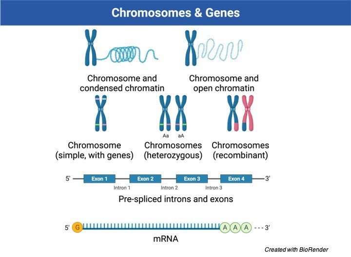
At the end of the day, it is absent as free straight strands. The chromatin comprises of DNA, RNA, and protein.
IV. Auxiliary Constriction
It is generally present for the nucleolar association.
V. Telomere
Telomere is the terminal locale of each side of the chromosome. Ach chromosome has 2 Chromonema. It is a threadlike snaked filamentous construction along which chromomeres are organized.
Chromonema controls the size of the chromosome, and it goes about as a site of gene bearing.
VI. Chromomeres and Chromonema
These are the dot like constructions present on strings or chromonema. These are orchestrated in succession along the length of chromonema.
The quantity of chromosomes is steady, and it is liable for conveying the genes during cell division to the future.
VII. Network
Pellicle is the film encompassing every one of the chromosomes. Lattice is the jam like substance present inside pellicle. It is shaped of non-genetic materials.
Elements of Chromosomes
Interestingly, Sutton and Bover recommended role of chromosomes in heredity in 1902. The main capacity of chromosomes is to convey the fundamental genetic material – DNA.
DNA gives genetic data to different cell capacities. These capacities are fundamental for development, endurance, and generation of the life forms.
Histones and different proteins cover the Chromosomes. These proteins shield it from compound (e.g., chemicals) and actual powers. Hence, chromosomes additionally play out the capacity of ensuring the genetic material (DNA) from harm during the interaction of cell division.
During cell division, shaft filaments joined to the centromeres contract and play out a significant capacity.
The compression of centromeres of chromosomes guarantees exact dispersion of DNA (genetic material) to the girl cores.
Chromosomes contain histone and non-histone proteins. these proteins control gene activity. Cell molecules that control genes work by enacting or deactivating these proteins. This initiation and deactivation extend or contract the chromosome.
Types of Chromosomes
I. Metacentric Chromosomes
Metacentric chromosomes have the centromere present precisely in the middle. Both the areas are metacentric chromosomes are thusly of equivalent length.
Model: Human chromosome 1 and 3 are metacentric.
II. Submetacentric Chromosomes
In Submetacentric chromosomes, the centromere is absent precisely at the middle. The centromere is marginally balanced from the middle. Both the areas are in this manner not of equivalent length or are unbalanced.
Model: Human chromosomes 4 to 12 are submetacentric.
III. Acrocentric Chromosomes
Acrocentric chromosomes have a centromere which is profoundly balanced from the middle. Consequently, one of the strands is extremely long and one exceptionally short.
Model: Human chromosomes 13,15, 21, and 22 are acrocentric.
IV. Telocentric Chromosomes
In telocentric chromosomes, the centromere is available at the finish of the chromosome. Telocentric chromosomes are available in species like mice. People don’t have telocentric chromosomes.
Variations in Chromosomes
The chromosome set of an animal groups remains moderately stable throughout extensive stretches of time. Notwithstanding, inside populaces there can be discovered irregularities including the design or number of chromosomes.
These adjustments emerge immediately from blunders in the ordinary cycles of the cell. Their results are normally injurious, leading to people who are unfortunate or sterile, however in uncommon cases modifications give new versatile freedoms that permit developmental change to happen.
Truth be told, the disclosure of noticeable chromosomal contrasts between species has led to the conviction that extremist rebuilding of chromosome engineering has been a significant power in development.
Changes in Chromosome Structure
Two significant standards direct the properties of a huge extent of underlying chromosomal changes. The main standard is that any deviation from the typical proportion of genetic material in the genome brings about genetic irregularity and strange capacity.
In the typical cores of both diploid and haploid cells, the proportion of the individual chromosomes to each other is 1:1.
Any deviation from this proportion by expansion or deduction of either entire chromosomes or portions of chromosomes results in genomic unevenness.
The subsequent standard is that homologous chromosomes put everything on the line to match at meiosis. The firmly matched homologous locales are joined by a ladderlike longitudinal construction called the synaptonemal complex.
Homologous areas appear to have the option to track down one another and structure a synaptonemal complex whether they are important for typical chromosomes.
Accordingly, when primary changes happen, not exclusively are the subsequent blending arrangements profoundly normal for that kind of underlying change yet they additionally direct the bundling of ordinary and unusual chromosomes into the gametes and therefore into the descendants.
Chromosome Citations
- Chromosome crosstalk in three dimensions. Nature . 2009 Sep 10;461(7261):212-7.
- Organization of Chromosomal DNA by SMC Complexes. Annu Rev Genet . 2019 Dec 3;53:445-482.
- B-chromosome evolution. Philos Trans R Soc Lond B Biol Sci . 2000 Feb 29;355(1394):163-78.
- Chromosome structures. Sci Prog . 1992;76(301-302 Pt 3-4):425-50.
- Mitotic chromosome structure. Exp Cell Res . 2012 Jul 15;318(12):1381-5.
- Mitotic Chromosome Mechanics: How Cells Segregate Their Genome. Trends Cell Biol . 2019 Sep;29(9):717-726.
Share
Similar Post:
-

Plasma Membrane: Definition, Structure, and Composition
Continue ReadingWhat is Plasma Membrane?
The plasma membrane, or the cell membrane, gives security to a cell. It gives a set climate inside the cell. What’s more, that membrane has a few unique capacities.
One is to move supplements into the cell and furthermore to move poisonous substances out of the cell. Another is that the membrane of the cell, which would be the plasma membrane, will have proteins on it which associate with different cells.
Those proteins are often glycoprotein, which suggests a sugar and a protein moiety, or they could be lipid proteins, which means there’s a fat and a protein.
Furthermore, those proteins which stick outside of the plasma membrane will consider one cell to interface with another cell. The cell membrane additionally offers some underlying help for a cell.
Furthermore, there are various sorts of plasma membranes in various kinds of cells, and the plasma membrane has in it in everyday a ton of cholesterol as its lipid segment.
That is not the same as certain different membranes inside the cell. Presently, there are various plants and various microorganisms, like microscopic organisms and green growth, which have diverse defensive systems.
Indeed, they have a cell divider outside of them, and that cell divider is a lot harder and is fundamentally more solid than a plasma membrane is.
The plasma membrane of a cell is an organization of lipids and proteins that frames the limit between a cell’s substance and the outside of the cell. It is likewise essentially called the cell membrane.
The fundamental capacity of the plasma membrane is to shield the cell from its general climate. It is semi-porous and manages the materials that enter and leave the cell.
Structure of Plasma Membrane
Phospholipids: The membrane is somewhat comprised of molecules called phospholipids, which unexpectedly orchestrate themselves into a twofold layer with hydrophilic (“water adoring”) heads outwardly and hydrophobic (“water abhorring”) tails within. These associations with water are what permit plasma membranes to frame.

Proteins: Proteins are wedged between the lipids that make up the membrane, and these transmembrane proteins permit molecules that couldn’t enter the cell in any case to go through by shaping channels, pores or doors.
Thus, the cell controls the progression of these molecules as they enter and exit. Proteins in the cell membrane assume a part in numerous different capacities, for example, cell flagging, cell acknowledgment, and compound movement.
Sugars: Starches are additionally found in the plasma membrane; explicitly, most carbs in the membrane are important for glycoproteins, which are framed when a sugar connects to a protein.
Glycoproteins assume a part in the connections between cells, including cell bond, the cycle by which cells join to one another.
Liquid Mosaic Model of Plasma Membrane
Actually, the cell membrane is a fluid. At room temperature, it has about a similar consistency as vegetable oil. Lipids, proteins, and sugars in the plasma membrane can diffuse unreservedly all through the cell membrane; they are basically skimming across its surface.
This is known as the liquid mosaic model, which was begat by S.J. Vocalist and G.L. Nicolson in 1972.
The extracellular segments of plasma membrane proteins are for the most part glycosylated. Moreover, the sugar segments of glycolipids are uncovered on the external substance of the plasma membrane.
Thusly, the outside of the cell is covered by a starch coat, known as the glycocalyx, shaped by the oligosaccharides of glycolipids and transmembrane glycoproteins.
Part of the job of the glycocalyx is to secure the cell surface. Also, the oligosaccharides of the glycocalyx fill in as markers for an assortment of cell-cell collaborations.
A very much considered illustration of these connections is the bond of white platelets (leukocytes) to the endothelial cells that line veins—a cycle that permits the leukocytes to leave the circulatory framework and intercede the incendiary reaction in harmed tissues.
The underlying advance in attachment among leukocytes and endothelial cells is interceded by a group of transmembrane proteins called selectins, which perceive explicit sugars on the cell surface.
Two individuals from the selectin family (E-selectin and P-selectin), communicated by endothelial cells and platelets, tie to explicit oligosaccharides communicated on the outside of leukocytes.
An alternate selectin (L-selectin) is communicated by leukocytes and perceives an oligosaccharide on the outside of endothelial cells.
The oligosaccharides uncovered on the cell surface subsequently give a bunch of markers that assist with recognizing the unmistakable cell sorts of multicellular creatures.
Importance of Plasma Membrane
I. Plasma Membrane as a Physical Barrier
The plasma membrane encompasses all cells and truly isolates the cytoplasm, which is the material that makes up the cell, from the extracellular liquid external the cell.
This ensures every one of the segments of the cell from the external climate and permits separate exercises to happen inside and outside the cell.
The plasma membrane offers underlying help to the cell. It ties the cytoskeleton, which is an organization of protein fibers inside the cell that hold every one of the pieces of the cell set up.
This gives the cell its shape. Certain life forms, for example, plants and organisms have a cell divider notwithstanding the membrane.
The cell divider is made out of molecules like cellulose. It offers extra help to the cell, and it is the reason plant cells don’t blast like creature cells do if an excessive amount of water diffuses into them.
II. Specific Permeability
Plasma membranes are specifically penetrable (or semi-porous), implying that lone certain molecules can go through them.
Water, oxygen, and carbon dioxide can without much of a stretch travel through the membrane.
For the most part, particles (for example sodium, potassium) and polar molecules can’t go through the membrane; they should adhere to explicit procedures or pores in the membrane rather than uninhibitedly diffusing through.
Along these lines, the membrane can handle the rate at which certain molecules can enter and leave the cell.
III. Endocytosis, Exocytosis and Plasma Membrane
Endocytosis is the point at which a cell ingests generally bigger substance than the single particles or molecules that pass-through channels.
Through endocytosis, a cell can take in enormous amounts of molecules or even entire microorganisms from the extracellular liquid.
Exocytosis is the point at which the cell delivers these materials. The cell membrane assumes a significant part in both of these cycles. The state of the actual membrane changes to permit molecules to enter or leave the cell.
It additionally frames vacuoles, little air pockets of membrane that can move numerous molecules without a moment’s delay, to ship materials to better places in the cell.
IV. Plasma Membrane and Cell Signalling
Another significant capacity of the membrane is to work with correspondence and motioning between cells. It does as such using different proteins and carbs in the membrane.
Proteins on the cell “mark” that cell so different cells can recognize it. The membrane likewise has receptors that permit it to do certain assignments when molecules, for example, chemicals tie to those receptors.
Plasma Membrane Citations
- Caveolae as plasma membrane sensors, protectors and organizers. Nat Rev Mol Cell Biol . 2013 Feb;14(2):98-112.
- Toward a new picture of the living plasma membrane. Protein Sci . 2020 Jun;29(6):1355-1365.
- The Lateral Organization and Mobility of Plasma Membrane Components. Cell . 2019 May 2;177(4):806-819.
- Plasma membrane repair. Curr Biol . 2018 Apr 23;28(8):R392-R397.
Share
Similar Post:
-

What is Gene? Definition, Structure, Expression, and...
Continue ReadingWhat is Gene?
The term ‘gene’ was authored by Danish botanist Wilhelm Johannsen in 1909. It is the essential physical and utilitarian unit of heredity.
Heredity is the exchange of characters from guardians to their posterity that is the reason youngsters take after their folks.
An innate unit comprises of an arrangement of DNA (besides in some infections that contain RNA, all things being equal) that possesses a specific area on a chromosome and decides a specific trademark in a living being.
DNA is a tremendous substance data information base that conveys the total arrangement of directions for making every one of the proteins that a cell will at any point need.
Every gene contains a specific arrangement of instructions, normally coding for a specific protein.
Genes accomplish their belongings by coordinating protein combination. The grouping of nitrogenous bases along a strand of DNA decides the genetic code.
At the point when the result of a specific gene is required, the segment of the DNA molecule that contains that gene parts, and a corresponding strand of RNA, called courier RNA (mRNA), structures and afterward passes to ribosomes, where proteins are integrated.
A second kind of RNA, tRNA, coordinates the mRNA with explicit amino acids, which consolidate in series to shape polypeptide chains, the structure squares of proteins.
Experiments have shown that a significant number of the genes inside a cell are dormant much or even constantly, however they can be turned here and there.
There are around 30000 genes in every cell of the human body. DNA present in the gene involves just 2% of the genome.
Numerous investigations have been made on the very that discovered the area of almost 13000 genes on every one of the chromosomes.
Attributes Controlled by Genes
The human cell contains 23 sets of chromosomes. The quality is one of the attributes controlled by at least one gene. Unusual genes and genes that are shaped because of new changes additionally bring about specific characteristics.
Genes differ in size contingent upon the code or the protein they produce. All cells in the human body contain a similar DNA. The distinction between the cells happens because of the diverse sort of genes that are turned on and, in this way, produce an assortment of proteins.
Genes come two by two similarly as the chromosomes. Each parent of an individual conveys two duplicates of their genes, and each parent passes one duplicate of genes to their kid.
This is the motivation behind why the youngster has numerous qualities of both the guardians like hair tone, same eyes and so on
Elements of Genes
Genes control the elements of DNA and RNA. Proteins are the main materials in the human body which not just assistance by being the structure blocks for muscles, interfacing tissue and skin yet additionally deals with the creation of the compound.
These catalysts assume a significant part in directing different synthetic cycles and responses inside the body. Along these lines, protein union is answerable for all exercises carried on by the body and are essentially constrained by the genes.
Genes comprise of a specific arrangement of guidelines or explicit capacities.
For instance, the globin gene was told to create haemoglobin. Haemoglobin is a protein that assists with conveying oxygen in the blood.
The genetic plan contained in the nucleotide succession can decide the aggregate of a person. The inherited units, which are sent from one generation to the cutting edge are called genes.
A gene is a basic organic unit like molecule which is the key actual unit. Mendel was the primary researcher who proposed genes as particulate units and called them inherited components or variables.
In any case, the idea of gene has gone through a impressive change since Mendel’s time.
Present Day Concept of Gene
A gene can be depicted as a polynucleotide chain, which is a fragment of DNA. It is a practical unit controlling a specific attribute, for example, eye tone.
Beadle and Tatum closed by different investigations that gene is a portion of DNA that codes for one chemical.
They proposed one gene-one protein speculation. In any case, as certain genes code for proteins that are not chemicals, the meaning of gene was changed to one gene-one protein speculation.
Protein Hypothesis
The idea of gene has gone through additional progressions as the new realities became exposed. Since proteins are polypeptide chains of amino acids deciphered by mRNA, gene was characterized as one gene-one polypeptide relationship.
A few proteins have at least two various types of polypeptide chains, each with an alternate amino corrosive grouping.
They are results of various genes.
For instance, hemoglobin has two sorts of chains an andβ chains, which vary in amino corrosive arrangement what’s more, length.
They are encoded by various genes. In this manner, gene is characterized as one geneone polypeptide relationship.
Primary and Regulatory Genes
Indeed, even the one gene-one polypeptide definition isn’t finished as it does exclude gene which codes for rRNA and tRNA. Just mRNA is converted into proteins.
Thusly genes which code for polypeptides and RNAs are called underlying genes. Notwithstanding primary genes, DNA additionally contains a few successions that have just administrative capacity.
These administrative genes establish signals, which “turn on” and “turn off” the record of primary genes and perform different other administrative capacities.
In this manner the meaning of gene incorporates underlying genes just as administrative genes.
As indicated by Lodish and others, gene is characterized as the whole nucleic corrosive grouping that is vital for the combination of a practical gene item, which might be polypeptide or any sort of RNA.
Notwithstanding primary genes (coding genes) it additionally incorporates all the control arrangements and non-coding introns.
Most prokaryotic genes interpret polycistronic mRNA and generally eukaryotic genes interpret monocistronic mRNA.
Number of Genes on a Single Chromosome
Absolute number of genes on a solitary chromosome is distinctive in various living beings. Bacteriophage infection R17 comprises of just three genes, SV40 comprises of 5-10 genes.
E. coli microorganisms have in excess of 3000 genes on single 1 mm long chromosome.
Size of a Gene
In E. coli there are multiple million sets of nucleotides (4638858 base sets). It has been assessed that there are around 3000 genes in E. coli.
The base size of a gene that encodes a protein can be straightforwardly assessed. Every amino acids of a polypeptide chain is encoded by a grouping of three back to back nucleotides in a solitary strand of DNA.
Thusly by estimating the size of the polypeptide chain, the size of a gene can be straightforwardly estimated.
The normal polypeptide chain has around 450 amino acids, which are encoded by 1350 nucleotides.
Hence, in E. coli the quantity of genes will be around 3000 (4000000/1350=3000). Human genome contains around 30000 genes,
Structure of a Gene
A gene is available just in one strand of DNA, which is a twofold abandoned helix. A gene comprises of a few distinct districts.
The fundamental district is the coding arrangement which conveys data in regards to amino corrosive succession of polypeptides.
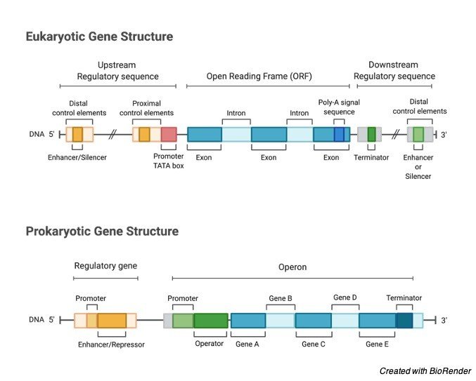
The locale on the left half of coding succession (upstream or less area) and on the right side (downstream or in addition to area) comprises of genuinely fixed administrative groupings.
Administrative arrangements comprise of advertisers which are distinctive in prokaryotes what’s more, eukaryotes.
Types of Genes
1. Straightforward Genes: Basic genes have a coding arrangement of bases in a single DNA strand. Upstream the coding area, the advertiser is available. Downstream, the end locale is available.
2. Split Genes: In the majority of eukaryotes, numerous non-coding successions are available between coding arrangements.
The coding successions of DNA of the genes are called exons. In the middle exons are available non-coding arrangements called introns.
Exons substitute with introns. Typically, introns don’t have any genetic data and are not deciphered. Such genes are called divided genes or intruded on genes.
3. Covering Genes: Most genes comprise of DNA arrangements that code for one protein. In any case, there are a few arrangements that code for more than one protein.
Fredrick Sanger found this marvel in bacteriophage φ x 174. Covering genes are normal in numerous infections.
Here the little length of viral DNA is misused for integrating unique proteins. This is accomplished in an unexpected way.
Now and again, one gene generates two proteins by having diverse beginning stages. Additionally, a similar gene generates two proteins by ending the articulation at various focuses.
In different cases, an arrangement of DNA sees no difference amongst exons and introns. This arrangement of DNA, which utilizes just exons for articulation, likewise employments bordering introns at different occasions for articulation.
The differential joining of a solitary stretch of mRNA prompts covering and, in this way, various proteins. Thusly, numerous proteins can be generated from a solitary stretch of DNA.
4. Jumping Genes or Transposons: Prior it was believed that genes are static and have clear and fixed locus. Be that as it may, as of late it has been found that sections of DNA can leap to new areas in the same or diverse chromosome.
Gene Citations
- Overlapping genes: a window on gene evolvability. BMC Genomics . 2014 Aug 27;15(1):721.
- The origin of new genes: glimpses from the young and old. Nat Rev Genet . 2003 Nov;4(11):865-75.
- Genes, genes and more genes in the human major histocompatibility complex. Bioessays . 1992 Aug;14(8):565-71.
- Origins, evolution, and phenotypic impact of new genes. Genome Res . 2010 Oct;20(10):1313-26.
Share
Similar Post:
-

Vernalization: Definition, Mechanism, and Factors
Continue ReadingWhat is Vernalization?
Light and temperature are two primary external factors which directly influences the plant growth and development; inducing changes at cellular, molecular levels; with distinctive mechanisms: Photoperiodism, Vernalization, Seed dormancy, Climate acclimation, bolting etc.,
All these mechanisms and responses are in accordance with light and temperature. Light and temperature; external determinants; are inevitable in determining the transition of vegetative phase to reproductive phase ensuring the progeny.
Where photoperiodism induces flowering and classify plants based on their photoreception as Short-Day Plants, Long – Day Plants, Day neutral plants, long short-day plants and Short long day plants.
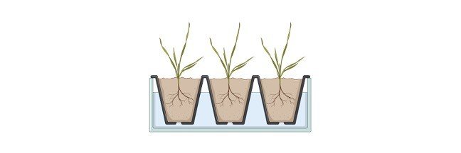
However, temperature also induces the transition and Seed dormancy, Vernalization are evident of the fact. One of the temperature induced growth or mechanism is the vernalization, a quantitative process where the temperature is modified to obtained better yield from plant.
Vernalization is a process of quantitative yield, where plants undergo cold treatment or chilling period during winter, without hindering the vegetative phase of the plants which continues throughout the winter.
The cold treatment prevents the development of premature reproductive phase in early winter preventing poor growth of the plant which eventually might fail during the course of development.
Not all plants require such a treatment for producing reproductive phase, many species are capable of flowering without cold treatment and inducing cold treatment might increase their productivity thereby accelerating the flowering capacity of the plant.
Vernalization is more specific characteristic feature in temperate plants where the sunlight is insufficient for optimal growth.
The plants in this region have developed many modifications to withstand the climatic variations of the region and to yield better.
The more prevalent plants of the region are Biennials and Winter annuals where they grow well in 2 growing season and annuals have one growing season respectively.
History of Vernalization
The phenomenon was first observed at temperate regions of the world. Early agriculturists of the region started sowing cereal seeds that are winter annual type at the end of winter.
This prevented the early development of the plant accompanied by stunted growth and prevents poor yield. The plants made germinating in this period; so that the vegetative phase progress slowly along the reduced growth and on optimal conditions; can support flowering.
Systematic research on vernalization begun by 1858 by Klippart were he found that cold temperature is the determining factor producing qualitative flowering in the following photoperiodic season.
In 1918, Gassner worked extensively on the concept of vernalization and identified plants sensitized for such growth. They are: Biennials and Winter annuals. He also highlighted the importance of germinated seedling which are capable of receiving “chilling treatment” signals and promote growth.
Lysenko in 1928, a Russian genetist who clearly proposed the water imbibition in winter seedling is the key feature which increases the sensitivity to initiate germination and the vernalizing capacity of the plant.
The name vernalization was initially termed as ” Jarovization” were JAR means spring or fire god in Russian. Hence, spring plants are called JAROVOE and he termed the process as Jarovization.
Jarovization is a phenomenon where winter plants behave as a spring plant to produce healthy crops. Later, Lysenko translated Jarovization as “vernalization” derived from Latin.
Concept laid out by Lysenko was scientifically disapproved because he considered vernalization as an inherited acquired characteristic passed on from parent to offspring.
But this is the main lag for the Soviet biology as a whole. Later, an accurate study was taken up by 2 groups at different region of the world with two plants.
Gregory and Purvis studied Petkus Rye a winter annual in London. Melchers and Lang, Tubigen studied on Henbane a biennial plant separately.
These two studies had led a great and detailed understanding of vernalization and its application in agriculture.
Characteristic Features of Vernalization
1. Effects of vernalization is visible after the process had taken place, where the efficiency is seen in reproductive phase of the plant in flowering.
2. It does not induce flowering but retains the capacity under unfavourable condition and to flower during favourable condition.
3. It is a supportive feature for plants during winter, sustains the vegetative growth and produces reproductive efficiency for the plants. Vernalization and breaking of dormancy are two varied phenomenon involving cold temperature.
The main difference during dormancy is the leaf primordia and other organs stops their growth with very minimal or no metabolic activity.
Dormancy is oriented towards the growth phase. But vernalization is substituting cold exposure to prevent unfavourable expression of reproductive phase and at the same time the growth phase remains unaffected.
4. It is species specific and observed only during selective climatic time period. Temperate plants exhibit the phenomenon more clearly than other plants.
Not all plants require cold treatment for productive yield. Few plants on exposure to cold treatment may cause death to the plant.
5. Vernalization is a preparatory process, where essential growth factors and hormones are manufactured and stored. On favourable condition, provided an appropriate photoperiodic stimulation will induce flowering.
6. Vegetative phase retention by vernalization is limited and species specific. The phenomenon employs short time retention of vegetative phase.
A vernalized plant must be exposed to appropriate condition within a time period. When appropriate conditions are unavailable within the limit the plant may not grow well and die earlier.
7. It occurs naturally and can be induced artificially under laboratory conditions. Vernalizing temperature range lies between 1°C – 15°C for most of the plants.
Natural vernalization takes place but not under all circumstances as it will be influenced by various other factors. But under artificial condition factors can be maintained constantly to induce efficient growth in vernalized plant.
8. The vernalized state can be reversed by subjecting it to higher temperature, Low irradiance, darkness immediately after vernalization; will remove the effects of vernalization; hence called as the phenomenon of devernalization.
9. The phenomenon provides competence for the plant to transform from vegetative phase to reproductive phase.
10. Vernalization is followed by photoperiodic period for flowering; is a mandatory process.
11. The response to vernalization is inherited to the dividing cells during vernalization. The dividing cells are present in the flowering parts of the plant, the cell division and DNA replication provides mitotic stability and mitotic inheritance of the response.
Types
Vernalization does not have any types. But based on the requirement to vernalization, plants are divided into Obligate, Facultative plants and certain plants are unresponsive for vernalization.
Obligate Plants: vernalization is essential for flower production (i.e.) transition from vegetative to reproductive phase of the plant without which plant remains in vegetative phase or may die.
Example: henbane – Hyoscyamus niger, sugar beet – Beta vulgaris.
Facultative Plants: Facultative plants does not completely depend on vernalization for flowering but on exposure to vernalization will accelerate and becomes efficient in flowering. The transition from vegetative to reproductive phase does not require the vernalizing phenomenon.
Example: Arabidopsis thaliana, Wheat, Barley.
Vernalization Perception Site
The receptor for vernalization is present in the shoot apex for most of the plant species. In grass family of the temperate zones the receptor sites are present at both leaves and shoot of the plant.
Another specificity of the site of reception is the site must have mitotic division to receive the external environmental cues. The site was identified from various experimental procedures.
Transplantation procedures involving transfer of vernalized apex to non – vernalized plant will induce efficient flowering.
Melchers also found a substance named vernelin in which transverse across different tissues carrying the information of flowering induction and are in coordination with gibberellins or gibberellins will produce vernelin like activity.
Vernalization Genes
Genes such as VRN1, VRN2, VRN3 are essential in determining the vernalization requirements of cereals. Vernalization process involves gene expression regulation of FLOWERING LOCUS C during vernalization.
Mechanism of Vernalization
The mechanism of vernalization is not fully identified. At each step of discovering the process of vernalization, many scientists had laid many hypothesis on their way of discovering. Formally, many theories were given regarding vernalization.
The theories are:
1. Phasic Development Theory
Lysenko, 1934 proposed the theory of phasic development. Vegetative to reproductive transition takes place over a period of time phase after phase involving 2 different exposure of environmental factors.
The exposed factors start after the preceding factor. According to the theory, the plants are initially exposed to the lowest temperature undergoes vernalization.
Plants are then exposed to change in light and temperature which initiates the transition to flowering phase of plant’s life cycle.
Phasic development theory hence has 2 stages: Thermostage and Photostage.
Thermostage depends on the low temperature to accelerate the vernalization. The length of low temperature exposure depends on the plant’s capacity to withstand and maintain the metabolic process.
Photostage requires high temperature to transverse from Vegetative stage to Reproductive stage. The hormone vernelin helps in plants inflorescence. For a winter plant, longer low temperature and appropriate high temperature and long light exposure to produce qualitive flower and seeds.
As a whole, the theory states that vernalization must follow photoperiodic induction for healthy progeny.
2. Hormonal Theory
In 1939, Melcher and Lang proposed this theory and many other authors from their studies revealed a similar mechanism of the theory.
The theory states that the chilling treatment makes the plant to secrete hormones which are responsible to produce reproductive growth in plant body.
The hormone is named as Vernelin and after recent findings and improvement in plant physiology research it is found that Gibberellins has similar action of Vernelin.
To prove the action of vernelin Transportation taking place, Melcher united a vernalized and non – vernalized plant. The flower bloomed in both vernalized and non – vernalized plant.
Chailakhyan also identified the presence of vernelin in plants exposed to vernalization. He also identified the presence of Anthesin in LDP to initiate growth and Absence of Anthesin SDP.
Vernelin and Anthesin produces Gibberellin which induces plant growth. In SDP absence of Anthesin does not induce flowering.
In 1961, Purvis identified that “A” substance is formed from a precursor. On cold temperature A converts to “B”.
B an unstable component during cold environment produces “D” a substance functioning similar to Vernelin or Vernelin itself, on light exposure or photoperiodic induction produces Florigen – a hormone responsible for flowering induces the reproductive phase of the plant.
Factors Affecting Vernalization
1. Age of the Plants: Different types of plants has different receptive capacity of the stimulus. Winter Annuals are capable of producing shoot at the stage of Seedling where it is imbibed with water. on the other hand, Perennials and Biennials require at least 5-week-old to be effective to receive cold stimulus.
2. Temperature: The vernalizing stimulus is effective and receptive under an optimal temperature around 1-6C and the temperature above and below the optimum levels will decrease the receptive capacity of the plants.
3. Site of the Plant: the phenomenon requires continuously dividing mitotic apical meristem to transit the stem or leaf primordia to flower primordia. Hence, the stimulus is receptive at the apices and not induced on lateral side of the plants.
4. Exposure to light: Following vernalization, the plant must be exposed to long light before the effect of vernalization wears off to support the production of efficient reproductive organ.
5. Oxygen: Vernalization takes place under aerobic condition. When devoid of oxygen the plants get devernalized and develops the reproductive phase and the production may or may not be successful in virilization dependent plants.
6. Water: In Winter annuals vernalization has effects on germinating seedling imbibed with water. Dry seeds never respond to vernalization.
Significance of Vernalization
1. Increase the production efficiency of the crop in a year as it reduces the vegetative phase of the plant.
2. Long – Day Plants and Long summer crops raised in tropical regions can be grown in temperate zones by vernalization.
3. Winter stunted growth can be prevented from freezing by introducing vernalized plants in spring.
Vernalization Citations
- Vernalization – a cold-induced epigenetic switch. J Cell Sci . 2012 Aug 15;125(Pt 16):3723-31.
- The transition to flowering in winter rapeseed during vernalization. Plant Cell Environ . 2021 Feb;44(2):506-518.
- Vernalization in cereals. J Biol . 2009;8(6):57.
- Vernalization-mediated chromatin changes. J Exp Bot . 2012 Jul;63(12):4343-8.
- Vernalization-Triggered Intragenic Chromatin Loop Formation by Long Noncoding RNAs. Dev Cell . 2017 Feb 6;40(3):302-312.e4.
- Experiencing winter for spring flowering: A molecular epigenetic perspective on vernalization. J Integr Plant Biol . 2020 Jan;62(1):104-117.
Share
Similar Post:
-

Heterochromatin: Definition, Function, and Structure
Continue ReadingChromosome, Heterochromatin, and DNA
We all know that our body is made up of millions of cells, each cell makes up the structural and functional unit of life. Each cell consists of many cellular organelles such as nucleus, mitochondria, lysosome, etc.
Chromosomes are one of the important components which are present in the nucleus of the cell and contains an organized package of DNA, which is very much important in carrying out the body’s metabolic and enzymatic activities and also has a vital i role in carrying out the hereditary characters.
Chromatin is one of the complexes of the DNA and proteins which forms the chromosomes. Chromatin generally occurs in two forms, namely euchromatin and heterochromatin.
Euchromatin is less condensed and can be transcribed, whereas the heterochromatin is highly condensed and is not typically transcribed.
What is Heterochromatin?
Heterochromatin is a condensed or a tightly packed form of DNA, which comes in various forms. These forms stay in a continuous manner in between the two ends of the constitutive heterochromatin and facultative heterochromatin.
These two play an important role in expressing the genes. As it tightly packed, it was said to be inaccessible to the polymerases and therefore it cannot be transcribed.
If much of this DNA, is transcribed. It is continuously turned over the RNA induced transcriptional silencing, recent studies suggest that electron microscopic staining reveal that the dense packing is not due to the chromatin.
Constitutive heterochromatin affects the genes near itself. It occurs in the repetitive manner and it performs structural functions such as centromeres and telomeres.
It acts as an attractor along with expressing the genes and also in repressing signals.
Facultative heterochromatin is due to the result of genes which are silenced through an activity of the histone deacetylation or Piwi-interacting RNA, which is commonly called as PiRNA through RNAi.
This does not occur in a repetitive manner and it also shares the compact structure of the consecutive heterochromatin.
Under specific developmental or environmental signaling of cues, it can sometimes lose its condensed structure and it becomes transcriptionally active.
Heterochromatin is also associated with the di and tri methylation of H3K9 in some certain portions of the genomes. Here H3K9me3 related methyl transferases appears to have pivotal role in modifying the heterochromatin during the lineage commitment which occurs at the onset of organogenesis and it also maintains the lineage fidelity.
Structure of Heterochromatin
Chromatin usually occurs in two varieties such as euchromatin and heterochromatin. These two forms are distinguished cytologically by assuming how intensely they are getting stained; The euchromatin is stained lightly, whereas the heterochromatin is stained intensely such that it looks dark in color, which indicates that it is a tight packaging.
Heterochromatin is usually located at the end of the nucleus. Inspite of this early dichromia, recent evidences in both animals and plants suggest that there were more than two different states of heterochromatin.
But it has also been said that it exists in four or five states which are marked with different combinations in the epigenetic marks.
Heterochromatin usually has its genetically inactive satellite sequences and many of the genes which represses the various extents.
Even though some of them are not able to express their genes in euchromatin at any of the time. Where as both centromeres and telomeres are heterochromatic, as such as how it is present in the Barr body to the second inactivated X-chromosome n the female.
Function of Heterochromatin
Heterochromatin is usually associated with several functions which forms the regulation of gene to protect the integrity of chromosome.
Some roles of heterochromatin help in attributing the dense packaging of DNA, which helps in making it less accessible to the protein factors to bind DNA or its other associating factors.
Some regions of chromatin are very densely packaged along with the fibers which displays a condition compared to that of the chromosomes at its mitosis stage.
Heterochromatin is usually inherited by cloning, During the process of cell division the two daughter cells are produced which contains heterochromatin, in the same regions of DNA which results in epigenetic inheritance.
Variations in the heterochromatin encroaches on the adjacent genes or recedes from the genes to the extreme of the domains.
Transcribable material can be expressed by positioning at the boundary domains. This gives rise to the levels of expression that are varying in the cells from one cell to the other.
Constitutive Heterochromatin
Usually, all cells in many of the species has the same regions of the DNA as their packaging sites, especially in a constitutive heterochromatin and as a result any genes which is present within the constitutive heterochromatin in all the cells are poorly expressed.
In most of the organisms this constitutive heterochromatin occurs around the region of chromosomal centromere and near the telomeres.
Facultative Heterochromatin
The regions of the DNA which are packaged in the facultative heterochromatins are not consistent in all the cell type within the pieces, and hence the sequence on one cell is packed in facultative heterochromatin, which means that the genes are expressed poorly so that it can packed in a euchromatin of another cell.
However, the formation of this facultative heterochromatin is regulated often and it is associated with the morphogenesis or differentiation.
Yeast Heterochromatin
One of the species of yeast, such as Saccharomyces cerevisiae, which is also commonly called as budding yeast is considered as one of the eukaryotes and it also has a definite structure of heterochromatin.
Most of its genome are characterized as euchromatin where the regions of the DNA are transcribed very poorly, this type of loci are called as silent mating type loci, the rDNA, sub telomeric regions.
Where as the fission yeast uses another mechanism for the formation of heterochromatin in its centromeres.
Heterochromatin Citations
Share
Similar Post:
-

Euchromatin: Definition, Function, and Acetylation
Continue ReadingChromosomes, Euchromatin, and DNA
We all know that our body is made up of millions of cells, each cell makes up the structural and functional unit of life. Each cell consists of many cellular organelles such as nucleus, mitochondria, lysosome, etc.
Chromosomes are one of the important components which are present in the nucleus of the cell and contains an organized package of DNA, which is very much important in carrying out the body’s metabolic and enzymatic activities and also has a vital role in carrying out the hereditary characters.
Chromatin is one of the complexes of the DNA and proteins which forms the chromosomes.
Chromatin, a complex of DNA and proteins which forms the chromosomes with the nucleus of the eukaryotic cells, where the Nuclear DNA does not appear in the form of free strands, rather it is highly condensed and winded around the nuclear proteins to get fit inside the nucleus.
Chromatin generally occurs in two forms, namely euchromatin and heterochromatin.
Euchromatin is less condensed and can be transcribed, whereas the heterochromatin is highly condensed and is not typically transcribed.
What is Euchromatin?
Euchromatin is one of the types of chromatin, which is slightly packed in a condensed form’ which contains structural genes along with it and it is usually transcriptionally active.
It is the genetically active region of the chromosome, which contains structural genes that are replicated during G1 phase and S phase of the interphase which allows the polymerases to access the genes.
It also gives accesses to the RNA gene regulatory proteins and the complexes of RNA polymerases which gives access to the genes to be transcribed into mRNA, the euchromatin regions are always accessible for the process of transcription for the housekeeping genes.
These genes are very much essential for the basic function and the survival of the cell. G-band techniques helps in enabling the visualization and distinction between the euchromatin and heterochromatin regions which are present in the chromosomes.
Euchromatin is always stained lightly where as the heterochromatin stains darkly. The lighter staining of the Euchromatin is due to their slightly packed structure of chromatin.
It is a lightly packed form of chromatin which is enriched with genes and is often active but it is not always active.
It has the most active portion of the genome present within the cell nucleus. Humans contain almost 92% of their cells as euchromatin.
Structure of Euchromatin
Euchromatin has a reminiscent structure which has an unfolded set of beads on a string, where as those beads represents the nucleosomes.
Nucleosomes has eight proteins which are known as histones, which are made up of approximately 147 base pairs of DNA winds around them, in euchromatin this wrapping is loose, so the raw DNA may get accessed.
Each core histone posses a tail like structure, which varies in several ways, it is said that these variations act as a master control switches which determines the over all arrangement of the chromatin.
Particularly it is believed that the presence of methylated lysine four on the tail of the histone acts a general marker. The euchromatin is generally found with its appearance that it has a light color bands which when stained with G-banding techniques, and observed under an optical microscope, it appears opposite to that of the hetero chromatin as heterochromatin have a darker stain.
This lighter staining of the euchromatin is due to the less compact structure which is due to the loosely condensed fibers.
The basic structure of euchromatin is an elongated and fiber which has a size of about 10nm, which looks like microfibrils; when observed under an electron microscope.
Usually in prokaryotes, chromatin is present only in the form of euchromatin and hetero chromatin is absent.
It is also said that heterochromatin structure evolved lately in the nucleus of the cell, which acts as a mechanism to handle the increasing size of the genome.
Function of Euchromatin
Generally, Histone modifications contributes the regulation of DNA transcription. Whereas the genes present in the heterochromatin are not accessible for transcription.
Acetylation promotes the formation of the euchromatin which allows the transcription of the genes.
It helps in active transcription of DNA to mRNA products.
The unfolded structure of euchromatin helps in allowing the gene regulatory proteins and RNA polymerase complexes to bind the DNA sequences.
This initiates the process of transcription. It is also important to know that not all euchromatins are necessarily transcribed, but in general the genes which are non-transcribed are transformed into heterochromatins, these heterochromatins protect the genes when they are not in use.
This is how there is a direct link between the actively producing cell and the amount of euchromatin which can be found in the nucleus.
It is said that the cells uses transformation for converting euchromatin into heterochromatin as method to control the process of expression of genes and in replication, because these processes behave differently on a densely packed or compacted form of chromatin which is known as accessibility hypothesis.
One such hypothesis of the consecutive euchromatin is that it always turns the housekeeping genes, which helps in coding the proteins which are needed for the basic functions of the survival of the cell.
Euchromatin Citations
- Nuclear dynamical deformation induced hetero- and euchromatin positioning. Phys Rev E Stat Nonlin Soft Matter Phys . 2015 Sep;92(3):032709.
- Nuclear dynamics of radiation-induced foci in euchromatin and heterochromatin. Mutat Res . 2013 Oct;750(1-2):56-66.
- Phosphorylated Lamins in Euchromatin: New Clues to Progeria. Dev Cell . 2020 Mar 23;52(6):676-678.
Share
Similar Post:
-

Chromatin: Definition, Function, and Condensation
Continue ReadingChromosome, Chromatin, and DNA
We all know that our body is made up of millions of cells, each cell makes up the structural and functional unit of life. Each cell consists of many cellular organelles such as nucleus, mitochondria, lysosome, etc.
Chromosomes are one of the important components which are present in the nucleus of the cell.
It contains an organized package of DNA, which is very much important in carrying out the body’s metabolic and enzymatic activities and also has a vital role in carrying out the hereditary characters.
Chromatin is one of the complexes of the DNA and proteins which forms the chromosomes.
What is Chromatin?
Chromosomes are made up of complex substances named as chromatin which is composed of DNA and proteins. When it is observed under a microscope, chromatin, looks like a bead present on a string.
The bead like structure are the nucleosomes. Which are composed of DNA, wrapped around with eight proteins which are called as histones.
These nucleosomes are then further wrapped into a spiral coil of about 30nm which is known as solenoid.

Where the additional histone proteins support the structure of chromatin.
During the process of cell division, the structure of chromosomes and chromatin are clearly visible under a light microscope, and the shape gets change when the DNA is duplicated or separated into two cells.
Chromatin, a complex of DNA and proteins which forms the chromosomes with the nucleus of the eukaryotic cells, where the Nuclear DNA does not appear in the form of free strands, rather it is highly condensed and winded around the nuclear proteins to get fit inside the nucleus.
Chromatin generally occurs in two forms, namely euchromatin and heterochromatin. Euchromatin is less condensed and can be transcribed, where as the heterochromatin is highly condensed and is not typically transcribed.
Structure of Chromatin
Usually, chromatins are the complex macromolecules found inside the nucleus and it is composed of DNA and proteins and in few cases, it also has RNA.
It is found inside the nucleus of the eukaryotic cells. Chromatin mostly exists in two forms as mentioned above, as euchromatin and heterochromatin where euchromatin is found in the extended form and heterochromatin as condensed form.
Chromatin is composed of primary proteins called as histones which helps in organizing the DNA into a structure of beads known as nucleosomes, which provides a base for the DNA to get wrapped around.
Commonly a nucleosome consists of 147 base pairs of DNAs, which is wrapped around a set of eight proteins; named as octomer.
This nucleosome can be further folded to produce the fibers of chromatin. Chromatin fibers are coiled and then condensed to form chromosomes.
Chromatin helps in numerous cell functions such as DNA replication, transcription, DNA repair, genetic recombination of DNA, cell division etc.
Function of Chromatin
Chromatin undergoes various processes like Packaging of DNA, regulation of transcription, and also in the process of repairing the DNA.
These functions are discussed below.
I. Chromatin and DNA Packaging
Packaging of DNA, is considered as one of the important and fundamental function of Chromatin. Here the long strands of DNA are compressed.
Usually the length of DNA, in the nucleus is very large compared with the other DNAs and it is far greater than the compartment of the cell structure.
To fit this in a particular compartment, packaging of DNA is being done, it must be packed in the perfect ratio, it cannot be stuffed such as , it is present in a structure of chromatin.
There is a certain procedure and several hierarchies of organisation. The first step involved in packing is winding the DNA around the nucleosome, it gives a packing ratio of about six.
This structure is invariant in both euchromatin and in heterochromatin in all the chromosomes. The second level involved in packaging is winding the beads which has fibre of size 30nm which is found in both interphase chromatin and in mitotic chromosomes.
Thus this structure increases the packing ratio to forty. The final step involved in this process is the fibres are organised in the form of loops and scaffolds or in the form of domains which gives a final packing ratio of about 1000 in the interphase chromatin and about 10,000 in mitotic chromosomes.
II. Chromatin and Transcription Regulation
Generally, transcription is the process in which the genetic information stored in a DNA, are read by the proteins and are transcribed into the functional proteins.
If the chromatin gets strengthened and restricts the access for the proteins to being read, there will be no transcription. Where as the euchromatin which is in the extended form conducts the transcription; on the other case the heterochromatin which is in the condensed form pack the DNA so tightly for the proteins to be read.
The fluctuations which occur between these closed and open chromatin structures leads to the discontinuity of transcription.
Other factors such as association and disassociation of the transcription factor with the chromatins.
This phenomenon opposes the simple models of transcription which accounts for the high variability in expressing the genes which occurs between the cells in case of isogenic population.
III. Chromatin and DNA Repair
Packaging the DNA into the chromatin results as a barrier for all the functioning’s of the DNA-based processes. Due to this highly dynamic arrangement of proteins and DNA in the chromatin; leads to the change in structure and shape of it.
At this time chromatin relaxation occurs Rapidly at the site of DNA damaging, which allows the repaired proteins to bind with the DNA and repairs it.
Differences Between Chromatin, Chromosomes and Chromatids
We often feel that all these three terms; chromatin, chromosomes and chromatids are similar, because we come across all these terms when we study the same chapters ahead.
However, these three are the structures which composes our genetic material, DNA and the other proteins and which are present inside the nucleus.
Each of these terms can be defined in a unique manner. As said above, chromatin is composed of DNA and histones which are packed as a thin, stringy fibers, Chromatin often undergoes condensation to form the chromosome.
So, it can be said that chromatin serves as a basic unit for chromosomes and it as well forms the lower order in organization of DNA and chromosomes are consider as the higher order in the DNA organization.
Chromosomes are the group of single stranded condensed chromatin. During the process of cell division chromosomes replicates and forms a new daughter cells such that each cell receives the same number of chromosomes.
This duplicated form of chromosome is double stranded and it is of X – shaped, where the two strands are identical to each other and are connected to a central region known as centromere.
Chromatid is the name of the strand in a singular form of the replicated chromosome.
Chromatids are often called ad sister chromatids as they are similar in structure and they are connected by the centromere. At the end of the cell division, sister chromatids separate and become as daughter chromosomes in the newly formed daughter cells.
Chromatin Citations
- Formation of Chromatin Subcompartments by Phase Separation. Biophys J . 2018 May 22;114(10):2262-2270.
- Chromatin’s physical properties shape the nucleus and its functions. Curr Opin Cell Biol . 2019 Jun;58:76-84.
- Chromatin and nucleosome dynamics in DNA damage and repair. Genes Dev . 2017 Nov 15;31(22):2204-2221.
Share
Similar Post: