Author: Admin

Author: Admin

  • Reactive Oxygen Species: Source, Types, and Examples

    What are Reactive Oxygen Species?

    During the previous many years, clearly reactive oxygen species (ROS) apply a huge number of natural impacts covering a wide range that reaches from physiological administrative capacities to harmful modifications taking part in the pathogenesis of expanding the number of infections.

    ROS are metabolic items emerging from different cells; two cell organelles are personally engaged with their creation and digestion, specifically, the endoplasmic reticulum and the mitochondria.

    Updates on research that massively helped in affirming the major parts of the two organelles in the redox guidelines will be examined.

    Since the time, the presentation of oxygen by the oxygen delivering photosynthetic life forms Reactive oxygen species (ROS) have been the unwanted visitors of high-impact life.

    Being free radical, O2 contains two unpaired electrons sharing the same twist quantum number that makes it desirable over acknowledge electrons, producing the alleged Reactive oxygen species.

    Most overwhelming ROS incorporate Hydrogen peroxide (H2O2), Hydroxyl (OH*), nascent oxygen (O2), Superoxide anion (O2*-), etc.

    A vast number of metabolic pathways working in different cell desk areas are ceaselessly creating ROS as their results.

    Chloroplast, Mitochondria and peroxisomes are the prevailing cell organelles delivering ROS as a result of their higher oxidizing metabolic exercises and quick paces of the electron stream.

    ROS levels should be stringently controlled to ensure the flagging elements of these particles and to forestall poisonousness.

    Diminished ROS rummaging effectiveness would consequently be required to increment oxidative harm and cell passing

    Reactive Oxygen Species vs Redox Stress

    ROS are profoundly reactive particles that begin primarily from the mitochondrial electron transport chain (ETC).

    Practically all cells and tissues constantly convert a little extent of atomic oxygen into superoxide anion by the equivalent decrease of sub-atomic oxygen in the ETC.

    The ROS are delivered by different pathways too, remembering the respiratory burst occurring for initiated phagocytes, ionizing radiation’s harming impact on parts of cell layers, and as results of a few cell catalysts including NADPH oxidases (Nox), xanthine oxidase (XO), and uncoupled endothelial nitric oxide synthase (eNOS).

    Because of the expected advantageous part of ROS showed by a few lines of exploration, going from their job as flagging particles to the more sudden function in the progress of certain malignancy, the expression “redox guideline” may end up being more precise than “redox stress”; there have been even a few circumstances where cancer prevention agents are portrayed to be “awful”.

    In any case, the expression “redox stress” is all the more generally utilized. Both the ER and the mitochondria take part in keeping up with typical cell homeostasis.

    It is through the ER job in keeping up with appropriate protein collapsing that this organelle is unpredictably associated with the general ROS guideline.

    The ER detects signs of changed cell redox states and afterward acts in like manner to reestablish and keep up with typical homeostasis. During the UPR of the ER, ROS will be grouped either because of genuine creation of ROS or because of utilization of the cell reinforcements like GSH.

    Since the ER can be a piece of an endless loop, where oxidative pressure prompts ER stress, and the last will additionally demolish the redox status, there are a few defensive systems to restrict the expected harm.

    A solid affiliation and a potential reason impact relationship exist between faulty mitochondria and metabolic illnesses. As in the ER case, a few defensive instruments exist to shield the mitochondria from oxidative harm. The cancer prevention agents, as superoxide dismutase, catalase, and glutathione peroxidase/reductase framework, are not in the extent of this survey.

    UCPs are normal controllers for mitochondrial ROS, reacting to and controlling the ROS production by decreasing the mitochondrial huge proton inclination. As of late, UCP2 has been connected to different capacities too.

    Reactive Oxygen Species vs Oxidative Stress

    Oxidative pressure suggests any divergence between reactive oxygen species and framework ability to detoxify these side-effects of digestion.

    Disappointment of ordinary rodox state prompts harmful impacts through creation of free revolutionaries and peroxides harming cell segments like lipids, proteins and DNA.

    Abiotic stresses like saltiness, dry season, and limits of temperature, UV radiations, synthetic poisonousness, and so forth are among the solid up-and-comers that hamper plant development and result in horticultural efficiency worldwide.

    This load of sort of stresses are joined by the overproduction of ROS. Microorganism attack is among one of the causes that makes ready for the age of exceptionally oxidizing ROS.

    Overproduction of ROS and its side-effects being toxic outcomes in oxidative pressure, it is, accordingly, the important to keep the amicability between the age and digestion of ROS and its results so that plant can play out its indispensable cell metabolic capacities easily.

    During the typical life cycle plants are consistently delivering Reactive oxygen species from different cell compartments with the serious paces of electron stream.

    It is a result of this reality that plants are completely outfitted with various enzymatic and non-enzymatic cancer prevention agent apparatus for searching ROS and to monitor them during ideal states of development.

    Then again during upsetting conditions, the unevenness between ROS creation and their searching may prompt oxidative pressure.

    Reactive Oxygen Species vs Cancer

    An alternate face of the ROS coin has been uncovered dependent on considering the impact of changes actuating the record atomic factor, nuclear factor-erythroid 2-related factor 2 (Nrf2).

    Nrf2 is a redox stress-delicate record factor that actuates a few cells reinforcement and detoxification qualities.

    Reactive Oxygen Species - research tweet 1

    Without redox stress states, Nrf2 is kept idle by restricting to another protein, Kelch-like ECH-related protein 1 or KEAP1, guaranteeing successful Nrf2 constraint.

    Substantial transformations in either Nrf2 or KEAP1 that forestall their limiting will result in constitutive Nrf2 enactment and record of Nrf2 target qualities.

    Such transformations have been segregated from patients with cellular breakdown in the lungs recommending a protumorigenic job of Nrf2.

    Moreover, drug obstruction in some antitumor treatment may happen because of such substantial transformations. All the more as of late it has been exhibited that in mice a few endogenous oncogenes, for example, Kras, Braf, and Myc effectively incite Nrf2 articulation, advancing a ROS detoxification program and subsequently making a more “decreased” intracellular climate, a program that the creators propose to be needed for tumor commencement.

    Thusly, the 10,000 foot view mirroring the commitments of different go amid in addition to nearby natural elements is by all accounts the real determinant for ROS-instigated results in both physiology and pathology, and subsequently it is fundamental to unwind the not-yet-surely knew portions of this complicated picture for better comprehension of the ROS incited modifications.

    There is developing proof that redox controllers, related dynamic intermediates, cell organelles works, and general conditions are altogether integrated in multifaceted organizations influencing the entire body, digestion, condition of wellbeing and illness and even life expectancy.

    Despite the fact that at present the utilization of cell reinforcements appears to be disillusioning in forestalling the movement of the ROS-related sicknesses, flow research discoveries have proposed novel focuses that may end up being more fitting cancer prevention agents.

    Further exploration is expected to examine the conceivable preventive or potentially restorative upsides of these particles.

    Reactive Oxygen Species Citations

    Share

    Similar Post:

    Continue Reading
  • Turner Syndrome: Symptoms, Causes, Diagnosis, and Treatment

    What is Turner Syndrome?

    Turner syndrome is one of the chromosomal conditions which affects only females and males are not affected mostly by this condition.

    It is considered as one of the genetic disorders which occurs due to the partial or complete loss of one of the X chromosomes.

    It is also referred to as monosomy of X.

    This condition leads to various developmental abnormalities and they are also suspected to many diseases.

    It was first observed by Henry. H. Turner, so this syndrome is commonly known as Turner syndrome.

    Turner Syndrome - research tweet 1

    Characteristics of Turner Syndrome

    Turner syndrome affects the primary and secondary sexual characteristics in females.

    The person with these conditions are usually sterile in condition, they are also short in stature and webbing of skin occurs and also webbing of neck is a common symptom.

    The person with this syndrome usually have other disorders such as hearing problems, cardiac abnormalities, vision loss, etc.

    There is no absolute cure for this disorder but they can be given speech and other intellectual trainings to improve their motor areas of the brain.

    There are usually short in stature and there will be inadequate progress in their sexual development.

    It is usually diagnosed by antenatal tests at the time of pregnancy.

    Types of Turner Syndrome

    Turner syndrome is one of the most occurring genetic disorder which is caused due to an aneuploidy of sex chromosomes.

    Where one X chromosome is found missing in all cells or few cells in our body. This condition occurs almost one in 2500 female births.

    They are classified into types depending on how they affect the body cells.

    I. Monosomy of X Chromosome

    This is the most common type of syndrome occurring in most of the cases.

    In this case there is a complete loss of one X chromosome. This occurs due to an abnormal cell division at the time of gamete formation.

    In this condition egg or sperm cell losses one chromosome which results in only 45 chromosomes in all the cells, this result in XO where there will be no bar bodies.

    Which means that X chromosome is not inactivated, but for the genes to get activated they must be present in pairs for the proper growth and development.

    II. Mosaicism

    The sex chromosomes are lost during mitosis after fertilization. During the development of zygote, it results in 45 chromosomes in some cells and where as the other cells have normal 46 number of chromosomes. This also results in Turner syndrome.

    Where as if the loss of chromosomes occurs late in the development, the number of aneuploidy cells are less and it results in reduced severity. When considering somatic mosaics, there will be a presence of 45X cells in few of the cells and in other cells it will be present in the extra X chromosome(47XXX).

    In this condition girls are taller. In some cases, the female with Turner Syndrome has an extra Y chromosome along with that 46X chromosome which leads to the condition of developing a cancer known as gonadoblastoma.

    III. X- Chromosome Abnormalities

    During this condition the 20% of Turner Syndrome cases results in two X chromosomes which are present in all the cells, but only one chromosome has an abnormal structure.

    This is usually due to the circular shaped chromosome which is present along the joint ends and forms a ring like structure which is known as ring chromosome.

    Sometimes it may also result in a condition where one of the X chromosomes has one long arm instead of the q arm. Which results in two long arms in a single chromosome. It is known as Isochromosome.

    Turner Syndrome Symptoms

    The genetic make up of an individual varies accordingly depending upon the traits present in the chromosome.

    Mostly females with this condition have rudimentary ovary and they are mostly sterile individuals along with short stature.

    The most common symptoms of turner syndrome are Abnormal facial features like drooping eyelids, narrow palate along with short jaw line and abnormal earlobes which are set low than normal ones.

    Growth is irregular among them in most of the cases.

    Puberty is delayed, but the females with this condition have good intelligence and reading skills along with good verbal communication. But few of these people face learning difficulties as a results in difficulty in memorizing and also in solving the mathematical skills.

    These people face difficulties in understanding the others emotion. These people suffer from undeveloped or poorly developed breasts and there will also be delay in menstrual cycles and in most of the cases they remain sterile.

    There will also be other disturbances like murmuring of heart, due to the narrowing of aorta and other abnormalities in heart, liver and kidney which results in other symptoms.

    Some symptoms also result in developing fetus, like Lymphedema which is characterized by swelling of muscles due to fluid leakage in the body.

    It also causes swelling or thickness of neck which results in lower weight than normal.

    Diagnosis of Turner Syndrome

    These conditions can be diagnosed at the time of pregnancy by the process of amniocentesis or other tests like ultra soundwave therapies which helps us to find the karyotype of the fetus.

    Turner syndrome can also be identified in infants by swelling of hands and feet and also other problems with kidney and heart and also webbed neck with broad chest and nipples are widely placed.

    In some cases, it cannot be diagnosed till puberty.

    Turner Syndrome Treatment Options

    Though there is no permanent cure for turner syndrome, the other severe symptoms arising along with this such as vision and hearing problems can be treated.

    We can also keep a regular check at heart discomforts and thyroid issues.

    Hormone therapy is followed now a days to prevent short stature, which also helps in inducing the sexual development.

    Invitro fertilization can be followed to get pregnancies among this kind of people.

    Other intellectual activities can be improved by giving proper therapies.

    Turner Syndrome Citations

    Share

    Similar Post:

    Continue Reading
  • Klinefelter Syndrome: Symptoms, Causes, Diagnosis, and Treatment

    What is Klinefelter Syndrome?

    Every living organism in the environment are suspected to many diseases. But there is a lot of difference between disease and a disorder. A disease is a kind of illness to due to any abnormality in the physiology of a body, which can be treated under medications.

    But disorder is a condition where it cannot be treated easily with vaccines and medications where as it needs a special diagnosis of genetic material and other vital tests depending upon the symptoms. Klinefelter’s syndrome is a one type of genetic disorder due to an abnormality in a chromosome.

    Reason of Klinefelter Syndrome

    This is one of the types of genetic disorder which occurs in males. It occurs when there is extra X chromosome than usual.

    We know that males have two sex chromosomes namely X and Y, the people who are suffering from this condition have an extra X chromosome along with it.

    This condition can cause the affected male inappropriate physical characteristics that does not suit them.

    Klinefelter’s syndrome is mostly found in one of 1000 males. Where the unwanted additional X chromosome results in the random error of formation of eggs or sperm in the affected individual.

    It usually results in error in the formation of sperm and it also results in some complications in development of egg which leads to many abnormalities.

    Klinefelter Syndrome - research tweet 1

    Usually, this type of abnormalities in the chromosome can be found with the help of karyotype, Karyotype is usually done to identify the shape and size of the chromosomes with the help of the banding techniques.

    With the help of this technique missing or abnormal or any additional chromosomes can be identified. Klinefelter’s syndrome is one of the types of disorder due to the presence of extra X chromosome in boys.

    Cause of Klinefelter Syndrome

    This condition is due to the presence of one extra X chromosome in males this occurs due to the splitting of genetic material unevenly during the formation of egg.

    Women those who are getting pregnant above the age of 35 can have high probabilities of having the child with this condition.

    Although it is said to be as genetic disorders this syndrome is not always carried between families.

    Klinefelter Syndrome Characteristic

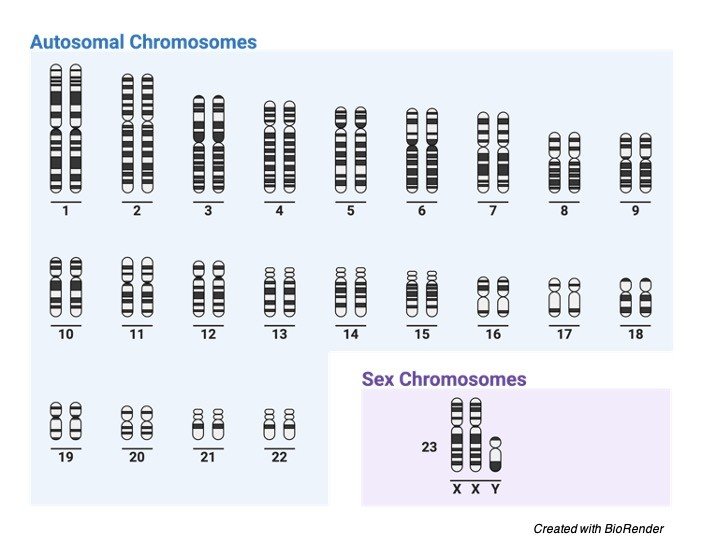
    Klinefelter’s syndrome is one of the conditions which affects only male. The individual with this condition is affected with many intellectual activities.

    Mostly the affected ones are taller comparing with the other individuals of the same group. They are also found to be infertile.

    Anyhow there will be a similarity between boys and men in this syndrome, where as in some cases, the symptoms will be mild and cannot be diagnosed until puberty.

    So, these types of syndromes cannot be diagnosed mostly at early stages or through amniocentesis.

    The individual with this condition usually has smaller testes which also produces only smaller amount of testosterone.

    This condition is also refereed to as primary testicular insufficiency. Testosterone is one of the most important hormones in males; which instructs the body cells to direct the primary and secondary sexual developments in male and also the other developments during the time of puberty.

    If this is not diagnosed at the early stages it leads to shortage of testosterone which leads to delayed puberty, increase in breast size, decreased muscle tone, lack of body and facial hairs, reduced bone density.

    Mostly the affected males will be sterile due to this condition but due to treatments and other therapies using reproductive technologies they can be improved.

    Some of the affected males have difference in their structures of genitalia such as undescended testes which is called as cryptorchidism and the opening of urethra on the underside of penis and also the decrease in size of penis.

    Children’s affected with this condition usually have other problems such as difficulty in brain co ordination which leads to delays in developing intellectual skills, and also in the development of motor skills such as sitting, standing and walking and also, they delay in other activities like speaking and learning.

    But the individuals with this syndrome have better skills of language and understanding and they are usually good at vocabulary.

    Along with this they also have other troubles like anxiety, depression, and other behavioral problems like emotional immaturity and impulsivity. In some cases, they will also be hyperactive.

    Mostly half of percent of people with this syndrome have other metabolic disorders which includes the condition of type 2 diabetes, increased blood pressure, enlarged belly fat, high levels of lipids in the body such as triglycerides and cholesterols.

    In other sense, on comparing with men adults with this syndrome have risks of developing cancers, weaking of bones, and other autoimmune disorders.

    Does Klinefelter Syndrome Inheritary?

    Generally, Klinefelter’s syndrome is not inherited, Because the addition of x chromosomes usually forms at the time of reproduction if any one of the parents has affected egg or sperm.

    During the process of non-disjunction during cell division X chromosomes fails to separate normally among cells which results in extra copy of X chromosome and leads to this type of syndrome.

    Considering the two results i.e., the egg cell having an extra chromosome gets fertilized with the sperm with Y chromosome causes this condition and similarly when the sperm cell having both X and Y chromosome gets fertilized with egg cell having one X chromosome can also leads to this condition.

    Whereas another type of this which is known as mosaic Klinefelter’s syndrome which occurs due to a random cell division in the fetal development which leads to the normal copy of chromosomes in some of the cells and abnormal (XXY) in other cells.

    Symptoms of Klinefelter Syndrome

    The people with this condition usually have thin dispersed hairs, wide hips, enlarged breasts, small testes and smaller penis, reduced masculine features.

    Treatment Options for Klinefelter Syndrome

    There are various kind of treatments practiced for this kind of syndromes, however based on the signs and symptoms the treatment varies.

    The treatments are as follows

    ï‚§ Fertility treatment

    ï‚§ Psychological counselling

    ï‚§ Removing the excess breast tissues

    ï‚§ Hormone replacement therapies

    ï‚§ Supporting them with educational evaluation.

    Klinefelter Syndrome Citations

    Share

    Similar Post:

    Continue Reading
  • Dog Down Syndrome: Symptoms, Causes, and Diagnosis

    Dog Down Syndrome

    Many disorders are common in all organisms sustaining in this world. Any change in structure or number of an organism result in genetic disorders, as a result of these mutations many severe symptoms occur which leads to a death of an individual.

    These syndromes are also common other animals as of humans. Downs syndrome is a condition where there is addition of one number of chromosomes in the cell. It often occurs in chromosome number 21.

    Does Dogs Have Down Syndromes?

    Yes! definitely; each and every living organism even plant is supposed to many diseases and all of us take medications and treatment as all, we want is to have a healthier life.

    Before knowing whether dogs have this syndrome it is necessary to understand about down syndrome.

    The addition or extra number of chromosomes present in the chromosome number 21 causes down syndrome.

    In humans, it is generally categorized by flattened nose, protruded tongue and mental retardation. Where as they also suffer with other disorders like hearing loss, or eye diseases like cataract.

    As given above there are so many ways to find a Down syndrome in humans but it is not so easy to find whether the dog is suffering from any syndrome unless any other syndromes show the physical characteristics.

    We all know that the number of chromosomes present in the cells differ accordingly with each species, where we; the humans contain 23 pairs of chromosomes but dogs contain 39 pairs of chromosomes.

    Downs syndrome in humans can be diagnosed by having 47 number of chromosomes but where as the dogs contains 78 number of chromosomes.

    Dog Down Syndrome Diagnosis

    These kind of chromosomal abnormalities leads to early death of dogs. Usually, genetic tests are followed to identify this syndrome. But usually, this condition does not truly exist in dogs.

    Does Dog Down syndrome Really Exist?

    Downs syndrome does not truly exist in dogs. On the other hand, other congenital and developmental conditions occur in dogs where the symptoms are similar to that of downs syndrome, Congenital hypothyroidism is one of the good examples of this type of congenital disease.

    Some of the symptoms that resemble as such of down syndrome is slow growth which eventually results in small stature, protrusion of tongue and also in large tongue than usual.

    Short limbs, poor muscle tone, delaying in opening of eyes and ears etc.

    The other symptoms which relate the congenital diseases with downs syndrome is pituitary dwarfism, congenital hypo Cephalus, growth hormone deficiencies and portosystemic shunt.

    Symptoms That Mimic Dog Down Syndrome

    If we find a dog by struggling with growth delays or any other cognitive diseases, apart from genetic diseases there is chances of having other congenital heart or thyroid issue which are due to pituitary dwarfism or any other growth hormone deficiencies where all these disorders and symptoms that results as such of down syndrome.

    Dog Down Syndrome Symptoms

    Along with above said symptoms the other symptoms that doubts whether it is a down symptom are as follows.

    Poor Eye Sight: Hence in this condition dogs have poor health conditions which results in poor eyesight. They also have ahigh risk of developing cataract at least in any one of the eyes. In this case the cloudiness of the eyes should be checked with Veterinarian. Because this condition is the best symptom of forming a cataract.

    Unusual Physical Features: Normally dogs with this condition have different facial features and shortened growth. Other common symptoms include shortened neck, flattened face, small structure of head, slanting eyes with a flap of skin above the eyes. And ears are abnormal in structure. Along with these, the affected dogs also have dry noses, patches on skin, incomplete legs, and they start shedding their hair from the fur.

    Hearing Deficiencies: The affected dogs also have the hearing problems but which is very difficult to diagnose, because the dog having these conditions follow the commands later and they are always slow in reacting to the situations.

    Pain: The dog with these symptoms usually gets it whole body affected which also results in abnormal circulatory system and causes severe and random pains throughout the body of the dog,

    Unusual Discharges: These dogs usually with this condition has many diseases along with this congenital disease which results in abnormal blood discharge from their rectum. We can also notice this discharge coming out randomly and often during the later stages of their infection.

    Skin Problems: We can see many dogs along the streets that are suffering from skin disease. But the dogs with these associated symptoms forms missing and abnormal patches and causes irritant to the skin and it also led to other health problems.

    Thyroid Problems: Dogs with these down syndromic symptoms are usually associated with thyroid problems due to malfunctioning of the thyroid gland, which results in metabolic problems and other effects in controlling the temperature of our body. Generally, the dogs with these symptoms have low temperature when compared to normal healthy dogs.

    Behavioral Issues: Dogs who are suffering with this syndrome show odd behaviors, howling or wailing unnecessarily than usual times. So, it finds very difficult to feed and care like other usual dogs. They also cannot control their bladders as well. So, they feel very discomfort and howl deeply.

    However, it is still not yet confirmed that downs syndrome is also present in dogs. 

    Care to be Taken

    The dog showing all these symptoms can be taken for a veterinarian regularly once in a week and they had to given with proper medications.

    They should also be taken for a walk twice in a day. It is also important to observe the emotional and social skills of the dog.

    It is also vital to keep in touch with their body languages and to find out the emotion that they want to conquer us.

    However, the other symptoms associated with down syndrome with the dogs leads them to death, in many cases it is said that mother itself will kill the dog which is having abnormality.

    Or in other cases the mother dog also isolates the affected puppy to prevent the infection passing through other puppies through the affected one’s litter.

    But mostly this condition is spelled out as down syndrome because it is till now not clear whether dogs obtain down syndrome.

    Down Syndrome Citations

    Share

    Similar Post:

    Continue Reading
  • Adenosine Triphosphate (ATP): Definition, Function, and Mechanism

    What is Adenosine Triphosphate (ATP)?

    Adenosine triphosphate, otherwise called ATP, is a molecule that conveys energy inside cells.

    It is the primary energy source of the cell, and it is a finished result of the cycles of photophosphorylation (adding a phosphate gathering to a molecule utilizing energy from light), cell breath, and maturation.

    All living things use ATP. As well as being utilized as a fuel source, it is likewise utilized in signal transduction pathways for cell correspondence and is joined into deoxyribonucleic acid (DNA) during DNA combination.

    Structure of Adenosine Triphosphate (ATP)

    This is an underlying graph of ATP. It is comprised of the molecule adenosine (which itself is comprised of adenine and a ribose sugar) and three phosphate gatherings.

    ATP - Adenosine triphosphate Structure - research tweet

    It is dissolvable in water and has a high energy content due to having two phosphoanhydride bonds interfacing the three phosphate affairs.

    Features of Adenosine Triphosphate (ATP)

    ATP is the primary transporter of energy that is utilized for all cell exercises. At the point when ATP is hydrolyzed and changed over to adenosine diphosphate (ADP), energy is delivered.

    The expulsion of one phosphate bunch discharges 7.3 kilocalories per mole, or 30.6 kilojoules per mole, under standard conditions.

    This energy controls all responses that occur inside the cell.

    ADP can likewise be changed over once more into ATP, so the energy is accessible for other cell responses.

    ATP is delivered through a few unique techniques. Photophosphorylation is a methodology express to plants and cyanobacteria. It is the formation of ATP from ADP utilizing energy from daylight and happens during photosynthesis.

    ATP is additionally shaped from the interaction of cell breath in the mitochondria of a cell. This can be through vigorous breath, which requires oxygen, or anaerobic breath, which doesn’t.

    High sway produces ATP (close by carbon dioxide and water) from glucose and oxygen.

    Anaerobic breath utilizes synthetic substances other than oxygen, and this cycle is basically utilized by archaea and microscopic organisms that live in anaerobic conditions.

    Aging is another method of delivering ATP that doesn’t need oxygen; it is not quite the same as anaerobic breath since it doesn’t utilize an electron transport chain.

    Yeast and microbes are examples of life forms that utilization aging to create ATP.

    Adenosine Triphosphate (ATP) and Signal Transduction

    ATP is a flagging molecule utilized for cell correspondence. Kinases, which are catalysts that phosphorylate particles, use ATP as a wellspring of phosphate gatherings.

    Kinases are significant for signal transduction, which is the way a physical or compound sign is sent from receptors outwardly of the phone to within the phone.

    At the point when the sign is inside the cell, the cell can respond appropriately.

    Cells might be offered signs to develop, utilize, separate into explicit kinds, or even pass on.

    Adenosine Triphosphate (ATP) and DNA Synthesis

    The nucleobase adenine is important for adenosine, an molecule that is framed from ATP and put straightforwardly into RNA.

    The other nucleobases in RNA, cytosine, guanine, and uracil, are correspondingly framed from CTP, GTP, and UTP.

    Adenine is likewise found in DNA, and its consolidation is practically the same, with the exception of ATP is changed over into the structure deoxyadenosine triphosphate (dATP) prior to turning out to be essential for a DNA strand.

    Where Adenosine Triphosphate (ATP) Manufactured?

    Numerous cycles are equipped for creating ATP in the body, contingent upon the current metabolic conditions. ATP creation can happen within the sight of oxygen from cells respiration, beta-oxidation, ketosis, lipid, and protein catabolism, just as under anaerobic conditions.

    i. Cell Respiration

    Cell breath is the way toward catabolizing glucose into acetyl-CoA, creating high-energy electron transporters that will be oxidized during oxidative phosphorylation, yielding ATP.

    During glycolysis, the initial step of cell breath, one particle of glucose separates into two pyruvate atoms. During this interaction, two ATP are delivered through substrate phosphorylation by the compounds PFK1 and pyruvate kinase.

    There is additionally the creation of two diminished NADH electron transporter particles.

    The pyruvate atoms are then oxidized by the pyruvate dehydrogenase complex, shaping an acetyl-CoA particle. The acetyl-CoA atom is then completely oxidized to yield carbon dioxide and decreased electron transporters in the citrus extract cycle.

    After finishing the citric acid cycle, the complete yield is two particles of carbon dioxide, one likeness ATP, three atoms of NADH, and one atom of FADH2.

    These high-energy electron transporters then, at that point move the electrons to the electron transport chain in which hydrogen particles (protons) are moved against their inclination into the inward layer space from the mitochondrial framework.

    ATP particles are then incorporated as protons dropping down the electrochemical inclination power ATP synthase.

    The amount of ATP delivered changes relying upon which electron transporter gave the protons. One NADH particle produces more than two ATP, though one FADH2 atom produces one and a half ATP molecules.

    ii. Beta-Oxidation

    Beta-oxidation is another method for ATP blend in organic entities. During beta-oxidation, unsaturated fat chains are forever abbreviated, yielding Acetyl-CoA atoms.

    All through each pattern of beta-oxidation, the unsaturated fat is diminished by two carbon lengths, creating one particle of acetyl-CoA, which can be oxidized in the citrus extract cycle, and one atom every one of NADH and FADH2, which move their high energy electron to the vehicle chain.

    iii. Ketosis

    Ketosis is a response that yields ATP through the catabolism of ketone bodies. During ketosis, ketone bodies go through catabolism to create energy, producing 22 ATP particles and two GTP atoms for every acetoacetate atom that gets oxidized in the mitochondria.

    iv. Anaerobic Respiration

    At the point when oxygen is scant or inaccessible during cell breath, cells can go through anaerobic breath.

    During anaerobic conditions, there is a development of NADH atoms because of the failure to oxidize NADH to NAD+, restricting the activities of GAPDH and glucose utilization.

    To keep up with homeostatic degrees of NADH, pyruvate is diminished to lactate, yielding the oxidation of one NADH atom in a cycle known as lactic maturation.

    In lactic aging, the two particles of NADH made in glycolysis are oxidized to keep up with the NAD+ repository. This response delivers just two atoms of ATP for every particle of glucose.

    Molecules Similar to Adenosine Triphosphate (ATP)

    Different molecules are identified with ATP and have comparable names, for example, adenosine diphosphate (ADP), adenosine monophosphate (AMP), and cyclic AMP (cAMP).

    To stay away from disarray, know a few contrasts between these particles.

    i. Adenosine Diphosphate (ADP)

    Adenosine diphosphate (ADP), which is at times otherwise called adenosine pyrophosphate (APP), particularly in science, has effectively been referenced in this article. It differs from ATP since it possesses two phosphate groups. ATP becomes ADP with the passing of a phosphate gathering, and this response discharges energy. ADP itself is framed from AMP. Cycling among ADP and ATP during cell breath gives cells the energy expected to do cell exercises.

    ii. Adenosine monophosphate (AMP)

    Adenosine monophosphate (AMP), additionally called 5′- adenylic corrosive, has just a single phosphate bunch. This particle is found in RNA and contains adenine, which is essential for the hereditary code. It tends to be delivered alongside ATP from two ADP molecules, or by hydrolysis of ATP.

    It is additionally shaped when RNA is separated. It very well may be changed over into uric corrosive, which is a part of pee, and discharged through the bladder.

    iii. Cyclic Adenosine monophosphate (cAMP)

    Cyclic adenosine monophosphate (cAMP) is gotten from ATP and is another courier utilized for signal transduction and actuating certain protein kinases.

    It very well may be separated into AMP. cAMP pathways may assume a part in specific diseases like carcinoma. In microorganisms, it has a job in digestion.

    At the point when a bacterial cell isn’t delivering sufficient energy (from deficient glucose, for example), high cAMP levels happen, and this turns on qualities that utilization fuel sources other than glucose.

    Adenosine Triphosphate (ATP) Citations

    Share

    Similar Post:

    Continue Reading
  • Plant Growth Hormone Abscisic Acid: Definition, Mechanism,...

    Growth Hormone: Abscisic Acid

    Growth and development of an organism depends upon the internal and external factors supporting the growth of organism.

    The external environmental cues are essential for the stimulation of growth and development for reproduction and survival efficiency of these environmental cues (i.e.) stimulus process the internal response of the organism.

    The response is manifested and cell functions corresponding to the response is recorded. Such stimulus and response are well coordinated in the plant body by the chemical messengers – the hormones.

    Hormones acts as a mediator for carrying and transferring information for the coordination of physiological, metabolic and chemical activity.

    For example: the growth of coleoptile of Phalaris canariensis towards the light is the coordinated movement for external stimulus light (i.e.) the phototropism is mediated by the hormone auxin. This was the initial discovery for the presence of phytohormones in plant system regulating the function of whole plant body.

    These chemical compounds enhance cell communication and integrate the multicellular organism to organized as a single unit.

    Further studies were undergoing for the deducing the mechanism and variety of hormones coordinating plant body.

    Between 1950 – 1960 a group of five hormones were identified to maintain the plant homeostasis. These hormones were combinedly termed as ” Classical Five” they are: AUXIN, CYTOKININ, ETHYLENE, GIBBERELLIN, ABSCISIC ACID.

    Along with classical five there are brassanosteroids and jasmonic acid. These set of hormones are termed as “Plant Growth Regulators” as they have an active role in regulating growth and development rather than a broad action spectrum.

    Hormones are sensitive, specific, low concentration action and are naturally occurring in plant species. These important characteristic makes it an ideal small molecule chemical messengers and regulators.

    The mode of action is receptor mediated and are transported to different regions by vascular tissues(i.e.) xylem and phloem. Hormones like ethylene are volatile, hence they are diffused throughout the plant body.

    Abscisic Acid Discovery

    Discovery of ABA took place between 1950 – 1960, scientists had a hunch that when a growth stimulating endogenous hormones are present in the plant cell, growth inhibiting hormones which causes the senescence or abscission of fruits must be governed by other hormones namely Abscisic Acid (ABA).

    ABA does not cause abscission they just inhibit growth.

    Functions of Abscisic Acid

    1. Inhibits growth and metabolism based on the developmental stage

    2. Growth and inhibition of root is variable when ABA is present in them

    3. Increases stress tolerance

    4. Seed development and maturation enables seed to withstand desiccation.

    Abscisic Acid Biosynthesis

    ABA are synthesised from Xanthophylls namely Violaxanthin and neoxanthin.

    Epoxidation or the presence of epoxy – carotenoids is necessary for the synthesis of ABA.

    The synthesis however initiates from IPP forming GGPP further leads to the formation of Zeaxanthin produces violaxanthin. Violaxanthin forms cis – neoxanthin followed by cis – xanthin produces ABA Aldehyde leads to ABA.

    Site of Synthesis: In mature leaves and stems and in developing fruits, seeds, etc.,

    Regulation of Abscisic Acid Levels

    1. Synthesis

    2. Conjugation

    3. Oxidation to inactive forms

    Abscisic Acid Citations

    Share

    Similar Post:

    Continue Reading
  • Plant Growth Hormone Auxin: Definition, Mechanism, and...

    What is Auxin?

    Growth and development of an organism depends upon the internal and external factors supporting the growth of organism.

    The external environmental cues are essential for the stimulation of growth and development for reproduction and survival efficiency of these environmental cues (i.e.) stimulus process the internal response of the organism.

    The response is manifested and cell functions corresponding to the response is recorded. Such stimulus and response are well coordinated in the plant body by the chemical messengers – the hormones.

    Hormones acts as a mediator for carrying and transferring information for the coordination of physiological, metabolic and chemical activity.

    For example: the growth of coleoptile of Phalaris canariensis towards the light is the coordinated movement for external stimulus light (i.e.) the phototropism is mediated by the hormone auxin. This was the initial discovery for the presence of phytohormones in plant system regulating the function of whole plant body.

    These chemical compounds enhance cell communication and integrate the multicellular organism to organized as a single unit.

    Further studies were undergoing for the deducing the mechanism and variety of hormones coordinating plant body.

    Between 1950 – 1960 a group of five hormones were identified to maintain the plant homeostasis. These hormones were combinedly termed as ” Classical Five” they are: AUXIN, CYTOKININ, ETHYLENE, GIBBERELLIN, ABSCISIC ACID.

    Along with classical five there are brassanosteroids and jasmonic acid. These set of hormones are termed as “Plant Growth Regulators” as they have an active role in regulating growth and development rather than a broad action spectrum.

    Hormones are sensitive, specific, low concentration action and are naturally occurring in plant species. These important characteristic makes it an ideal small molecule chemical messengers and regulators.

    The mode of action is receptor mediated and are transported to different regions by vascular tissues(i.e.) xylem and phloem. Hormones like ethylene are volatile, hence they are diffused throughout the plant body.

    Auxin Discovery

    Auxins are naturally occurring Indole 3 – Acetic Acid that are abundant and can exist as non-indole component too.

    Auxin in Greek means – “to grow” or “to increase”.

    The main function of auxin is cell division.

    The Dutch biologist Frits Warmolt Went was the one who first described auxins and their role in plant growth.

    In the 1920s Kenneth V. Thimann (1904-1997) became the first to isolate one of these phytohormones and to determine its chemical structure as indole-3-acetic acid (IAA).

    Auxin Functions

    1. Growth from embryo to adult

    2. Cell division

    3. Stem elongation

    4. Apical dominance

    5. Fruit development

    6. Tropic responses

    Auxin Biosynthesis

    Conclusive research analysis or findings are not available for the biosynthesis of IAA because of minimal availability of hormones.

    Many studies have concluded that Tryptophan to be the precursor for IAA. The synthesis initiates from Erythrose – 4 phosphate of pentose phosphate pathway which on further degradation provides tryptophan.

    Tryptophan has many precursors from which many mechanisms are followed to produce Tryptophan.

    Tryptophan deaminates to Indole – 3 -pyruvic acid which on decarboxylation produce indole – 3 – acetaldehyde on oxidation yields Indole Acetic Acid.

    Though the synthesis and pathways deduced for tryptophan’s to be the precursor for IAA many bioassays and other techniques has proved that there are precursors other than Tryptophan from which IAA is synthesized.

    A conclusion can be laid that the IAA can be synthesized by tryptophan dependent process and Tryptophan Independent process.

    Site of biosynthesis: site of biosynthesis is dependent on endogenous precursor pools.

    The precursors are not well defined for IAA. But, based on the existing analysis it can be detected that tryptophan are rich in plastids and it also has a cytoplasmic pool in plant cell.

    Hence plastids and cytoplasm of plant cell can be assumed as site of biosynthesis.

    An affirmation regarding the site of synthesis of auxin can be provided considering the presence of Auxin in the Apical meristematic zones and are absent in mature cell.

    Regulation of Auxin Levels

    Hormones are regulated by variety of mechanisms. They are – Synthesis, Conjugation, Irreversible modification breakdown, transportation and compartmentation.

    Lesser the knowledge of biosynthesis of Auxin lesser the role of regulation identified for auxins. It is regulated by formation of conjugates and irreversible modification by breakdown of IAA.

    Conjugation: IAA forms conjugates with ester linkage to sugar or alcohol or to amino acids by amide linkage.

    Breakdown: IAA is decarboxylated with products: oxindole – 3 – methanol, 3 – methylene oxindole, 3 – methyl oxindole, indole – 3 aldehyde.

    Auxin Inhibitors

    IAA are inhibited by synthetic compounds. These compounds inhibit IAA and not the synthesis.

    These inhibiting molecules are called antiauxins. They inhibit by binding to the receptors of IAA.

    Auxin Citations

    Share

    Similar Post:

    Continue Reading
  • Plant Growth Hormone Cytokinin: Definition, Mechanism, and...

    Plant Growth Hormone: Cytokinin

    Growth and development of an organism depends upon the internal and external factors supporting the growth of organism.

    The external environmental cues are essential for the stimulation of growth and development for reproduction and survival efficiency of these environmental cues (i.e.) stimulus process the internal response of the organism.

    The response is manifested and cell functions corresponding to the response is recorded. Such stimulus and response are well coordinated in the plant body by the chemical messengers – the hormones.

    Hormones acts as a mediator for carrying and transferring information for the coordination of physiological, metabolic and chemical activity.

    For example: the growth of coleoptile of Phalaris canariensis towards the light is the coordinated movement for external stimulus light (i.e.) the phototropism is mediated by the hormone auxin. This was the initial discovery for the presence of phytohormones in plant system regulating the function of whole plant body.

    These chemical compounds enhance cell communication and integrate the multicellular organism to organized as a single unit.

    Further studies were undergoing for the deducing the mechanism and variety of hormones coordinating plant body.

    Between 1950 – 1960 a group of five hormones were identified to maintain the plant homeostasis. These hormones were combinedly termed as ” Classical Five” they are: AUXIN, CYTOKININ, ETHYLENE, GIBBERELLIN, ABSCISIC ACID.

    Along with classical five there are brassanosteroids and jasmonic acid. These set of hormones are termed as “Plant Growth Regulators” as they have an active role in regulating growth and development rather than a broad action spectrum.

    Hormones are sensitive, specific, low concentration action and are naturally occurring in plant species. These important characteristic makes it an ideal small molecule chemical messengers and regulators.

    The mode of action is receptor mediated and are transported to different regions by vascular tissues(i.e.) xylem and phloem. Hormones like ethylene are volatile, hence they are diffused throughout the plant body.

    Cytokinin Discovery

    Discovery of cytokinin was first started from Haberlandt, German plant physiologist in 1913 that the phloem extracts are used to cause cell division in tubers.

    In 1921, from another experiment he found that cell division is promoted by soluble factor.

    In 1940’s and 1950’s the cell culture techniques were practiced, where the addition of auxins did not yield cell division. but on addition of autoclaved herring sperm initiated rapid cell division.

    The substance that promoted the cell division was named Kinetin. Later naturally occurring zeatin were discovered.

    Functions of Cytokinin

    1. Promotes cell division in callus and tissue

    2. Auxin and Cytokinin combines together to form a shoot bud vs root growth in tissue culture and stem cuttings

    3. Regulate Apical Dominance and lateral root initiation

    4. Retard senescence and chlorophyll degradation

    5. Present in major parts of plant

    Cytokinin Biosynthesis

    IPP is the key for cytokine synthesis.

    IPP isomerize with DMAPP.

    DMAPP condense with AMP give rise to iso – pentenyl adenosine mono phosphate (iPMP).

    iPMP is precursor for all naturally occurring Cytokinins.

    iPMP is hydroxylated at C4 of the side chain and forms zeatin riboside.

    In further steps, Ribose and phosphate are cleaved to yield Zeatin.

    Site of synthesis: primarily synthesized in meristematic tissues.

    Regulation of Cytokinin Levels

    1. It is regulated by conjugation of Cytokinins

    2. Irreversible inactivation

    Cytokinin Citations

    Share

    Similar Post:

    Continue Reading
  • Plant Hormone Ethylene: Definition, Mechanism, and Function

    Ethylene: a Plant Growth Hormone

    Growth and development of an organism depends upon the internal and external factors supporting the growth of organism.

    The external environmental cues are essential for the stimulation of growth and development for reproduction and survival efficiency of these environmental cues (i.e.) stimulus process the internal response of the organism.

    The response is manifested and cell functions corresponding to the response is recorded. Such stimulus and response are well coordinated in the plant body by the chemical messengers – the hormones.

    Hormones acts as a mediator for carrying and transferring information for the coordination of physiological, metabolic and chemical activity.

    For example: the growth of coleoptile of Phalaris canariensis towards the light is the coordinated movement for external stimulus light (i.e.) the phototropism is mediated by the hormone auxin. This was the initial discovery for the presence of phytohormones in plant system regulating the function of whole plant body.

    These chemical compounds enhance cell communication and integrate the multicellular organism to organized as a single unit.

    Further studies were undergoing for the deducing the mechanism and variety of hormones coordinating plant body.

    Between 1950 – 1960 a group of five hormones were identified to maintain the plant homeostasis. These hormones were combinedly termed as ” Classical Five” they are: AUXIN, CYTOKININ, ETHYLENE, GIBBERELLIN, ABSCISIC ACID.

    Along with classical five there are brassanosteroids and jasmonic acid. These set of hormones are termed as “Plant Growth Regulators” as they have an active role in regulating growth and development rather than a broad action spectrum.

    Hormones are sensitive, specific, low concentration action and are naturally occurring in plant species. These important characteristic makes it an ideal small molecule chemical messengers and regulators.

    The mode of action is receptor mediated and are transported to different regions by vascular tissues(i.e.) xylem and phloem. Hormones like ethylene are volatile, hence they are diffused throughout the plant body.

    Ethylene Discovery

    Discovery of ethylene was done by Dimitry Neljubov. He was growing pea seedling in dark and the laboratory was gas lit, instead of elongating apical stem, the stem is shortened and had stunted growth. the gas lit lab had an ethylene leak which stunted the plant growth by inhibiting it.

    From the experiment, ethylene was identified to be reducing the growth rate of plants.

    In 1930 endogenous ethylene pools were identified in plant material especially in fruits that are ripened.

    Functions of Ethylene

    1. Fruit ripening

    2. Promotes senescence and abscission

    3. Root hair production

    4. Seed germination

    5. Sprout lateral buds

    Ethylene Biosynthesis

    Methionine is the precursor for ethylene on addition of Adenosine group from ATP forms Ado Met. Ado Met cleaves to produce Aminocyclopropanecarboxylic acid and Methylthioadenosine.

    ACC on enzyme catalyses yields ethylene. MTA is cleaved by adenosine and give rise to adenine and methylthioribose.

    MTR on further steps yields methionine for the cycle to proceed.

    Site of Synthesis: Ripening regions and in senescing leaves and other parts.

    Regulation of Ethylene Levels

    Ethylene synthesis is regulated by certain inhibitors

    The production of ethylene itself becomes an indicator to inhibit the production.

    Conjugation of ACC

    Oxidation of ethylene

    Inhibitors of Ethylene Synthesis

    Methyl cyclopropane, Dimethyl cyclopropane

    Ethylene Citations

    Share

    Similar Post:

    Continue Reading
  • Gibberellin: Definition, Mechanism, and Function

    Growth Hormone: Gibberellin

    Growth and development of an organism depends upon the internal and external factors supporting the growth of organism.

    The external environmental cues are essential for the stimulation of growth and development for reproduction and survival efficiency of these environmental cues (i.e.) stimulus process the internal response of the organism.

    The response is manifested and cell functions corresponding to the response is recorded. Such stimulus and response are well coordinated in the plant body by the chemical messengers – the hormones.

    Hormones acts as a mediator for carrying and transferring information for the coordination of physiological, metabolic and chemical activity.

    For example: the growth of coleoptile of Phalaris canariensis towards the light is the coordinated movement for external stimulus light (i.e.) the phototropism is mediated by the hormone auxin. This was the initial discovery for the presence of phytohormones in plant system regulating the function of whole plant body.

    These chemical compounds enhance cell communication and integrate the multicellular organism to organized as a single unit.

    Further studies were undergoing for the deducing the mechanism and variety of hormones coordinating plant body.

    Between 1950 – 1960 a group of five hormones were identified to maintain the plant homeostasis. These hormones were combinedly termed as ” Classical Five” they are: AUXIN, CYTOKININ, ETHYLENE, GIBBERELLIN, ABSCISIC ACID.

    Along with classical five there are brassanosteroids and jasmonic acid. These set of hormones are termed as “Plant Growth Regulators” as they have an active role in regulating growth and development rather than a broad action spectrum.

    Hormones are sensitive, specific, low concentration action and are naturally occurring in plant species. These important characteristic makes it an ideal small molecule chemical messengers and regulators.

    The mode of action is receptor mediated and are transported to different regions by vascular tissues(i.e.) xylem and phloem. Hormones like ethylene are volatile, hence they are diffused throughout the plant body.

    Gibberellin Discovery

    Gibberellins were discovered from a Japanese crop disease “Bakanae” where the crops are subjected to elongated and weak growth caused by an ascomycetes Gibberella fujikuroi. the extracts from the fungi are collected and termed as Gibberellin by Yabuta and Hayashi.

    In 1950’s, Europe and American scientists started to standardise methods of isolating hormones. On learning about Gibberellin, its function to elongate the stem was related to higher plants which have taller stems and branches.

    When extracted the responding hormone scientists named it as GIBBERELLIN. Gibberellins are tetracycline diterpenoids prevalent in two forms. C20 – GA’s and C19 – GA’s out of this C19 GAs are found abundant in plant cell.

    The variety of GA’s are increasing, presently there are about 125 GAs identified.

    Functions of Gibberellins

    1. they promote elongation in stem and root. Their role in cell division is nil but they elongate both root and shoot

    2. During Seed germination, the stored starch, proteins and lipids are broken down by hydrolysis. GA’s promote hydrolysis during seed germination.

    3. Floral development

    4. Phloem tissue differentiation

    5. Cambial Reactivation

    Gibberellin Biosynthesis

    Out of all Plant Growth Regulators, GA’s are very well studies and biosynthesis are well known.

    Terpenes are secondary products of plants is defensive in function prevents plant from plant feeding Insects.

    Apart from the defence terpenes helps in photosynthesis, membrane stability and signalling Isoprene a C5 structure is the base for all terpenes.

    The isomerization of isopentenyl diphosphate yields dimethylallyl diphosphate following condensation yields C10 terpenes, C15 terpenes on further condensation gives C30 and C40 terpenes.

    IPP was synthesized by mevalonic acid dependent pathway or by Mevalonic Acid Independent pathway by Pyruvate and Glyceraldehyde.

    Synthesis takes place in 3 stages:

    Stage 1: Geranylgeranyl Diphosphate (GGPP) undergoes cyclation to form ent – kaurene followed by enzyme catalysation takes place in Plastids.

    Stage 2: Oxidations leads to the formation of ent – kaurenoic acid hydroxylated to form ent -7alpha kaurenoic acid later yields G12 Aldehyde

    Stage 3: GA12 – aldehyde yields GA12. 13 – Hydroxylation leads to the formation of GA53. Oxidations at C20 leads to lactone formation.

    Regulation of Gibberellin Levels

    Levels of GA are maintained by plant tissues

    Their inactivation is done by conjugation or catabolic breakdown

    Irreversible inactivation, transport to different parts of the plant, storage in compartments such as vacuoles.

    Gibberellin Citations

    Share

    Similar Post:

    Continue Reading