Author: Admin

Author: Admin

  • Cell Plating, Media Change, and Culture Maintenance...

    About Cell Plating or Cell Passaging

    Most types of cells stop or slow down their growth at a characteristic population density in any particular medium.

    Cell to cell contact, localized or generalized depletion of nutrients or production of cell generated inhibitors leads to density dependent inhibition or sometimes called as contact inhibition of cell growth.

    Cells at this stage are said to be confluent.

    This is important to avoid Contact Inhibition of Proliferation (CIP), exhibited by most cancerous cell lines.

    The phenomenon of a cell ceasing to proliferate after contact with other cells is called as CIP.

    Adherent cells cover the entire growth surface available to them is referred to as confluence.

    Hence they need to be subcultured, passaging, cell plating (indicated by passage number).

    The passage number is the number of times that the culture has been subcultured.

    Why Cell Plating or Cell Passaging

    Cells are often passaged at semiconfluency when they are in log phase.

    Failure to subculture or cell plating, the proliferating cells results in reduced mitotic index and eventually cell death.

    The most important step in subculturing or cell plating of monolayers is to detach cells from the surface of the primary culture vessel by either enzymatic means such as trypsinization or mechanical means such as scraping of monolayer cells.

    The resultant cell suspension is then reseeded, into fresh cultures.

    Secondary cultures are checked for growth, fed periodically, and may be subsequently subcultured to produce tertiary cultures, etc.

    The time between passaging cells depends on the growth rate and varies from cell line to cell line.

    Detachment of cells by mechanical means is accomplished by scraping monolayer of cells with commercially available silicon rubber spatula known as a scrapper.

    Whereas proteases such as trypsin, collagenase and pronase etc. are used for enzymatic dissociation.

    Prolonged enzymatic dissociation may damage cell membrane.

    Thus, it is wise to decrease the incubation period and physical injury when in enzyme solution.

    Cell Plating - Research Tweet 1

    Cell Plating or Cell Passaging Overview. Images are created with BioRender

    Cell Plating or Cell Passaging Requirements

    1. Confluent culture of cells

    2. Appropriate culture media

    3. Antibiotics and antifungal agent

    4. Sterile PBS (Phosphate buffer saline)

    5. Trypsin/EDTA solution (0.25% Trypsin in 1mM EDTA in PBS)

    6. Sterile pipettes, culture vessels, sterile beakers

    7. Cotton swabs, 70% IPA,

    8. 15 ml & 50 ml Centrifuge tubes

    Cell Plating or Passaging of Adherent Cells

    Check all the cell lines under the microscope for contamination, cell morphology, density and presence of clumps or detachment.

    1. The protocol described here is for a typical confluent culture in a 25 cm2 flask

    2. Remove medium with a sterile pipette without disturbing the cells.

    3. Wash adhering cell layer with sterile PBS (Phosphate buffer saline) to remove traces of serum that can inhibit trypsin.

    4. Add 2 ml trypsin/EDTA at 37ºC to cover the cell layer.

    5. Incubate for 2 minutes at 37ºC in CO2 incubator. Tap occasionally to verify that the cells are releasing. Check in microscope to visualize detachment of cells.

    6. Remove trypsin-EDTA. Add fresh 2 ml of medium and rinse cell layer two or three times to dissociate cells and to dislodge any remaining adherent cells.

    7. Transfer the required number of cells to a new labelled flask containing pre-warmed medium. Maintain the split ratio as recommended.

    8. Incubate cells in humidified incubator at 37ºC, 5% CO2.

    Cell Plating or Passaging of Suspension Cells

    In suspension culture cells are suspended in complete growth medium rather than attached to a culture flask surface, it is not necessary to disperse them enzymatically before passaging.

    However, before passaging, cells must be maintained in culture by feeding every 2 to 3 days until they reach confluency (i.e. until the cells clump together in the suspension and medium appears turbid when the flask is swirled).

    1. With a pipette, mix the cells thoroughly by pipetting up and down and rinsing the side of the flask.

    2. Decant the cell suspension into the centrifuge tube, spin at 1300 rpm for 7 minutes at 4 °C temperature to pellet down all the cells.

    3. Add 1 ml of fresh media.

    4. Split into two flasks with 0.5 ml of cell suspension and add 6.5 ml of fresh media.

    5. Label new passage number on both the flasks.

    6. Incubate cells in humidified incubator at 37 C, 5% CO2.

    Precautions During Cell Plating or Passaging

    Cell degeneration may lead to change in morphology i.e. rounding up of cells and their detachment from the surface of the culture vessel.

    The most frequent reasons for rapid cell degeneration include use of too high seeding density, use of poor quality or too high concentration of fetal calf serum when preparing cultures.

    Whenever rapidly growing, continuous cell lines are maintained in a laboratory there is a risk of cell line cross-contamination.

    The problem of cell degeneration can usually be overcome by appropriate adjustment of the cell count or foetal calf serum concentration.

    Only one cell line should be used in at any one time.

    After removal of the cell cultures from the cabinet, the cabinet should be swabbed down with a suitable disinfectant and the cabinet UV run for five minutes before introduction of another cell line.

    Bottles or aliquots of medium should be dedicated for use with only one cell line.

    Regularly return to frozen stocks — never grow a cell line for more than three months 15 passages from stock passage level, whichever is the shorter period.

    All culture vessels must be carefully and correctly labelled. (including name of cell line, passage number and date of transfer)

    Some cultures whilst growing as attached lines adhere only lightly to the flask called as fragile cells, thus it is important to ensure that the growth medium is retained and the flasks are handled with care to prevent any cells detachment prematurely.

    Although majority of cells will detach in the presence of trypsin alone the EDTA is added to enhance the activity of the enzyme.

    Presence of serum inactivate Trypsin activity. Therefore, it is essential to wash the monolayer of cells with PBS without calcium and magnesium in order to remove all traces of serum from the culture medium.

    Cells should only be exposed to trypsin/EDTA long enough to detach cells.

    Prolonged exposure could damage cell surface receptors.

    In case of cells lost in trypsin removal, trypsin should be neutralised with serum prior to seeding cells into new flasks otherwise cells will not attach.

    Trypsin may also be neutralised by the addition of soyabean trypsin inhibitor, where an equal volume of inhibitor at a concentration of 1mg/ml is added to the trypsinised cells.

    The cells are then centrifuged, resuspended in fresh culture medium and counted as above.

    This is especially necessary for serum-free cell cultures.

    If the cells harvested are at too low a cell density to re-seed at the appropriate cell density into fresh flasks it may be necessary to centrifuge the cells e.g. 5 mins at 1300 rpm, and resuspend in a smaller volume of medium.

    Precautions During Cell Plating or Passaging Ciations:

    Share

    Similar Post:

    Continue Reading
  • Codominance: an Overview, Definition, and Examples

    Law of Dominance: Codominance

    In hybridization techniques, two alleles are considered one as dominant character and other as recessive character when these alleles are let to undergo fertilization by crossing techniques the expression of dominant allele will be high compared to that of recessive allele phenotypically.

    Hence the dominant character will be expressed Hence it is known as law of dominance. Law of dominance is also said to be Mendel’s first law of inheritance.

    Mechanism of Dominance

    On undergoing various experiments Mendel evidenced himself that there will be a difference in their genetic characters, even though the phenotypic character resembles as such as their parents.

    Because the character does not remain the same as it is being for their parents.

    Therefore, there will be something which controls all these characters which are later found as genes which are the units of DNA (Deoxyribo nucleic acid) or RNA (Ribo nucleic acids) accordingly which are present in the chromosomes of an individual and are passed on to the next generation through parents via gametes of male and female.

    Hence the individual has 23 pair of chromosomes each pair from one of the parents, each chromosome consists of genes as their functional unit which consists of contrasting characters made up of alleles.

    If there are two or more contrasting pair of alleles then it is said to be as allelomorph.

    These alleles are produced as the effect of mutation in a wild gene. For example, let us consider a pea plant, where homozygous tall plant has two alleles such as TT on their gene loci in their homologous chromosomes and homologous dwarf plants is represented by the allele tt.

    During the process of gametogenesis, the two homologous alleles TT and tt are separated and each chromosome contains a single allele as T and t and it is passed via gametes.

    These alleles which are passed through gametes of both the parents (father and mother) combine together during fertilisation.

    Thus, the new individual in the F1 generation has two different alleles and it is referred to as heterozygous in condition.

    The dominant character is being expressed and the recessive character of the individual gets suppressed this the mechanism why only dominant characters are expressed though the individual has both the alleles.

    Variation in Dominance

    Mendel studied the dominant and recessive characters in pea plants which helped him to identify seven pairs of genes showing different phenotypes in homozygous and heterozygous condition with lot of variation.

    Variation in dominant character is further classified into two types as Incomplete and complete dominance.

    Codominance

    In some cases, both the alleles in a heterozygote lacks the character of being both dominant and recessive.

    Which means that each trait is capable of obtaining some degree of phenotypic expression from their parents, hence it can also be considered that there will no dominance between two alleles, and they be in equilibrium condition to express their traits.

    1. Example of Codominance

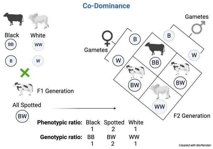
    Here the coat colour of cattle breeds is taken into account, Where the coat colour of the cattle is chosen as Black and white.

    The cattle with Black colour coat have its allele as BB and the white cattle’s coat is denoted as WW.

    When these two alleles are crossed the resultant allele obtained is considered as BW, where the coat colour obtained is Spotted (mixture of both).

    Where the white colour hair is spread throughout the coat and black patches is scattered on the coat.

    In the filial 2 generation the coat colour of the cattle appears to be spotted and also the parental characters of black and white also appears.

    CoDominance-Incomplete Dominance - Definition and Examples - research tweet 1

    2. Example of Codominance

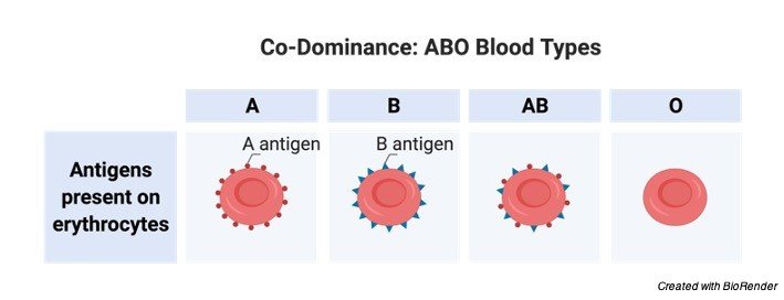
    The best example of co-dominance in humans is ABO blood group, it was first discovered by Landsteiner and Levine.

    The alleles here are represented as A and B accordingly.

    Here three groups are possible which are denoted as A, B and AB which have their alleles as AA, BB and AB accordingly.

    The genotype and its characteristic antigen and antibody are listed below.

    Allele, Allele Definition, What is Allele, Allele Examples,
    Codominance Citations

    Share

    Similar Post:

    Continue Reading
  • Micronucleus Assay: a Widely Used Assay for...

    About Micronucleus Assay

    Genotoxicity is a word in genetics defined as a destructive or adverse effect on a cell’s genetic material either DNA or RNA, affecting its integrity.

    Genotoxins are mutagens (compounds that changes nucleotide); they can cause mutations.

    Genotoxins include both radiation and chemical genotoxins.

    A substance (radiation and chemical compounds) that has the property of genotoxicity is known as a genotoxin.

    Micronucleus Assay Principle

    The purpose of in vitro testing is to determine whether an environmental factor, substrate, or any product induces damage to genetic material.

    One technique entails cytogenetic assays using different animals cells.

    The aberrations detected in cells affected by a genotoxic substance are chromosome gaps, chromatid, complex rearrangements, chromosome breaks, fragmentation, translocation, chromatid deletions, and many more.

    One such example is testing for the formation of micronuclei.

    The micronucleus test is used as a tool for genotoxicity assessment of various chemicals.

    Chromosomal aberration test is easy to conduct in order to evaluate genotoxicity.

    A micronucleus is the erratic (third) nucleus that is formed during the anaphase of mitosis or meiosis.

    Micronuclei (the name means ‘small nucleus’) are cytoplasmic bodies having a portion of acentric chromosome or whole chromosome which was not carried to the opposite poles during the anaphase.

    Micronuclei formation results in the daughter cell lacking a part or all of a chromosome.

    These fragments of chromosome or whole chromosomes normally develop nuclear membranes and form as micronuclei as a third nucleus.

    After cytokinesis, one daughter cell ends up with one nucleus and the other ends up with one large and one small nucleus, i.e., micronuclei.

    There is a chance of more than one micronucleus forming when more genetic damage has happened.

    Micronucleus Assay Requirements

    1. Cancer cell line

    2. Media (DMEM or alpha MEM)

    3. FBS

    4. 12 well or 24 well or 4 well plate or 6 well plate

    5. Coverslips

    6. 1X PBS (pH-7.2)

    7. Triton-X 100- 0.05%

    8. Ethanol-70%

    9. Paraformaldehyde (PFA)-4%

    10. Dapi stain (0.5 ug/ml)

    11. Bright field microscope

    Procedure of Micronucleus Assay

    1. MDA-MB-468 cells were grown on coverslips in 12 well plates.

    2. After treatment period (24 hour or 48 hour), media was removed and cells were washed twice with PBS. Further the cells were fixed in 4% PFA (1 hour at RT or overnight to 1 week at 4oC).

    3. After fixation PFA was removed and the cells were washed with PBS (pH 7.2)

    4. After washing add 0.05% triton –X 100 (it should cover the cells grown on coverslip) and incubate for 1 hour.

    5. After incubation triton-X 100 was removed and the cells were washed gently with PBS (pH-7.2).

    6. After washing Dapi stain was added (it should cover the surface and make sure it should not get dried during incubation) and incubate for 30 minutes. Observe under fluorescence microscope.

    Note: For better result cells should be 70%-80% confluent.

    Micronucleus Assay Citations:

    Share

    Similar Post:

    Continue Reading
  • Transition Words And Phrases: Why And How...

    Transition Words

    If you are writing an assay that is full of idea and perfectly written but what if its not clear to your readers.

    That’s where transition words come in. Transitions are words, phrases, sentences, that link one idea to another showing the connection between them.

    What are a Transition Words?

    A transition means a change from one idea to another. There are many transitions in English.

    We use transitions to add information, to show contrast, to show condition, to show conclusion, etc.

    There are a lot of transition words out there, and they play very important role in English because they can join idea within the sentences and more importantly they can join two sentences.

    transition words - Research Tweet 1

    Why Transition Words are so Important?

    They create flow in your writing.

    Flow makes easier to understand your assay or writing and also makes your content a little bit more interesting to your reader.

    Transition words also act as a connecting bridge within the sentences and also between the sentences.

    Categories of Transition Words

    Causation: These words and phrases are used to connecting instigator(s) to consequence(s) and used after a sentence or paragraph that describes a cause.

    • Accordingly
    • As a result
    • And
    • So
    • Because
    • Consequently
    • For that reason
    • Hence
    • On account of
    • Since
    • Therefore
    • Thus

    The plant is growing very fast. [Accordingly / Consequently / As such] it is necessary to cut new branches.

    Because he failed to respond, John and Steve offered coffee.

    The speaker’s flight has been cancelled. [Therefore / As such / As a result] we will reschedule the conference lecture.

    Monday is a national holiday. [As a result / Accordingly / Thus / Hence / Consequently] classes will be canceled.

    transition words - Research Tweet 3

    Chronology:It is widely used to connect what issues in regard to when they occur.

    • After
    • Afterwards
    • Always,
    • At length
    • During
    • Earlier
    • Following
    • Immediately
    • In the meantime
    • Later
    • Never
    • Next
    • Now
    • Once
    • Simultaneously
    • So far
    • Sometimes
    • Soon
    • Subsequently
    • Then
    • This time
    • Until now
    • When
    • Whenever
    • While

    I went to the school after he gave me bike.

    He almost always wins lottery.

    During the morning, you can play.

    First, we are going fishing. Then, we will get ready for movie.

    transition words - Research Tweet 4

    Combinations: It connects multiple events or numerous elements that create long sentences.

    • Additionally
    • Again
    • Also
    • And
    • Or
    • Not
    • As a result
    • Besides
    • Even more
    • Finally
    • First,
    • Firstly
    • Further
    • Furthermore
    • In addition
    • In the first place
    • In the second place
    • Last
    • Lastly
    • Moreover
    • Next
    • Second
    • Secondly
    • Too

    Steve promised to respect, love and also obey his wife.

    Moreover, he will do all the cleaning and cooking while he cooks at home.

    Furthermore, he is grateful that his girlfriend earns enough money his groceries.

    transition words - Research Tweet 6

    Contrast: It connect two sentences by focusing on their differences.

    • After all
    • Although
    • And yet
    • At the same time
    • But
    • Despite
    • However
    • In contrast
    • Nevertheless
    • Nonetheless
    • Notwithstanding
    • On the contrary
    • On the other hand
    • Otherwise
    • Though
    • Yet

    Jones argues for the Oxford comma, although everyone doesn’t agree.

    I ran home, but I was still late.

    Despite the last fight, it is also very important to think about dogs.

    During my Chicago trip, I went to see my friend. However, I wasn’t sure.

    transition words - Research Tweet 5

    Example: It connects a general idea to a particular instance of this idea.

    • As an illustration
    • e.g., (from a Latin abbreviation for “for example”)
    • For example
    • For instance
    • Specifically
    • That is
    • To demonstrate
    • To illustrate

    Importance: It connect what is critical to what is more inconsequential.

    • Chiefly
    • Critically
    • Foundationally
    • Most importantly
    • Of less importance
    • Primarily

    Location: It connect elements according to where they are placed in relationship to each other.

    • Above
    • Adjacent to
    • Below
    • Beyond
    • Centrally
    • Here
    • Nearby
    • Neighboring on
    • Opposite to
    • Peripherally
    • There
    • Wherever
    transition words - Research Tweet 7

    Similarity: It connect to things by suggesting that they are in some way alike.

    • By the same token
    • In like manner in Similar fashion
    • Here
    • In the same way
    • Likewise
    • Wherever

    Share

    Continue Reading
  • Polycistronic mRNA: Definition, Examples, Types, Advantage

    What is Polycistronic mRNA?

    Polycistronic mRNA is a mRNA that deciphers various proteins and is an attribute of many prokaryotic bacterial and chloroplast mRNAs.

    For instance, if a bacterial cell desires to consume lactose as a source of energy, it will duplicate an mRNA molecule that encrypts several protein products needed for lactose metabolism.

    In contrast, eukaryotes possess monocistronic mRNA that only encrypts for a single protein product per mRNA molecule.

    Polycistronic mRNA comprises a leader sequence which pave the way for the first gene.

    The gene is trailed by an intercistronic zone and then towards another gene. A tail end of the amino acids follows the terminal gene in the mRNA.

    Example of Polycistronic mRNA

    Instances of a polycistronic records are found in the chloroplast.

    One area that displays various polycistronic messages from a similar area is the psbb/ psbH/ petB/ petD locale.

    The accompanying points records the qualities, their items and the complex of which the item is a section.

    Gene psbB leads to production of 51 kilo Dalton chl a binding protein of complex PSII.

    Gene psbH leads to production of 10 kilo Dalton phosphoprotein of complex PSII.

    Gene petB leads to production of cytochrome b6 of complex Cytochrome.

    Gene petD leads to production of subunit 4 of cytochrome b6/f of Cytochrome.

    Albeit the transcripts are co-deciphered, the proportion of the two complex differs in the lit and unlit just as between the mesophyll and the bundle sheath cells.

    In this way some kind of guideline should exist. Something like 15 distinct mRNAs are created from this gene group.

    Polycistronic mRNA: One mRNA, Multiple Polypeptides

    A mRNA atom is supposed to be monocistronic when it contains the hereditary data to decipher just a solitary protein chain (polypeptide).

    This is the situation for a large portion of the eukaryotic mRNAs.

    On the other hand, polycistronic mRNA conveys a few open reading frame (ORFs), every one of which is converted into a polypeptide.

    These polypeptides normally have a connected capacity (they frequently are the subunits creating a last unpredictable protein) and their coding succession is gathered and controlled together in an administrative locale, containing a promoter and an operator.

    The majority of the mRNA found in microorganisms and archaea is polycistronic, just like the human mitochondrial genome.

    Dicistronic or bicistronic mRNA encodes just two proteins.

    Virtually all positive-sense RNA viruses have genomic RNAs that encode different protein items as forerunner polyproteins that are then prepared to the utilitarian polypeptides utilized by the virus during contamination.

    A segment of these virus like human rhinovirus, hepatitis C infection, cricket paralysis virus possesses no less than one open reading frame (ORF) anteceded by IRES structures.

    In any case, there is developing proof that some cell and vertebrate mRNAs likewise have IRES-like designs further downstream of the 5′- UTR, inside or after the 5′ proximal ORF, empowering the expression of proteins from pair or overlapping ORFs.

    As with prokaryotic polycistronic qualities, standard interpretation normally starts close the 5’ends of mRNAs of vertebrate bicistronic genes.

    In any case, in a couple of cases it has been recorded that the production of a subsequent protein is started through an IRES component found downstream of or inside the first open reading frame.

    Polycistronic mRNA: a Cellular Genes

    Because of different systems of elective gene expression and translation in eukaryotic cells, the recognition of mRNAs holding onto true blue IRES sequences requires various tough models that should be fulfilled.

    Cryptic promoters in columnist plasmids and elective splicing occasions that can prompt unmistakable records and optional protein items should be precluded.

    Also, various systems of secondary protein translation involving ribosomal scanning, re-initiation, stop codon read-through, or translational frameshifting might be found in a similar gene.

    Indisputable proof for the presence of an IRES requires prohibition of these systems and a various utilitarian examination.

    Thirteen polycistronic genes recognized through the writing.

    Genes were excluded that simply communicated shortened types of a similar protein with basically a similar capacity.

    Genes for which a cap-independent expression mechanism had not been upheld tentatively were likewise eliminated.

    In the following segments, we give instances of the four useful classes of polycistronic genes and the biology/ gene expression designs associated with them.

    1. 2 subunits of a multi-subunit complex where the expression is directed in one transcript.

    2. Functionally same gene outcomes that are distinctively co-expressed.

    3. Functionally different gene outcomes that have programmatically-connected expression

    4. Signaling proteins produced by stimulus-coupled protease severe or by cap-independent translation.

    Benefits of Polycistronic mRNA

    Polycistronic mRNA or genes have various benefits for co—ordinated gene expression, 4 particular classes have been arranged showing expression mechanism of each polycistronic gene.

    Besides, while polycistronic gene association permits an exceptional and specific component for control of protein expression, within the sight of hereditary transformations or dysregulation of the IRES this hereditary methodology has a various possible antagonistic clinical results.

    Definite mutations of polycistronic genes lead to intricate and various phenotypes, potentially on account of their consequences for either ORF or the IRES sequence itself.

    Thus, polycistronic gene could likewise make the way for novel treatments.

    Polycistronic mRNA Summary

    The organization and expression of particular protein from vertebrate polycistronic mRNAs appears to give a same layer of coordinated expression control to that used fundamentally by invertebrates and protozoans.

    Therefore, an additional comprehension of the control of polycistronic gene expression in mammalian tissues should dispense new understanding into several human genotype-phenotype correlations along with therapies of human disorder and disease.

    Polycistronic mRNA Citations

    Share

    Similar Post:

    Continue Reading
  • Respiratory Quotient: Definition and Examples I Research...

    What is Respiratory Quotient?

    Energy is the driving force for all living organism. Energy in biosystem is present in the form of ATP.

    Energy is obtained when the biomolecules are broken down. The process of oxidizing the biomolecules for the energy is termed as respiration.

    Internal and External respiration are two types of respiration involving the interaction of external environment with the internal environment through responsible organs.

    The air filled with oxygen is transported to the internal tissues which is absorbed by the cell to drive the metabolic pathway that yield carbon dioxide and energy as the end products.

    This carbon dioxide is released from the tissues to the external environment.

    The amount of O2 consumed and CO2 released will provide the metabolic rate of any organisms along with the nature of component can also be detected.

    To measure the metabolic rate of any organism the oxygen consumption and carbon dioxide release is experimentally determined.

    The ratio between the O2 consumption and CO2 release is the respiratory quotient of the particular substrate uptake. Expressed as:

    RQ = volume of CO2 released / volume of O2 consumed.

    The quotient lacks dimension and unit. The respiratory quotient is also termed as Respiratory Ratio.

    Features of Respiratory Quotient

    1. RQ is substrate specific and species specific

    2. RQ is dimensionless hence lacks unit.

    3. RQ changes according to the external environmental factors such as pH, Temperature etc.,

    4. RQ determines type of respiration – aerobic or anaerobic

    5. Basal Metabolic Rate of the body can be determined.

    Plants and animal utilize oxygen at different rate according to their needs and external environment to produce energy. But the basic concept of respiratory quotient and substrate specificity remains the same.

    Condition and Interpretation of Respiratory Quotient

    There are certain condition and interpretation for RQ.

    1. When, RQ = 1

    Considering Glucose molecule,

    C6H12O6 + 6 O2 → 6 CO2 + 6 H2O

    RQ = 6/6 = 1

    Interpretation: Respiration is aerobic.

    2. When, RQ < 1

    Considering Triolein,

    C57H104O6 + 80 O2 → 57 CO2 + 52 H2O

    RQ = 57/80 = 0.7

    Interpretation: Respiration is still aerobic but the substrate is either fat or protein

    3. When, RQ = 0

    Carbohydrates are transformed to organic acids consuming Oxygen with no release of CO2.

    2C6H1206 + 302 → 3C4H605 + 3H20

    RQ = 0/3 = 0

    Interpretation: carbon dioxide is not released This takes place in succulents during night.

    4. When, RQ > 1

    C4H60 + 3 02 → 4C02 + 3H20

    RQ = 4/3 = 1.3

    Interpretation: Organic acids breakdown under aerobic

    C6H12O6 → C4H5OH + 2 CO2

    RQ= 2/0 = ∞

    Interpretation: Anaerobic respiration taking place

    The respiratory quotient of any animal is of the average 0.8. this is because any organism will not consume either one of the biomolecules at a particular time. The substrate hence remains mixed this yields the reduced RQ.

    Factors Affecting Respiratory Quotient

    1. Role of diet: From the above conditions the RQ for carbohydrate remains is one indicating the O2 consumption and CO2 release are the same. In fats they consume a lot oxygen and reduce the CO2 release rate. Hence diet influences the RQ.

    2. Effect of Interconversion: In interconversion of glucose to fat and fat to glucose. The interconversion increases High CO2 and Low O2

    3. Alkalosis and Acidosis: In alkalosis reduced less CO2 is released. Acidosis will increase the O2 consumption

    4. Rise of Body Temperature: Results in excessive loss of CO2

    Importance of Respiratory Quotient

    1. Low value of RQ means

    a. Aerobic respiration takes place

    b. CO2 is absorbed in them.

    2. High value of RQ means

    a. Anaerobic respiration

    b. Carbohydrates converts to fats

    c. Food storage process takes place.

    3. Respiratory Quotient is used to determine the BMR – Basal Metabolic Rate

    4. Quality of respiratory organ

    Respiratory Quotient Citations

    Share

    Similar Post:

    Continue Reading
  • Citric Acid: Description, Properties, and Health Benefits

    What is Citric Acid?

    Citric acid is a natural compound with the substance equation HOC(CO2H)(CH2CO2H)2.

    Typically experienced as a white strong, it is a powerless natural acid.

    It is seen normally in citrus fruits.

    In natural chemistry, it is a transition in the citric acid cycle, which happens in the function of every single cell.

    Multiple million tons of citric acid are produced each year. It is utilized generally as an acidifier, as an enhancing, and a chelating agent.

    A citrate is a subsidiary of citric acid; that is, the salts, esters, and the polyatomic anion found in arrangement. An illustration of the previous, a salt is trisodium citrate; an ester is triethyl citrate.

    At the point when part of a salt, the equation of the citrate anion is composed as C6H5O3−7 or C3H5O(COO)3−3.

    Citric Acid Occurrence

    Citric acid exists in an assortment of products from the soil, most remarkably fruits.

    Lemons and limes have especially high convergences of the acid; it can comprise as much as 8% of the dry load of these organic products (around 47 g/L in the juices).

    citric acid - research tweet 1

    The groupings of citric acid in citrus organic products range from 0.005 mol/L for oranges and grapefruits to 0.30 mol/L in lemons and limes; these qualities shift inside species relying on the cultivar and the conditions where the organic product was developed.

    Citric acid was first separated in 1784 by the scientist Carl Wilhelm Scheele, who solidified it from lemon juice.

    Industrial Production of Citric Acid

    Mechanical scale citric acid creation initially started in 1890 dependent on the Italian citrus organic product industry, where the juice was treated with hydrated lime (calcium hydroxide) to hasten calcium citrate, which was disconnected and changed over back to the acid utilizing weakened sulfuric acid.

    In 1893, C. Wehmer found Penicillium form could create citric acid from sugar. Nonetheless, microbial creation of citric acid didn’t turn out to be modernly significant until World War I upset Italian citrus sends out.

    In 1917, American food scientific expert James Currie found certain strains of the shape Aspergillus niger could be proficient citric acid makers, and the drug organization Pfizer started mechanical level creation utilizing this method two years after the fact, trailed by Citrique Belge in 1929.

    In this creation procedure, which is as yet the major modern course to citric acid utilized today, societies of A. niger are benefited from a sucrose or glucose-containing medium to deliver citric acid.

    The wellspring of sugar is corn steep alcohol, molasses, hydrolyzed corn starch, or other modest, sweet solution.

    After the form is sifted through of the subsequent arrangement, citric acid is secluded by encouraging it with calcium hydroxide to yield calcium citrate salt, from which citric acid is recovered by treatment with sulfuric acid, as in the immediate extraction from citrus natural product juice.

    In 1977, a patent was conceded to Lever Brothers for the substance blend of citric acid beginning either from aconitic or isocitrate/alloisocitrate calcium salts under high tension conditions; this delivered citric acid in close to quantitative transformation under what had all the earmarks of being an opposite, non-enzymatic Krebs cycle reaction.

    Features of Citric Acid

    Worldwide creation was more than 2,000,000 tons in 2018. More than half of this volume was delivered in China. Over half was utilized as an acidity controller in refreshments, some 20% in other food applications, 20% for cleanser applications, and 10% for applications other than food, like makeup, drugs, and in the synthetic industry.

    Citric acid can be acquired as an anhydrous (without water) structure or as a monohydrate.

    The anhydrous structure solidifies from high temp water, while the monohydrate structures when citric acid is solidified from cold water.

    The monohydrate can be changed over to the anhydrous structure at around 78 °C.

    Citric acid additionally breaks up in supreme (anhydrous) ethanol (76 pieces of citric acid per 100 pieces of ethanol) at 15 °C. It disintegrates with loss of carbon dioxide above around 175 °C.

    Citric Acid in Krebs Cycle

    Citrate is a transitional in the TCA cycle (also known as TriCarboxylic Acid cycle, or Krebs cycle), a focal metabolic pathway for creatures, plants, and microorganisms.

    Citrate synthase catalyzes the buildup of oxaloacetate with acetyl CoA to frame citrate.

    Citrate then, at that point goes about as the substrate for aconitase and is changed over into aconitic acid.

    The cycle closes with recovery of oxaloacetate. This series of compound responses is the wellspring of 66% of the food-determined energy in higher life forms.

    Hans Adolf Krebs got the 1953 Nobel Prize in Physiology or Medicine for the revelation.

    Citric Acid in Food and Drink

    Since it is one of the more grounded palatable acids, the prevailing utilization of citric acid is as a seasoning and additive in food and refreshments, particularly soda pops and candies.

    Within the European Union it is indicated by E number E330. Citrate salts of different metals are utilized to convey those minerals in an organically accessible structure in numerous dietary enhancements.

    Citric acid has 247 kcal per 100 g. In the United States the immaculateness prerequisites for citric acid as a food added substance are characterized by the Food Chemicals Codex, which is distributed by the United States Pharmacopeia (USP).

    Citric acid can be added to frozen yogurt as an emulsifying specialist to hold fats back from isolating, to caramel to forestall sucrose crystallization, or in plans instead of new lemon juice.

    Citric acid is utilized with sodium bicarbonate in a wide scope of bubbly formulae, both for ingestion (e.g., powders and tablets) and for individual consideration (e.g., shower salts, shower bombs, and cleaning of oil).

    Citric acid sold in a dry powdered structure is regularly sold in business sectors and food as “harsh salt”, because of its actual likeness to table salt.

    It has use in culinary applications, as an option in contrast to vinegar or lemon juice, where an unadulterated acid is required.

    Citric acid can be utilized in food shading to adjust the pH level of a regularly fundamental dye.

    Citric acid is utilized as an acidulant in creams, gels, and fluids. Utilized in food sources and dietary enhancements, it very well might be delegated a handling help on the off chance that it was added for a specialized or practical impact (for example acidulent, chelator, viscosifier, and so on).

    Citric acid is an alpha hydroxy acid and is a functioning fixing in substance skin peels.

    Citric acid is ordinarily utilized as a cushion to expand the solvency of brown heroin.

    Citric acid is utilized as one of the dynamic fixings in the creation of facial tissues with antiviral properties.

    Citric Acid in Research

    The buffering properties of citrates are utilized to control pH in family cleaners and drugs.

    Citric acid is utilized as a scentless choice to white vinegar for home colouring with acid colours.

    Sodium citrate is a part of Benedict’s reagent, utilized for recognizable proof both subjectively and quantitatively of decreasing sugars.

    Citric acid can be utilized as an option in contrast to nitric acid in passivation of pure steel.

    Citric acid can be utilized as a lower scent stop shower as a feature of the interaction for creating photographic film.

    Photographic engineers are soluble, so a gentle acid is utilized to kill and stop their activity rapidly, however regularly utilized acidic acid leaves a solid vinegar scent in the darkroom.

    Citric acid/potassium-sodium citrate can be utilized as a blood acid controller. Welding motion.

    Citric acid is a superb welding flux, either dry or as an amassed arrangement in water.

    It ought to be taken out in the wake of fastening, particularly with fine wires, as it is somewhat destructive.

    It disintegrates and washes rapidly in steaming hot water.

    Citations

    Share

    Similar Post:

    Continue Reading
  • qPCR: Real-Time Polymerase Chain Reaction (RT-PCR)

    qPCR Objective

    To estimate the copy number of a target gene

    About qPCR

    Polymerase chain reaction (PCR) is a biochemical process that copies and amplifies DNA using a thermally stable DNA polymerase.

    Real Time Quantitative PCR (qPCR) is a major development of PCR technology that enables reliable detection and measurement of products during each cycle of PCR reaction which are directly proportional to the amount of template prior to start of PCR process.

    qPCR Principle

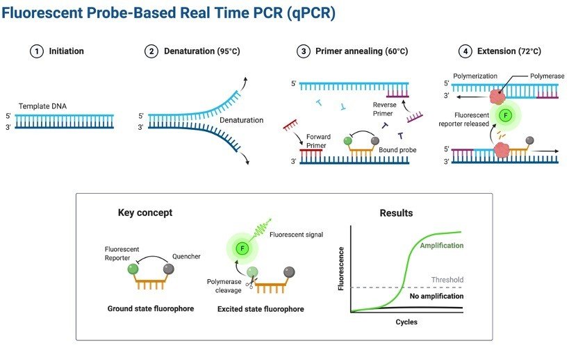
    PCR is a technique for amplifying DNA. Generally, the PCR reaction consists of a series of temperature changes that are repeated 25 – 45 times.

    These PCR cycles normally consist of three stages: the first, at around 95 °C, allows the separation of the nucleic acid’s double strands; the second, at a temperature of around 50-60 °C, allows the primer binding with the DNA template; the third, at between 68 – 72 °C, facilitates the polymerization carried out by the DNA polymerase.

    This principle of amplification is utilized in real-time PCR also widely known as qPCR (quantitative PCR) but instead of looking at bands on a gel at the end of the reaction, the continuous reaction is monitored in “real-time”.

    The reaction is placed in to a PCR machine that watches the reaction in real-time manner with a camera or fluorescence detector.

    There are a lot of techniques out there that are used to allow the progress of a PCR process to be monitored but they all have one thing in common.

    The DNA amplification to the generation of fluorescence which can be detected with a camera or sensor during each PCR cycle.

    Hence, as the number of PCR copies increases during the reaction, so the fluorescence increases.

    qPCR - research tweet 1

    Adopted from BioRender

    SYBR Green (or other intercalating dye)

    SYBR® Green is by far the most commonly used intercalating dye. This dye operate via a simple mechanism.

    The dye is fluorescent in its own but in the presence of double stranded DNA, the dye intercalates with the DNA double helix and thus alters the structure of the dye and causes it to fluoresce more.

    An increase in DNA product during PCR therefore leads to an increase in fluorescence intensity measured at each cycle.

    Nomenclature Commonly Used in qPCR

    Baseline is defined as PCR cycles in which a reporter fluorescent signal is accumulating but is beneath the limits of detection of the instrument.

    Threshold is an arbitrary level of fluorescence chosen on the basis of baseline variability.

    A signal that is detected above the threshold is considered a real signal that can be used to define the threshold cycle (Ct) for a sample.

    Threshold can be adjusted for each experiment so that it is in the region of exponential amplification across plots.

    Ct is defined as the fractional PCR cycle number at which the reporter fluorescence is greater than the threshold.

    The Ct is a basic principle of real time PCR and is essential component in producing accurate and reproducible data.

    qPCR Applications

    1. Quantitative mRNA expression studies.

    2. DNA copy number measurements in genomic or viral DNAs.

    3. Allelic discrimination assays or SNP genotyping.

    4. Verification of microarray results.

    5. Drug therapy efficacy.

    6. DNA damage measurement.

    Requirements for qPCR Assay

    1. Genomic DNA (2-20 ng per reaction)

    2. Primers for endogenous control and target gene

    3. SYBR green

    4. DNAse free Milli-Q water 5. Plasticware

    6. Real time instrument

    qPCR Procedure

    1. Make a mastermix for 10 reactions.

    ComponentsVol. per Reaction (ul)Vol. for 10 reaction (ul)
    SYBR Green550
    Forward Primer0.3 (300 nM-500 nM)3
    Reverse Primer0.3 (300 nM-500 nM)3
    cDNA1 (100 ng-100 fg)10
    Water3.434
    Total10100

    2. Mix well and centrifuge briefly

    3. Aliquote 9 μl of the mastermix in the wells of the 96-well plate

    4. Add 1 μl of the DNA ( 50 ng/ μl) in each well and then seal the 96-well plate with

    5. Setup the PCR reaction in the STEP ONE real-time PCR

    6. Cycling condition 

    7. Total number of cycle varies but usually 40.

    StageTemperature (C) Min
    Stage 1 (Denaturing)953:00
    Stage 2 (Annealing)950:10
    Stage 3 (Melt Curve)55-600:30
    55-950:05

    8. Introduce a hold at 4 ºC at the end of cycling if you need to store samples for some time (max. overnight) before the plate read.

    9. Take reading in STEP ONE.

    Formula for Calculating Relative Level of mRNA Expression

    The relative level of mRNA Expression were calculated using the cycle threshold (Ct) method:

    2−ΔΔCt = 2{ΔCt (treated samples) − ΔCt (untreated control)} where ΔCt = Ct (Gene of interest) − Ct (Endogenous Gene)

    qPCR Citations

    Share

    Similar Post:

    Continue Reading
  • Incomplete Dominance: Definition and Examples

    Incomplete Dominance: Introduction We all know that Mendel’s contribution is very important…

    Continue Reading
  • Krebs Cycle: Cellular Respiration, Full Steps With...

    Krebs Cycle Have you ever imagined why living system (plants) synthesize food?…

    Continue Reading