A compound falls into the category of organic when it is attached to another group which is either carbon- carbon or carbon hydrogen is said to be organic compound. When 2 or more than 2 groups are attached to each other chemically it is called as chemical compound. A single atom could also be referred to an element. An element forms a compound when there is a bond holding them up together.
In chemistry, compounds are broadly categorized into organic and inorganic compound. If organic compound is defined as the one containing carbon, then a compound is said to be inorganic if it does not possess carbon atom.
Organic Compounds Vitalism
The roots of vitalism has been seen coming from Greek philosophy and ancient Egypt. The basic thought behind vitalism is that living things differ from non-living, thus making the living things to produce chemicals. As living organism are in the influence of vitalism, they have the ability to make such compounds.
Scientist such as Louis Pasteur who was working on pasteurization and spontaneous generation, assumed that there is some life which is responsible in the fermentation process, as only something living had the ability to make a process such as fermentation possible and referred to them as vitalism and the followers were called vitalist.
The reactions which required a bit of alterations to be made were called as inorganic compound. However, the theory was latter rejected by various experiments conducted further. In 1828, a scientist Friedrich Wohler identified that using inorganic salts urea could be formed. However, the vitalism theory considered urea as organic compound, produced from living person only.
Organic Compounds Ambiguities
A compound is said to be organic if it possess carbon atoms bounded via a covalent bond. This is the definition of organic compound according to the modern era. However, it was difficult for the scientist to come up with a proper definition as with carbon atom in an organic compound, inorganic compound could also come into picture.
Example of inorganic compound containing carbon are cyanides, carbonate, carbon monoxide, carbon dioxide, thiocyanates, and structurally different forms of carbon such as coal, diamond, graphite are all composed of only carbon, therefore cannot be categorized into the class of organic compound.
Organic Matter
Although organic compounds on the earth’s crust form a small percentage, they are significant and are of great value. Organic compound examples are proteins, nucleic acids and carbohydrates. As living things are made up of carbon, thus when they die it can be degraded as these organic compounds can be broken to smaller ones.
Organic material is also secreted by living beings and this matter is found in the environment such as in soil, water where it could provide nutrients to other living organisms.
Natural vs Synthetic Organic Compounds
Organic compounds can be divided in various ways such as on the basis of its synthesis.
a) Natural Organic Compound: As the name suggests organic compound which are produced through natural sources such as plants and animals. Making them artificially increases the overall cost. Natural compound example are sugars, enzyme, antigens, lipids, neurotransmitters like protein, vitamins, amino acids, lectins, alkaloids and terpenoids.
b) Synthetic Organic Compounds: From the name itself it can be said that it is artificially prepared by making some chemical alterations in the reaction. To obtain synthetic compounds they might take place in a chemical reaction. They could use natural compounds as well in the process. Example plastics are synthetically produced.
Small Organic Molecules vs Large Organic Compounds
On the basis of the size or the length of the molecule it can be broadly categorized into small organic molecules and large organic molecule.
a) Small Organic Molecule: A molecule is said to be small if it has a molecular weight of less than 900 Daltons. Small molecules play a role in various processes example pharmaceutical drug. However, small molecules should not be misinterpreted with biomolecules such as protein, DNA, RNA and amino acids as they are huge.
b) Large Organic Compound: It is a macromolecule, or in simpler terms a huge molecule. This polymer is formed when many repeating subunits complete a large organic compound.
The net flow of solvent molecules through a semipermeable membrane is referred to as osmosis. It’s comparable to diffusion in that it moves downhill, from a higher to a lower concentration. However, in osmosis, the movement must take place over a semipermeable barrier.
Osmosis cannot be called osmosis without this component. While diffusion refers to the net movement of solutes between two solutions, osmosis is concerned with the net movement of solvent molecules, such as water molecules. The difference in water molecule concentration between the two sides of the membrane is what causes the water to migrate in order to bring the concentrations of the two sections closer together.
Osmosis Definition
Osmosis is defined in biology as the net transfer of water molecules from a higher to a lower water potential area through a semipermeable membrane (e.g., the cell membrane).
Other osmosis definitions are as follows:
1. The process of a solvent diffusing from a low-solute-concentration area to a high-solute-concentration area via a semipermeable barrier.
2. The ability of water to flow through a semipermeable barrier from a hypotonic solution (low concentration of dissolved chemicals) to a hypertonic solution (high concentration of dissolved substances).
Osmosis is characterized similarly in chemistry. When two solutions are separated by a membrane that selectively inhibits the passage of solute molecules while allowing the passage of solvent molecules. It is the passage of a pure solvent from one with a lower concentration of solutes to another with a higher concentration of solutes.
Osmosis Etymology
The word osmosis is a Latinized version of the now-defunct osmose. Osmotic is a derived word that means “pertaining to or of the character of osmosis.” Osmotic pressure, for example, is a pressure that occurs as a result of osmosis.
How Osmosis Works?
(1) net downhill flow of water molecules, (2) a selectively permeable membrane, and (3) an osmotic gradient are all required for osmosis to occur. Water molecules tend to migrate downhill, from a high-water concentration (or fewer solutes) to a low water concentration (or vice versa) (or greater solutes).
It cannot be considered osmosis if there is no net flow of water. It should also include a semipermeable barrier to allow for movement. Without it, the process is only diffusion rather than osmosis. Because water molecules are polar, channel proteins are required for them to travel along their concentration gradient.
These channel proteins are implanted in the cell membrane and create a hydrophilic conduit for water to flow through. The osmotic (pressure) gradient, or the difference in osmotic pressures between the two solutions, is what causes the water molecules to migrate.
Water potential is a measurement of the relative tendency of water to migrate from one location to another. The Greek letter Ψ is often used to symbolise it (Psi). Different tonicities of solutions produce a net flow of water across the cell membrane.
A solution consists mostly of the solute (material to be dissolved) and the solvent (the component that dissolves the solutes). The concentrations of components in two solutions will decide whether one is isotonic, hypotonic, or hypertonic in comparison to another.
Isotonic Solution
An isotonic solution is one in which the number of solutes in one solution is about equal to the number of solutes in another solution. A cell that is isotonic to the outside solution, for example, indicates that the internal fluid and the outside fluid have the same osmotic pressure and water potential. There will be no net flow of water molecules between the cell and the surrounding fluid in this instance.
Hypotonic Solution
A hypotonic solution is one that has a lower osmotic pressure (or contains fewer solutes) than the solution it is compared to. To dilute the solution, water flows toward the area with less water concentration or towards the more concentrated portion. Water will flow across the membrane and into the cell’s more concentrated solution if the fluid around the cell is hypotonic, for example.
Hypertonic Solution
A hypertonic solution is one that appears to be the polar opposite of a hypotonic solution. In comparison to the other solutions, a hypertonic solution contains more solutes and less water. Water will exit a cell submerged in a hypertonic solution to dilute the solution outside.
Osmosis Examples
i. Osmosis in Animal Cells
Osmosis is important in biological systems because many biological membranes are semipermeable, and it has a variety of physiological consequences. When animal cells are exposed to a hypertonic (lower water concentration) environment, the water leaves the cells, causing the cells to shrink. Crenation is the medical term for this ailment. When animal cells are put in a hypotonic environment (i.e., one with greater water content), water molecules migrate into the cells, causing them to expand. Cells will eventually rupture if osmosis persists and becomes extreme.
ii. Osmosis in Plant Cells
Plant cells do not rupture owing to an excessive amount of water input. Plants use their cell walls and vacuoles to protect themselves against excessive osmosis. The plant cell is stabilised by osmotic pressure exerted by the cell wall. Osmotic pressure, in fact, is what keeps plants upright. The big vacuole inside the plant cell aids osmoregulation, a regulatory process in which water potential is controlled to keep the osmotic pressure inside the cell within the optimal range.
Water efflux, on the other hand, does not protect plant cells. When a plant cell is put in a hypertonic environment, the cell wall is unable to prevent water loss. Cells shrink or become flaccid as a result of this.
Decomposers are the organisms that have the ability to decay or break down the dead organisms. Decomposers are also defined as those organism that have the ability of decomposition.
What is a Decomposer?
The decomposers break down dead material of both animals and plants in the ecosystem. Decomposition is the process of breaking down the complex organic matter into simpler substances.
The examples of decomposers are bacteria and fungi. These organisms feed upon the dead organic material and convert the matter into simpler substances. They break down the nutrient matter of the ecosystem and play an important role in the food chain. This decomposed organic matter is recycled and absorbed by plants and other primary producers.
In ecological pyramid, they found in lowest position. Decomposers are heterotrophic organisms and get nutrients from the dead organic material.
Decomposers are mainly saprophytic in nature. The word saprophyte is made up of two words, sapro meaning “rotten material” and phyte meaning “plant”. Saprophytes are those organisms which feed upon the dead plant material or plant litter. The break the plant litter into molecular elements like carbon, calcium, and nitrogen etc. They convert the organic dead mass into simpler substances with some digestive enzymes.
For example, proteins are broken down into amino acids and carbohydrates are converted into simple sugars like glucose and fructose. They can survive in high humid conditions, and in presence of oxygen. Some common features of saprophytes include-
• Have filaments
• Heterotrophic in nature
• Reproduce by means of spores
Importance of Decomposer
There are various crucial functions carried out by the decomposers. In ecosystem, they have some major functions like decomposition or disintegration of the dead organisms.
Ecological Cleansers and Balance Providers: Decomposers play an important role in cleansing the environment by decomposing the dead material and also help to create a biosphere for the new life. Thus they have an important role as ecological cleansers and also in balancing the ecosystem.
Recycling of Nutrients: Decomposers convert the dead material into molecular elements like carbon, nitrogen, phosphorus, etc. These elements are then absorbed by the primary producers like plants, algae etc., thus, decomposers helps in recycling the nutrients. The essential elements for the survival of producers are provided by decomposers.
Types of Decomposer
The decomposers are categorized into four types, namely fungi, insects, earthworms, and bacteria.
i. Fungi
Fungi are heterotrophic organisms that do not perform photosynthesis and feed on other organisms. Several fungi are saprophytic that feed upon dead material and principal decomposers in the ecosystem. Fungi secrete some enzymes in the environment to break down the plant litter into simpler molecules. These enzymes digest the material, which is absorbed by the fungi itself and thus, it get nutrient from the litter. Fungi grow in high humid conditions.
ii. Insects
Several insects like flies, maggots, dung beetles, and ants also help in decomposition. These insects are categorized as detritivores because they digest the dead material in their intestinal tract. Based upon the type of organic matter, the decomposers (insects) are classified into different categories-
• Dead plant tissue feeding insects
• Dead animal tissue feeding insects
iii. Earthworms
Earthworms are also considered as detritivores that orally feed upon the dead material. They are also known as friendly worms because of their ability to enrich soil by decomposition of litter. They release nutrients like phosphorus and calcium in the soil and helps to improve soil quality.
iv. Bacteria
Bacteria are microscopic, prokaryotic organisms that are ubiquitous in nature. They also involved in the process of decomposition. Bacteria also decompose the litter into carbon, nitrogen, etc. and helps in recycling of these nutrients. These nutrients are then absorbed by the producers of the food chain.
Decomposers and Detritivores
A group of organisms that break down the dead organic material into simpler molecules are called decomposers. The decomposers are categorized into two major groups: Detritivores and saprotrophs.
Detritivores are the type of decomposers including some animals like, worms and insects, whereas saprotrophs include fungi and bacteria. Sometimes, decomposers and detritivores are used as synonyms.
Decomposers
Detritivores
A group of organisms that decay the dead material.
A class of decomposers that feed orally in the dead material.
They act by secreting some digestive enzymes.
Act by digesting the matter in their digestive tract.
Cannot digest the large clumps of dead organic matter.
They can digest upon large clumps.
Examples- Fungi, bacteria, earthworms
Examples- Earthworms, insects
Decomposer vs Scavengers
Scavengers are the organisms that initiates the process of decomposition. There are some differences between Scavengers and decomposers-
Scavengers
Decomposers
The process of decomposition has initiated by scavengers.
They act on the particles that are decomposed by scavengers.
Act as initiators
Act as finishers
Example- Vultures, Flies
Examples- Fungi, Bacteria, earthworms
Decomposition Definition
Decomposition is the process of breaking down the dead organic material into simpler molecules. The process completed by some organisms called decomposers. Some common decomposers are fungi, bacteria.
Stages of Decomposition
The process of decomposition completes in five stages:
i. Fragmentation
The organic material is fragmented in this stage. The process initiates by detritivores to increase the surface area for decomposers. The detritivores feed upon the dead material and digest it with their digestive tract. Detritivores converts the matter into fragments for decomposers to act upon.
ii. Leaching
The fragmentation is followed by Leaching. In this stage, the water soluble nutrients present in the fragmented detritus are percolates through the soil.
iii. Catabolism
Third stage include secretion of enzymes by decomposers. These enzymes catabolize the decomposition process and convert the organic material into simpler substances.
iv. Humification
The formation of humus by dead matter is called humification. The amorphous substances form humus, which is very fertile.
v. Mineralization
It is the last step of decomposition. In this step the minerals are released in soil. The minerals or organic substances includes calcium, potassium, carbon, nitrogen etc.
Factors Affecting Decomposition
• Quality of Litter: Decomposition is affected by the structural and chemical properties of the litter.
• Temperature: The decomposition slower down with low temperature. Low temperature also decrease the microbial growth.
• Aeration: Aeration is essential factor for decomposition, thus oxygen is very important.
• Inorganic Chemicals: The process of decomposition decreases with presence of inorganic minerals.
• Moisture: moisture is essential factor that increases the rate of decomposition.
Decomposer Examples
i. Decomposers in Aquatic Ecosystem
1. Oceans / Seawater Decomposers: They are found in oceans and in seawater.
Some common examples are:
• Christmas tree worms
• Crabs
• Hagfish are the scavengers
• Sea urchin
• Tube worm
2. Freshwater Decomposers: Mostly found in fresh water bodies like rivers, ponds, etc.
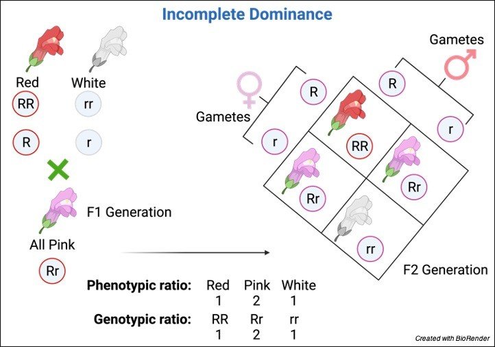
The term “Incomplete dominance” was first proposed by the German Botanist, Carl Correns (1864-1933). Based on his experiments conducted on the four o’clock plant, he described the concept of incomplete dominance.
Incomplete dominance can be defined as a condition in which none of the alleles shows complete dominance over others. The condition results in the phenotype that intermediates of the phenotype of both the alleles.
What is Incomplete Dominance?
Incomplete dominance is the phenomenon in which a heterozygous offspring produced by crossing two true-breeding parents intermediate the phenotype of both the parents. It is also described as partial dominance or intermediate inheritance.
The condition arises when any of the alleles are not expressed as dominant or recessive. In other words, the dominant allele cannot show complete dominance.
To understand the phenomenon of incomplete dominance, let’s understand some terms-
A set or version of gene expressions is called an allele. There are two alleles present in all organisms inherited from each parent for a specific gene. The allele can be categorized as dominant or recessive.
The prominent allele is called dominant whereas suppressed allele is called the recessive allele. The dominant allele shows its effect on the phenotype of the organism.
Based on different sets of alleles, an organism can be heterozygous or homozygous.
A genotype is a set of genes that is responsible for the features of an organism. The genotype determines the phenotype which refers to the physical appearance of the organism.
Defining incomplete Dominance
There are different ways to define incomplete dominance- The dilution of the dominant allele over the recessive allele is referred to as the incomplete dominance that results in the formation of a new heterozygous phenotype.
In heterozygous organisms, the intermediate trait appears between homozygous dominant and homozygous recessive alleles as termed as incomplete dominance.
The condition occurs due to a combination of both dominant and recessive alleles in an offspring. Whereas some definitions also show that incomplete dominance arises due to a specific trait in the offspring which is neither dominant nor recessive.
In that situation, the phenotype of the heterozygous offspring resembles intermediate characters. In incomplete dominance, an intermediate heterozygote is formed. This phenomenon plays a crucial role in the variation of an organism’s features.
Mechanisms of Incomplete Dominance
Mendel did his experiment in the pea plant and proposed laws of inheritance. He also described dominant and recessive traits. Carl Correns considered Mendel’s work and performed experiments in the four o’clock plant.
He crossed red flower-producing plants with white flower-producing plants and observed results. He got an intermediate pink-colored flower. Correns termed this phenomenon incomplete dominance.
How Does Incomplete Dominance Work?
The punnet square is used to understand the mechanism of incomplete dominance. It predicts the genotype of the offspring in the experiment. In this experiment, the red flower-producing plant is crossed with a white flower-producing plant. In the given picture, we can see that the result shows an intermediate phenotype that is a pink-colored flower.
It produces due to expressions of a dominant allele that cannot be expressed completely. So in incomplete dominance, the offspring contain both alleles but none of them are expressed completely therefore the phenotype gets intermediate between parent traits.
Incomplete Dominance vs Codominance
Mendel described the effects of alleles on the phenotype of an offspring. Based on phenotypic expressions, dominance can be categorized into different types. Codominance and incomplete dominance are also different types of inheritance. Mendel did not define both the term.
1. Incomplete Dominance
The phenomenon where the offspring show the phenotype that is intermediate between both the parents is called incomplete dominance.
For example- when a plant producing red flowers is crossed with the plant producing white flowers, the resulting offspring produces pink flowers, which is intermediate between red and white.
In incomplete dominance, the dominant allele cannot mask the recessive allele completely which results in the formation of a new phenotype. The garden pea plant does not show incomplete dominance therefore Mendel did not study this phenomenon. It can be seen in the plant of four o’clock flower
Codominance
The phenomenon where both of the alleles for a specific trait are expressed together in the offspring is called codominance. In this condition none of the alleles is dominant or recessive, therefore both alleles remain present and express in the phenotype.
In the results, the heterozygotic offspring produced by crossing two homozygotes produce a distinctive phenotype. There are several examples of codominance that can be seen in plants as well as in animals.
For example- When the plant with a white flower crossed with a red flower it produces flowers with red and white spots. Codominance was also not studied by Mendel. Another example of codominance is blood type in humans.
Incomplete Dominance
Codominance
The condition in which the dominant allele does not show complete dominance over the recessive allele.
The condition, in which none of the alleles show dominant and recessive characters.
The phenotype of the offspring is intermediate between both parents’ traits.
The phenotype of the offspring expresses traits of both alleles.
Mixing of traits occur.
No mixing of traits occur.
Examples- pink flowers of Mirabilis jalapa.
Examples- The blood type (A, B, and O) in humans.
Incomplete Dominance Examples
Several morphological and physiological variations occur due to incomplete dominance. Examples of incomplete dominance include the pink flower color trait in Mirabilis jalapa and other species. Incomplete dominance also occurs in some animals and humans.
Hair color, eye color, and skin color are some common examples of incomplete Dominance in humans.
In Plants
The common example of incomplete dominance is the four o’clock plant. In Mirabilis jalapa, the red and white flowers are crossed to produce pink flowers. The Carnation plant is also an example of incomplete dominance in which a cross between a red flowering plant and a white flowering plant produces pink flowers in its phenotype.
The phenomenon have also some significant roles in crop improvement. A corn plant has partially dominating traits that are high yielding than original ones therefore the crop is improved by using incomplete dominance.
In Humans
The phenomenon of incomplete dominance can also be seen in humans. The common examples are hair color, eye color, skin color, height, and sound pitch, etc.
For example, when a person having curly hair marries a female having straight hair, their offspring may be born with wavy hairs. The eye color of humans also shows incomplete dominance. Incomplete dominance can also be seen in height patterns and skin color. However, skin color is determined by genes that produce melanin therefore parents with dark and light skin produce offspring with intermediate skin color.
In Other Animals
The phenomenon is also visible in some other animals and birds. Chicken, rabbits, dogs, cats, and horses also show incomplete dominance. The common example of incomplete dominance is the Andalusian chicken. Feathers of Andalusian chicken show the intermediate color of both the parents.
When a white feathered male crosses with black feathered female chicken, the offspring results in blue and tinged feathers. Similarly, a dog’s tail is also an example of incomplete dominance. The length of the tail shows an intermediate phenotype when a long-tailed dog is crossed with a short-tailed dog. The spots in cats, dogs, and horses are also examples of incomplete dominance.
The state of balanced internal chemical and physical condition maintained by living organisms is called homeostasis. This ability is crucial for all living organisms.
Despite the dynamicity of the external environment, the physiological strategies adopted to maintain the proper functioning of a system, these strategies are called homeostasis.
Several regulatory mechanisms help to maintain homeostasis in humans. It consists of three general components: a receptor, a control centre, and an effector. A loop forming a homeostatic mechanism can either be positive or negative.
When the homeostasis mechanism stimulates the process, it is defined as positive feedback, whereas when it inhibits or decelerates the process, it is called negative feedback. Labor contraction during childbirth and blood clotting are some examples of positive feedback.
Thermoregulation, blood glucose regulation, and calcium homeostasis are some common examples of negative feedback.
Homeostasis Definition
The ability or tendency of any cell to maintain the condition of equilibrium is called homeostasis. It can also define as using feedback controls and other regulatory mechanisms to maintain a stable internal environment.
Homeostasis prevents the body or the cell from affecting its external environment. The process includes different multifarious physiological mechanisms to stabilize the functional stable of an organism.
Homeostasis Etymology
The term is made of two Greek words, homoios meaning “similar” and stasis, meaning “standing.” The term “homeostasis” was first coined by American physiologist Walter Bradford Cannon.
Homeostatic Processes
Various biological functions interconnect the needs of an organism with the help of a system. Higher animals, like humans, are made of complex bodily organs that need proper cellular functioning.
Although these organs are not similar, to sustain within the ideal range, they have to function alongside each other. There are several homeostatic processes, work by various regulatory mechanisms to stable the internal environment.
Homeostasis in Human Body
When there is a continuous imbalance in the internal stability of the human body, it cannot function properly. Various homeostatic mechanisms are used by the human body to sustain in extreme conditions and for proper functioning.
The homeostatic range consists of variables like pH, sodium level, potassium level, calcium level, and blood sugar level. Any particular variable has some upper and lower limits that are defined as the homeostatic range.
This range is crucial for proper functioning, beyond this range body will become dysfunctional. To maintain this range, various regulatory mechanisms are employed. The regulatory mechanisms are comprised of three components.
Components of Homeostasis
Components of Homeostasis are as follows-
i. A receptor
i. A control centre
ii. An effector
The information about the homeostatic range is received by the receptor. It passes this information to the control centre. Here this information is processed and then signal sent to the effector.
In the human body, several receptors are present, which are as follows-
a. Photoreceptors (stimulate in the presence of light)
b. Olfactory receptor cells (reacts to odours or smell)
c. Gustation receptors (reacts for taste)
d. Auditory receptors cells (present in the epithelium of organ, reacts to sound)
e. Thermo receptors (reacts to changes in temperature)
f. Mechanoreceptors (present in the skin and reacts to mechanical stimuli)
g. Interoceptors (reacts in stimuli inside the body) Peripheral chemoreceptors (respond to chemical changes)
In this process, all three components work by detecting and then responding to the stimulus. The processing for the detected stimulus is directed by the control centre. The control centre determines the response then the message is sent to the effectors. Later, a response is brought by effectors to revert the normal homeostatic range.
Homeostatic Mechanisms
The mechanism is also called a feedback mechanism, which can be positive or negative feedback. When the mechanism continues in the direction of stimuli, it is considered positive feedback.
Labor contraction, blood clotting are some examples of positive feedback. When the effector reverses the direction of the stimulus, it is called negative feedback. Examples are blood glucose level, thermoregulation, and osmoregulation.
Homeostasis Examples
a. Labor Contractions
It is an example of positive feedback. During childbirth, the muscles of the uterus further contract, that termed labor contractions. At the time, the body also stimulates to enhance the contractions by secretion of oxytocin.
b. Blood Clotting
Blood has the natural tendency to convert into solid, which is called blood clotting. It is also considered positive feedback because of the activation of clotting factors in series. It results in the formation of a fibrin clot.
c. Thermoregulation
It is an example of negative feedback. The thermoregulation reacts in the regulation of body temperature that helps to maintain the internal temperature of humans. The nervous system is responsible for the regulation of body temperature, particularly the anterior hypothalamus.
In our body, heat loss occurs when the ambient temperature is less than the skin temperature. In that condition, the thermoregulatory centre of the brain controls the mechanism to maintain normal conditions. The signal received by muscles to shake results in less heat degenerate.
The process also works in the condition of higher ambient temperature than the skin temperature. Here, the eccrine sweat glands secrete sweat to cool the body and reduce temperature. Thermoregulation is also crucial in other animals that maintain constant internal temperature.
d. Blood Homeostasis
The mechanism of regulating blood sugar levels in the body is called blood homeostasis. Our body regulates blood sugar levels by various hormones.
For example- the pancreas secretes glucagon when the blood sugar level is low that stimulates the conversion of glucagon into glucose. Similarly, insulin has secreted when blood glucose level increases in the body.
Insulin increases the absorption of glucose by skeletal muscles and fat tissues. Glucagon is produced by alpha cells of the pancreas whereas insulin is produced by beta cells. The pancreas is a glandular structure having two major types of cells.
The process of converting glucose into glycogen is called glycogenolysis, whereas the production of glucose from glycogen is called gluconeogenesis. Glycogen is the storage form of sugar in the human body, which is stored in the liver and muscles and absorbed by a carbohydrate-rich diet.
e. Blood Pressure Homeostasis
It is an example of negative feedback, which regulates blood pressure. The force exerted by blood circulation is called blood pressure. Our body regulates blood pressure through the cardiovascular center that has three distinct activities to regulate blood pressure.
(1) Nerve impulses sent by the cardiac center to increase cardiac output.
(2) Nerve impulses sent to decrease cardiac output.
(3) Regulation of diameter of blood vessels by the vasomotor.
f. Calcium Homeostasis
A parathyroid gland and parafollicular cells are sensitive to calcium ion levels in our body. When the level of calcium ions decreases in our body, the parathyroid hormone secreted calcitonin to increase calcium ion level.
A high level of calcitonin in the body causes bone resorption and excretion of phosphate ions through urine. The secretion of calcitonin stimulates the bone cells to absorb calcium.
g. Potassium Homeostasis
The potassium level in our body is balanced by the adrenal complex. High potassium concentration in our body causes membrane depolarization in the adrenal complex. Due to the high level of potassium, aldosterone stimulates the excretion of excess potassium from the body and normalizes the potassium level in plasma.
h. Osmoregulation
The human body consists of two major types of fluids- intracellular fluid and extracellular fluid. The body regulates the amount of water between these two fluids by the process of osmoregulation. The osmoreceptors present in the hypothalamus are sensitive to osmotic pressure changes.
In the case of hypertonicity or hyper-osmolality, these receptors secrete vasopressin. The vasopressin promotes water reabsorption by targeting the kidney. In the case of hyper-osmolality, the vasopressin level decreases in blood plasma and water excreted by the urine.
i. Action Potential Generation
It can be seen during membrane potential. The voltage-gated sodium channels open in series when the nerve impulse has relayed. It results in the influx of ions. Thus, it results in depolarization of the surrounding area and is considered positive feedback.
Biological Importance of Homeostasis
Homeostasis is a very crucial mechanism to maintain the optimal value of the innate variables. It is crucial to ensure sustain life and prevent instability in the body. The system cannot function properly without homeostasis which may lead to various health conditions or may face death due to organ failure.
A pair of chromosome having same gene sequence, loci, chromosomal length, and centromere location is called homologous chromosome. The pair consists of one paternal and one maternal chromosome.
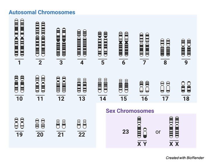
The human genome has total of 46 chromosomes. The offspring inherit half of the chromosome by its father and other half from the mother. During meiosis, the maternal chromosomes pair up with paternal chromosome. In humans, the female have all the 23 pairs of homologous chromosomes whereas male have 22 pairs of homologous and 23rd chromosome is not homologous.
At meiosis, the genetic recombination occurs between homologous pairs of chromosomes that is crucial for genetic variation. Due to this genetic recombination or crossing over, the offspring produced are genetically different from each other.
Homologous Chromosome Definition
Paired chromosome with same gene sequence, length, and centromere position are called homologous chromosomes. Homologous chromosomes may have different alleles that is composed of paternal and maternal chromosomes.
Homologous Chromosome Etymology
The word homologous is composed of two words, homos, meaning “same” and logos meaning “relation”. A scientist named as Wilhelm von Waldeyer – Hartz coined the term chromosome in 1889.
Homologous Chromosome vs Heterologous Chromosomes
The chromosomes can be of two types, homologous and heterologous chromosomes. The chromosomes that have similar gene sequences, chromosomal length, and centromere position are termed as homologous chromosomes whereas different chromosomes are termed as heterologous chromosomes.
During meiosis, exchange of genetic material occur in between homologous chromosomes whereas it is absent in heterologous chromosomes. Sometimes, due to any mutation, like translocation, heterologous chromosomes also exchange chromosomal parts.
Let’s compare the homologous chromosomes and heterologous chromosomes with this table-
Homologous Chromosomes
Heterologous Chromosomes
Pairing of chromosomes occur during meiosis
Pairing absent
Exchange of genetic material may occurs at synapse
Exchange of genetic material may occur during translocation
Homologous chromosome is composed of – Centromere Long arm Short arm
Heterologous chromosomes are composed of- Centromere Long arm Short arm
Examples- Autosomal chromosomes, sex chromosomes in female(XX)
Examples- sex chromosome (XY)
Homologous Chromosome Karyotype
Human nucleus consist 46 chromosomes. There are 22 pairs of autosomes having same length, same position of centromere that are considered as homologous chromosomes. The sex chromosomes have two XX chromosomes are considered as homologous chromosomes whereas X and Y chromosomes are considered as heterologous chromosomes. According to this, the female have all homologous chromosomes and male consists 22 pairs of homologous and one pair of heterologous chromosome.
Homologous Chromosomes vs Sister Chromatids
The centromere join two identical chromatids of homologous chromosomes. These identical chromatids are called sister chromatids. Chromatins are thread like structures that condense into chromosomes at time of cell division.
At interphase of cell division, the genetic material present in chromosome replicates by the process of DNA replication. These two copies of DNA are called chromatids that are joined by a centromere. The chromatids are commonly known as sister chromatids.
Homologous Chromosome Characteristics
In eukaryotic organisms, the genetic material or chromosome in located in the nucleus of the cell. There are two sets of the chromosomes present in the nucleus- male gamete called sperm and female gamete known as egg cell.
After fertilization, the egg cell develop into a diploid zygote by fusion of both male and female haploid gametes. All the organisms consist two sets of chromosomes, one set from each parent that can be similar or may be differ in gene sequence, chromosomal length, and position of centromere.
The pair of chromosomes exchange their genetic material during meiosis. Homologous chromosomes may have different alleles that carries genes for a specific trait. When both the alleles carry different genes for a particular trait, they are described as heterozygous and when they have same genes they are termed homozygous. The concept of dominance of alleles was described by Gregor John Mendel.
Homologous Chromosome Pairing Process
A cell can divide by two methods- mitosis and meiosis. The mitosis results in formation of two daughter cells, each having same no. of chromosomes as the parent cell whereas meiosis is described as reduction division where four daughter cells are formed with half of the chromosomes present in parent cell.
The gametes also undergo meiosis to produce haploid sperm cell and egg cell. These haploid gametes fuse in the process of fertilization. Meiosis completes in two stages- meiosis I and meiosis II. DNA replicates in S phase of interphase and sister chromatids are joined by centromere that exchange their genetic material at prophase I.
This process is called synapse or pairing. During anaphase I the homologous chromosomes separate and moves to the opposite poles of the cell. Telophase I results in formation of two daughter cells that undergo meiosis II Meiosis II results in formation of four daughter cells having reduced chromosomal number by half.
Homologous Chromosome Importance
All the cellular activities are regulated by genetic material i.e. chromosomes therefore chromosomes are crucial for all living beings. The trait of any organism is also determined by the chromosome that have the genetic information.
The homologous chromosomes also exchange their genetic material, which is essential to promote genetic variation. Because of the chromosomes, organisms can reproduce by sexual means to produce offspring that are not identical.
The differences between organisms increase their chances to survive in harsh conditions and prevent from wiped out by any viral disease or by any other method.
The transport of substances from their higher concentration to lower concentration with the help of any transport molecule is known as facilitated diffusion. In facilitated diffusion, the substances move across a biological membrane along the direction of their concentration gradient. It doesn’t require chemical energy.
There are some common examples of living processes that require facilitated diffusion for their completion. These biological processes include amino acid and glucose transport, gas transport, and ion transport.
Facilitated diffusion has great significance because it balances the transport of molecules from the plasma membrane. The plasma membrane or cell membrane is an outermost covering of cells that is selectively permeable hence it only allows selective molecules to transport through the membrane.
It can be defined as following- “The passive movement of substances, across the plasma membrane by the help of some transport proteins found in the cell membrane.”
Facilitated Diffusion Etymology
The term facilitated diffusion is consisting of two words where facilitated came from the Latin word facilis, meaning “make” and the word diffusion meaning “a pouring fort”. Facilitated diffusion is also known as passive-mediated transport.
Characteristic Features of Facilitated Diffusion
Cellular transport is of two types- Active transport and passive transport. Facilitated diffusion is a type of passive transport where substances move from their higher concentration to lower concentration. This concentration difference creates a gradient that encourages substances to constitutionally move and to distribute between two areas to achieve equilibrium.
Facilitated diffusion does not require chemical energy because it directs movement from higher to lower concentrations. So, the process completes with the kinetic energy of the cell. The process differs from other types of passive transport due to the assistance of transport proteins while other passive transport does not require any transport protein.
Facilitated Diffusion Diagram
Facilitated Diffusion Mechanism
The plasma membrane is composed of a lipid bilayer which regulates the passage of molecules across the membrane. The hydrophobic region of the membrane is located outwards and thus it prevents polar (hydrophilic) molecules to cross the membrane directly. However, the non-polar or hydrophobic molecules can easily diffuse through the membrane along with their concentration gradient.
Thus large polar molecules take on certain membrane proteins like channels and carriers to diffuse through the plasma membrane.
Now facilitated diffusion can be categorized into different types based on membrane proteins engaged.
E.g.- Facilitated diffusion by channel proteins- It is a type of diffusion that uses certain membrane proteins that act as a pore in the lipid bilayer. The most common example of channel proteins is transmembrane channels which form by protein complexes that span across the plasma membrane.
These channels help several charged ions to be transported across the plasma membrane. Besides channels, there are some other proteins like aquaporins, which are also integral membrane proteins involves in the transport of water molecules.
Another example of proteins involved in facilitated diffusion is carrier proteins. Carrier proteins can transport the larger molecules by changing their conformation during the transportation of molecules across the membrane (e.g. permeases).
Facilitated Diffusion Examples
i. Glucose and Amino Acid Transport
Glucose is a large polar molecule that is transported by the process of facilitated diffusion. Glucose transporters are types of carrier proteins that mediate the transport of glucose across the lipid bilayer. The glucose taken in the diet is absorbed by the small intestine by active transport.
In the bloodstream it is released via facilitated diffusion and other body organs, cells also take glucose vias facilitated diffusion. Similarly, amino acids are also a type of biomolecules that moves from the bloodstream into the cell by facilitated diffusion.
ii. Ion Transport
The charged ions cannot diffuse through the plasma membrane directly due to the charge of those ions. Thus, these ions are also to be transported by facilitated diffusion. Some common examples of proteins are sodium ions, calcium ions, potassium ions.
Facilitated Diffusion vs Active Transport
Active transport is a type of transport that also needs a concentration gradient to complete the process. Ions, sugars, and salts can be transported by any of the transportation methods. Both the methods use transport vehicles for transport. However, there are some differences between facilitated diffusion and active transport. The major difference is the direction of transport.
Facilitated diffusion transports the substances from their higher concentration to lower concentration whereas Active transport facilitates the flow of substances from lower to higher concentration. This against the concentration gradient movement of substances requires chemical energy in the form of ATP. facilitated diffusion does not require chemical energy.
Simple Diffusion vs Facilitated Diffusion
Both are the type of passive transport where substances move along with their concentration gradient. The processes differ in the way of transporting molecules across the membrane. There are some transport proteins required in the process of facilitated diffusion to transport substances whereas simple diffusion, does not require any transport proteins. The rate of the process also differs which tends to be affected by saturation limits in facilitated diffusion and the rate is more straightforward in simple diffusion.
Importance of Facilitated Diffusion
There is an unequal distribution of molecules between the intracellular region and extracellular region that enhances cellular transport. To establish equilibrium between these two regions, the movement of substances is very crucial. Facilitated diffusion plays an important role to transport certain polar and charged molecules that cannot transport via simple diffusion. It is also helpful in maintaining homeostatic optimal levels of molecules.
The process of creating polypeptide chains is called protein synthesis. The process completes with an implication of amino acid synthesis, transcription, translation, and post-translational events in a biological system.
The formation of amino acids from carbon sources like glucose is termed amino acid synthesis. It is a set of biochemical reactions. All organisms consist of several essential and non-essential amino acids. Some of the amino acids have been obtained from the diet whereas other amino acids are produced by the body.
Proteins are crucial biomolecules involved in various cellular functioning. These proteins are produced by the process of transcription and translation. The procedure of synthesizing mRNA templates from DNA molecules is called transcription. Later, these mRNA templates are used to translate them into amino acids.
The proteins or polypeptide chains are made by linking together these amino acids in a particular order based on the genetic code. After translation, these polypeptide chains undergo the process of post-translational modification, called protein folding.
Protein Synthesis Definition
The process of the creation of proteins is called protein synthesis. This process completes inside the cell in all organisms. In eukaryotic cells, the transcription takes place in the nucleus.
Later these mRNA templates move into the cytosol where it translated into polypeptides. The prokaryotic cells lack the nucleus since transcription completes in the cytoplasm of the cell.
Etymology
Protein synthesis is made up of two words, here the word protein is derived from the Greek word protos, meaning “first“. The word synthesis is derived from sunthesis, meaning “to put together“.
Prokaryotic Protein Synthesis vs Eukaryotic Protein Synthesis
All living organisms require several biomolecules to develop and proteins are one of the important biomolecules used by all cellular organisms. Numerous cellular activities in the cell need proteins in prokaryotic and eukaryotic cells.
These proteins are used in different biochemical reactions, act as a catalyst, and used for structural purposes. The mechanism of protein synthesis is different in prokaryotic and eukaryotic organisms.
Eukaryotes have a well-defined nucleus and the transcription completes in the nucleus of the cell whereas transcription in prokaryotes completes in the cytoplasm.
In a eukaryotic cell, the mRNA is transported into the cytoplasm where it is translated by ribosomes. Here the nucleotides are converted into amino acid chains.
Protein Synthesis and Genetic Code
A genetic code or codon is a trinucleotide sequence that identifies a particular amino acid. A codon is a group of three nucleotides so it is called a trinucleotide sequence. For example, a sequence GCC (guanine-cytosine- cytosine) codes for the particular amino acid alanine.
Similarly, 64 codons are present that encode different amino acids. These codons are present in the mRNA template that adjuncts to anticodons present in the tRNA molecule. These anticodons are also trinucleotide sequences.
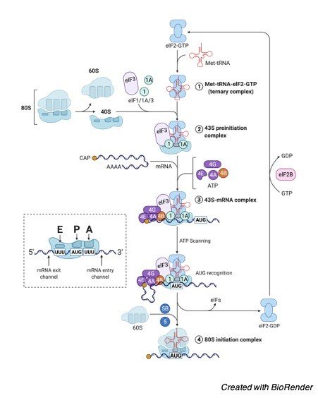
mRNA, tRNA, and rRNA
There are three types of RNA involved in the process of protein synthesis. The first type is mRNA (messenger RNA) which carries codons that are converted into an amino acid chain. These mRNA molecules are produced by a DNA template in the nucleus of the cell.
However, in prokaryotes, it synthesizes in the cytoplasm. The mRNA is consists of a 5′ cap, 5′ UTR region, coding region, 3′ UTR region, and a poly (A) chain. The coding region of mRNA is responsible for gene expression which has a start codon at 5′ end and a stop codon at 3′ end.
Another type of RNA is the tRNA molecule, also called transfer RNA. The transfer of particular amino acid to the ribosome is completed by a tRNA molecule. It has a cloverleaf resembling structure that has two major sites, named as- anticodon arm and accepter stem.
The anticodons are present in the anticodon arm and the accepter stem specifies the particular amino acid that needs to be attached. Now the ribosome translates the mRNA template into a polypeptide chain.
This ribosome is composed of rRNA (ribosomal RNA) and some proteins. It is a cytoplasmic organelle present in both prokaryotic as well as in eukaryotic cells. However, the structure and composition of the ribosome can differ in both the cell. In prokaryotes, the ribosome is composed of 70S subunit whereas eukaryotic organisms consist of 80S ribosomes.
There are three binding sites present in rRNA, named as- A, P, and E sites. The aminoacyl tRNA docks at the A site and peptidyl- tRNA binds at the P site. The site where the tRNA leaves the ribosome is termed as E (exit) site.
Protein Synthesis Steps
i. Transcription
The process of producing an mRNA template from a DNA molecule is termed transcription. The mRNA has trinucleotide sequences (codons) that encode for particular amino acids and provide a template for the process of translation.
Transcription completes in four major steps-
(1) Initiation
(2) Promoter Escape
(3) Elongation
(4) Termination
In the initiation phase, the RNA polymerase binds to the promoter of DNA with the intervention of any transcription factor. After the binding, the DNA starts unwinding and form a transcription bubble which is also termed as transcription start site.
In the next step, the RNA polymerase escapes the promoter to step into the elongation phase. The RNA travel across the DNA template and base pairs with the nucleotides on the noncoding strand in the elongation phase.
The RNA polymerase forms the sugar-phosphate backbone and thymines are replaced by uracils. The termination step starts with the breaking of hydrogen bonds of RNA- DNA helix.
The mRNA script in eukaryotes goes through polyadenylation, capping, and splicing but the prokaryotic mRNA does not undergo these modifications.
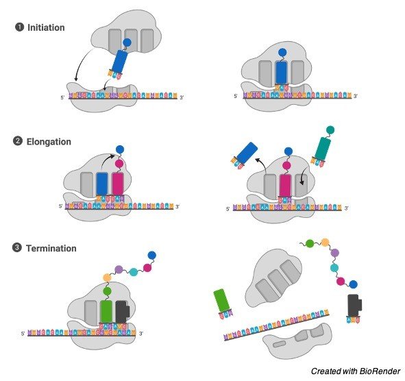
ii. Translation
The process of producing amino acids from the mRNA script is called translation. These amino acids are linked together and form a polypeptide chain.
Protein Synthesis Diagram
Translation completes in the cytoplasm of the cell and consist of four phases-
(1) Activation
(2) Initiation
(3) Elongation
(4) Termination
The initiation starts with the binding of the ribosome to 5’end of mRNA. In elongation, the binding of aminoacyl-tRNA to the ribosome occurs. When the A site of ribosome faces a stop codon, the process terminates.
iii. Post-Translation
In eukaryotic translation, proteins undergo some post-translational modifications like protein folding, proteolysis. These modifications are termed as several enzymatic actions of a polypeptide chain that occur after the formation of a polypeptide chain.
For example- to ensure proper cellular localization, the ends may be modified. The folding of the polypeptide chain to conclude secondary and tertiary structures is termed as protein folding.
A mode of reproduction in which the organism reproduces without mating and produces offspring by a single parent. It does not require the fusion of male and female gametes.
What is Asexual Reproduction?
The mode of reproduction where organisms produce offspring by the involvement of a single parent is called asexual reproduction.
In asexual reproduction, mating between male and female gametes is not necessary.
In asexual reproduction any organism can produce offspring in the absence of another parent, so the offspring produced by the asexual method resembles totally as its parent.
Asexual reproduction can be of different types like binary fission, fragmentation, sporogenesis, budding, parthenogenesis, and apomixis.
Asexual reproduction is a regular method of reproduction in microbes and primitive organisms; however, several plants, certain animals, fungi also reproduce by this method.
Reproduction is one of the crucial biological processes in which an organism produces biologically similar offspring. All organisms can reproduce in two significant modes- sexual and asexual.
Sexual Reproduction vs Asexual Reproduction
Asexual and sexual methods are major modes for reproduction adopted by most organisms. There are some differences between asexual and sexual reproduction which are listed below;
i. Asexual reproduction involves only one parent whereas sexual reproduction can only occur by the involvement of two parents which include paternal and maternal parents.
ii. In asexual reproduction, syngamy and meiosis are both processes that are not needed. Sexual reproduction cannot occur without syngamy.
iii. Meiosis is also present which is required to produce gametes. Asexual reproduction produces genetically identical off springs which are also termed clones of the parent, but in the case of sexual reproduction, off springs are not identical or different from their parents.
Advantages of Asexual Reproduction
The process of asexual reproduction is less complex and straightforward as compared to sexual reproduction. In this process, the organism produces offspring more quickly because of the participation of only one parent. Some courtship rituals seen in higher animals are also absent in organisms reproducing by asexual methods. Due to their quick reproducing ability, they can colonize fastly than higher animals.
Disadvantages of Asexual reproduction
Besides all the advantages, there are certain disadvantages also of this method. However, the method is less complicated and faster but apart from higher animals choose sexual reproduction commonly. Some higher animals which can reproduce asexually will only utilize it when sexual reproduction is not feasible.
According to researchers, 99.9% of eukaryotes reproduce by the sexual method. The major disadvantage of asexual reproduction is the lack of genetic diversity.
The organisms are a clone of its parent which leads to low genetic variation. In asexual reproduction, segregation during meiosis and crossing over is not present which is required for sexual reproduction.
These events are necessary for genetic variation, and therefore, their absence affects negatively evolutionary changes. Due to the same genetic material in all offspring, they may be similarly susceptible or resistant for a particular condition (e.g. viral disease) which increases the risk of getting wiped out by any disease.
The organisms which can reproduce only by asexual methods can get new genetic material through mutations.
Types of Asexual Reproduction
There are different types of asexual reproduction, like (1) binary fission (2) budding (3) vegetative propagation (4) spore formation (5) fragmentation (6) parthenogenesis (7) apomixis
i. Binary Fission
The process of dividing into two identical cells is called binary fission. Each of the cells can grow and reproduce also. Most prokaryotic organisms adopt binary fission to reproduce. Some archaea and protozoans also reproduce by this method.
There are three types of binary fission based on the manner of cell division, which is irregular type, longitudinal, and transverse- type. In irregular type, a cell divides along any plane. Longitudinal fission can be seen in Euglena and transverse-type is found in Paramecium.
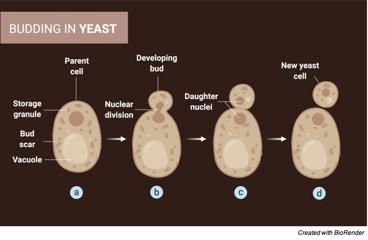
ii. Budding
Budding is a type of asexual reproduction; in which an outgrowth is formed by an organism that developed into a new individual.
Certain bacteria and other asexual organisms reproduce by budding. For example- Hyphomicrobium, Stella spp. Saccharomyces cerevisiae, hydra.
iii. Vegetative Propagation
Vegetative propagation is most commonly used by plants to reproduce asexually. Several plants cannot produce seeds, which can be propagated by vegetative methods. In this method, a new plant develops from vegetative parts of the plant, like roots, stems, and leaves.
Vegetative propagation is also used by gardeners and horticulturists to conserve and propagate rare and important plant species. Vegetative propagation is categorized into two types- natural and artificial propagation.
Artificial propagation can be done by the means of tissue culture methods and root hormones, whereas natural means include bulbs, runners, corms, tubers, etc.
iv. Spore Formation
Some organisms produce asexual spores to produce asexually. The method is called sporogenesis. Sporogenesis is made up of two words, spore, meaning ‘seed’, and genesis, meaning ‘origin’. As the name indicates they can disperse as seeds. They have thick-walled and highly resistant structures which differ from true seeds. Some fungi and vascular plants use sporogenesis to reproduce.
v. Fragmentation
When the parent organism breaks into several fragments and each fragment develop into a new individual, this process is called fragmentation. Several fungi (e.g. yeasts), vascular and non-vascular plants, and animals reproduce by the method of fragmentation.
vi. Parthenogenesis
When the female organism produces offspring without fertilization, it is termed parthenogenesis. It is divided into two types- apomictic parthenogenesis and automictic parthenogenesis. In this reproduction method, several methods are adopted to restore ploidy, e.g. doubling the chromosomes, fusion of meiotic products.
Examples- aphids, rotifers, nematodes, lizards, birds, etc.
vii. Plant Apomixis
It is the method of reproduction in plants where a plant reproduces without fertilization. Some bryophytes give rise to sporophyte-looking offspring having ploidy of gametophyte, known as apogamy. Similarly when a gametophyte is produced from the sporophyte but having ploidy of a sporophyte, is termed as apomixis or apospory.
It is the common method of reproduction in plants. Apomixis can be of two types- gametophytic apomixis and sporophytic apomixis.
Asexual Reproduction Examples
a) Bacteria
Bacteria reproduce mainly by binary fission. In the first step, the DNA is replicated into two identical copies, then the cell is divided into two identical cells by the method of binary fission. In this process, the chromosomes segregate towards the opposite poles.
Later, the cellular content is also divided by the process of cytokinesis. Meiosis is absent in this process whereas mitosis occurs which does not involve spindle apparatus formation.
b) Slime Molds
Slime molds are fungal species that produce stalked fruiting bodies called sporangia. These sporangia contain spores. The spores are haploid, that are produced by the process of meiosis. In favorable conditions, these spores germinate and produce new individuals.
Plasmodium species are called slime molds that consist of both sexual and asexual phases in their life cycle. At unfavourable conditions, they are termed pseudoplasmodium. These spores can disperse by wind and germinate into an amoeba-like cell in favourable conditions.
c) New Mexico Whiptail Lizards
The scientific name of New Mexico whiptail lizards is Aspidoscelis neomexicanus. These species have only female organisms therefore they cannot reproduce by sexual mode. They adopt the method of parthenogenesis to reproduce asexually.
At first, they double their chromosome number into eight copies of each chromosome, therefore after two rounds of cell division, each daughter cell gets two sets of chromosomes. New Mexico whiptail lizards behave like facultative parthenogenetic, thus they do not produce identical offsprings and show genetic diversity.
Apoptosis also known as cell suicide was discovered in the year 1972 by 3 scientists, Wyllie, Kerr and Currie. Its called cell suicide because the cell decides to kill itself as the cell has completed its function and, in the process, it will contract and the remaining broken pieces are engulfed with any effects to the other process.
Apoptosis is the planned death of a cell where 50-70 million cells die every day within a person. Thus, could also be called as cellular death as it is a highly specialized type of process for the extrusion of the cells within the body.
Why Apoptosis?
Along with the various organelles, cells also has a program fitted in it so that its aware when it has to initiate the apoptosis. This helps them to remove unwanted and harmful cells. Thus, apoptosis plays a pivotal role in processes such as cell cycle development, immune system operation, development of embryo and death of cell via chemicals.
The other roles of apoptosis in various fields are such as to segregate tissue in its respective classes. It also plays role in deleting cancerous cells, where the error in the cells can’t be resolved thus, undergoing apoptosis.
It is also seen to function when the antigen i.e the foreign particle is excluded so that it can make way for the necessary cells required, which could further prevent various sorts of diseases. A major motive of apoptosis is to maintain a healthy balance between the worn-out cells and the new cells.
Mechanism of Apoptosis
The mechanism of cell death process is highly organized, consisting of multiplexes steps and is energy dependent with 4 pathways to achieve the breakdown of the fragments, which is the ultimatum of apoptosis.
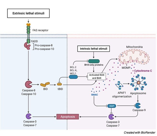
i. Extrinsic Pathway
For the apoptosis to begin, ligand and the receptor belonging to the tumor necrosis family initiate a transmembrane receptor mediated interaction. The TNF family has a death domain which allows the signal to reach the signaling pathway from the surface of the cell, with the death domain consisting of 80 amino acids loaded with cysteine.
There are 2 models in the extrinsic pathway and they are FasL /FasR and TNF ligand/TNFR 1 model both of them will interact by attaching the receptor to the respective ligand. Once the receptor is bound to the ligand, it will trigger cytoplasmic adapter proteins bringing the death domains.
The FasL FasR interaction will activate the FADD and the interaction between the TNF ligand and TNFR will activate RIP and FADD. Thus, the FADD will attach to procaspase 8 due to dimer formation of death domain and procaspase 8 will be switched on along with a death inducing signaling complex (DISC) being initiated. Once caspase 8 is triggered apoptosis process will start to proceed.
Apoptosis Pathway Diagram
ii. Intrinsic Pathway
In this pathway the signal will reach inside the cell and thus the name intrinsic/ intracellular which could be either positive or negative signals. Positive signals could be free radicals, toxins, radiations and others whereas negative signals could be lack of certain necessary factors such as hormones or growth factors which could initiate the apoptosis process.
The positive signals will release 2 various proteins in the cytosol from the transmembrane space due to the unlocking of the mitochondrial permeability transition pore. The first protein group poses cytochrome c which attaches to Apaf-1 (Apoptotic protease activating factor -1) and pro-caspases 9 leading to the establishment of apoptosome. After which the procaspase is nicked into its working form caspase 9 and then again nicked to form caspase 3 by the apoptosome.
There are also other proteins in the first group that initiate apoptosis and are HtrA2/Omi and SMAC (second mitochondria derived activator of caspases). The second group of protein would come into picture only when the cell has decided to die and thus leading to proteins secretion from mitochondria and will be broken into fragments and condensed.
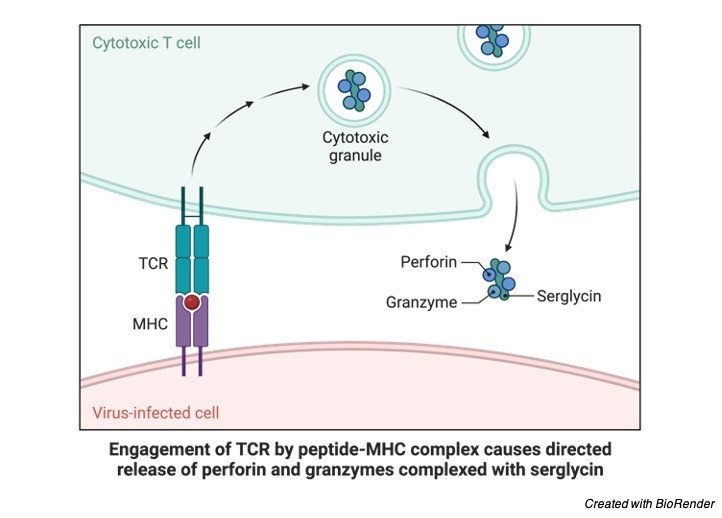
iii. Perforin Pathway
In this pathway, the toxic effect on the infected or cancerous cell is removed with the help of a molecule called perforin which secretes granules which are of 2 type, granzyme A and B which translocates the granule to the required cell and the pathway. Granzyme A will activate caspase and initiate apoptosis. Granzyme B will activate procaspase 10 at aspartate residues and nick factors such such as ICAD (Inhibitor of Caspase Activated Dnase).
To release cytochrome c granzyme B will use mitochondrial pathway for enlargening the death signal. Granzyme A nicks the Dnase when it’s in the vicinity of the cell to inhibit the apoptosis of tumor cell. Further granzyme will inhibit Dnase and nick the SET complex and this complex will further reassure that the DNA is intact and the inactivation of this complex will result in apoptosis and the DNA would be left that way.
iv. Execution Pathway
At the end of the intrinsic and extrinsic pathways begins the execution pathway. This pathway begins by triggering the caspase that will further trigger the proteases which will break proteins and endonucleases will break the nuclear material. Caspase 8, 9 or 10 will activate Caspase 3 which will further activate Caspase activated Dnase (CAD) which will further shrinken up the chromatid and degrade the DNA inside the nucleus.
Caspase 3 will cleave gelsolin, an actin binding protein to form apoptotic bodies. These apoptotic bodies will show the signs of phosphatidylserine on their outer coverings and the cell dies without any inflammatory reaction taking place.
Apoptosis Inhibitor
In order to prevent apoptosis from occurring, and the infected or dead cells to live, signaling pathways has to be inhibited. After which the inhibitors of apoptosis come into picture which are the anti-apoptotic protein factors called IAPs (Inhibitors of apoptosis) and Bcl-2. IAPs as their names suggest are the inhibitors prevent cell death and in human population this class poses 8 proteins, in which each one has a domain called the Baculovirus IAP Repeat which would link itself to the proteins involved along with caspases. Caspase 3 and Caspase 9 will attach to proteins such as XIAP to stop the process.
The other inhibitor called the Bcl – 2 is concerned with the movement of mitochondrial membrane. For the intrinsic pathway, Bcl-2, Bcl- x, BAG are some of the proteins stopping the cytochrome c and altering the mitochondrial membrane thus preventing the apoptosis to take place.
c- FLIP is a protein, which will attach to caspases and FADD to inhibit the extrinsic pathway of cell death.
The failure of the cells to apoptosis is the reason behind diseases such as myeloma, cancer and leukemia. It can further lead to mutation of the inhibitor protein XIAP causing a genetic disorder or even failure of immune system function.
Apoptosis Regulation
To regulate apoptosis, various proteins and other factors come together, however the mostly recruited one’s are Bcl-2 and IAP which has the authority to prevent or continue the cell death process.
In the extrinsic pathway, regulation takes place with the help of a protein called Toso which will not trigger caspase 8 as it has been blocked by the Fas. The intrinsic pathway of regulation requires Bcl-2 protein to regulate the movement of the mitochondrial membrane.
For the apoptosis regulation, cytochrome c is released from the mitochondria by the change in the mitochondrial membrane mobility. These proteins released along with Smac will regulate apoptosis by stop the IAPs. Thus, apoptosis takes place. To activate apoptosis, proteins like Puma and Noxa stop the anti-apoptotic protein.