We all know that cells form the structural and functional unit of the all the living organisms. Each and every cell in our body has its own structure. Have you all ever thinked how this stable structure is maintained.
Cytoskeleton plays an important role in maintaining the structure and shape of the cell. Our cell organelles start moving here and there and loses its appropriate shape thus losing its functions, which makes various critical conditions in our body.
If considering basically, what would happen if our internal organs move from its own places of origin, we find very difficult to move, speak and to do any kind of activities. In the same way, our cell also finds difficult to perform their activities.
We all think that each and every cell are just a bilobed round structures which does not have its own properties, but the truth is that these cells have their magnifies appropriate structure which contains a network of filaments known as Cytoskeleton, which is literally explained as Cells skeleton”.
It does not maintain the shape of the outer membranes like plasma membranes, it gives the overall structure of the cell and also plays an important role in placing the organelles in their correct positions and also aids in tracking the transport of the vesicles.
What is Cytoskeleton?
As mentioned above cytoskeleton is a group of fibers, which plays an important role in minting the structure and tracking the changes. Generally, in eukaryotes, there are about three types of protein fibers, which are named as follows:
i. Microfilaments
ii. Intermediate filaments
iii. Microtubules
i. Microfilaments
Microfilaments are one of the three types of protein filaments that are present in our cytoskeletons. Of the three, microfilament is the narrowest one having a diameter of about 7 nm which are being made up of many linked monomers of the protein known ad actin, these actions are present as a monomer and combines together to form a double helical structure.
Actin filaments usually have a directionality which means that they posses tow structurally different ends. Actin filaments usually consists of a variety of important functions in the cell.
Actin filaments also consists of numerous important roles in the functioning of the cell. It also serves as a track for the movement of a motor protein which is known as myosin.
As it has a relationship with the muscle protein myosin, actin is usually involved in performing many of the cellular functions. Microfilaments also aid in many functions of the cell like cytokinesis and in motility of the cell.
ii. Intermediate Filaments
These filaments are about 8 to 12 nm wide. They are called as intermediate filaments as their size is between the microfilaments and the microtubules. Intermediate filaments are generally made up of different kinds of proteins linked keratin, desmin, vimentin and lamin.
All the intermediate filaments are found in the cytoplasm of the cell, except those of lamins, as they are found in the nucleus and they help in supporting the nuclear envelope. Intermediate filaments present in the cytoplasm maintains the shape of the cell, cell tension and also provides structural support to the cell.
iii. Microtubules
Among these three protein filaments, microtubules are considered as the largest cytoskeleton fibers which is about 23nm in size. These are hallow tubes that are made up of alpha and beta tubulin. Microtubules forms a structure like flagella, which are the tails that propels forward.
Most of the microtubules in the cell usually comes from the cell organelles, the centrosome which is generally called as the organizing center of the cell.
Cytoskeleton Function
As mentioned above, cytoskeleton consists of several functions. The first and fore most function of the cytoskeleton is to give its cytoskeleton the appropriate shape.
Cytoskeleton is very much important in the cells without cell walls, as such animal cells do not get their shape from the thick outer layer. It provides the cell a good movement.
he microfilaments and the microtubules help in reassembling and disassembling the cells. It also helps the cell to crawl and migrate. The cytoskeleton also plays an important role in keeping the cell organelles in the place and it also helps in movement of the organelles throughout the cell.
Many of the compounds are found in the natural resources like fruits and its products which gives abundant of nutrient to our body and whereas few of them can also be used as the source of the medicines. On the other hand, few has its own side effects.
Resveratrol is one of the compounds which is found in red grapes and its products like juice or wine, which is very helpful for hay fever and it also deals with weight loss. Resveratrol has many effects in the bogy including those of expanding the blood vessels and in clotting the blood.
This also helps in treating the pain and swelling and also helps in reducing the sugar levels in the blood and it also helps the body to fight against infections. This compound, Resveratrol is usually used in dealing with many conditions like high cholesterol, cancer and heart disease and also in curing many abnormal conditions in the body.
What is Resveratrol?
Resveratrol is one of the natural compounds that is found in the skin of the red grape, in peanuts, and in berries like blue berries. Resveratrol is considered as one of the most powerful antioxidants which is produced by some species of plants to protect them against the environmental stresses.
These antioxidants help in neutralizing the free radicals, that are believed to decline the symptoms of aging. Japanese knotweed is one of the species of plant which has the highest source of resveratrol content. Large amount of the resveratrol is produced in the skin of the red grapes which helps the plant to protect it from various kind of fungal diseases and protecting the plant and fruits from skin diseases and sun radiations. It is therefore considered that grapes have a higher amount of resveratrol in it comparing it with other natural food materials.
However, red wine consists of a small amount of resveratrol which is less than 1 to 2 milligrams per eight ounces of red wine. But on the other hand, red wine contains more resveratrol comparing with the white wine as these red wines are fermented along with the skin of the red grapes than that of the white wine.
Thus, a greater number of antioxidants that includes resveratrol which are naturally present in the skin of the grapes are extracted to form a wine. This is also found in seeds and in pomace of the grapes. Grapes that have been grown in humid environments contains more amount of resveratrol than in grapes which are grown in arid areas. Resveratrol is also been very responsible for lowering the rate of the heart diseases in French populations comparing to that of the other populations.
Resveratrol is also found in many of the dietary supplements which are extracted from the grape seeds and the Japanese knotweed to form the red wine. Where as the most of the supplements that are found in the market is usually derived from the Japanese knotweed as this plant is considered as one of the highest concentrations of the resveratrol, found naturally.
However, the amount and the purity of the resveratrol in the supplements varies widely. Now-a days micronized resveratrol is available which is found in the form of powder or in pills. Resveratrol has a low systemic bioavailability and it is not well absorbed orally. This process of microionisation greatly reduces the average particle size of the compound. Resveratrol is also available in the form of solution or as transdermal patch.
How Resveratrol Works?
First of all, Resveratrol protects the DNA of the cell, it is considered as one of the powerful antioxidants, which prevents the cell damage that are caused by free radicals. Free radicals are one of the unstable atoms that are caused due to pollution, sunlight and where as the bodies, burning fat leads to cancer and other aging problems.
Resveratrol Benefits
Resveratrol has many of the health benefits like protecting the heart and the circulatory system, lowering the cholesterol and it also helps to protect the body from heart attacks, stroke, clots.
It also acts as an antioxidant and helps us to lower the blood sugar levels and it also reduces the risk of various cancers.
Resveratrol greatly increasing on the heart disease, people who consume higher amounts of the dietary resveratrol have a lower risk of heart diseases.
People who take resveratrol orally does not improves the levels of cholesterol or the blood cholesterol which are known as triglycerides.
The grouping of symptoms sometimes increases the risk of diabetes and the heart diseases.
They also build up a fat in the liver, when drinking little or no alcohol.
Resveratrol Side Effects
Patients having disorders in the blood, results in bleeding on consuming resveratrol.
Women who are pregnant or under breast feeding should not consume resveratrol.
Resveratrol has its mild estrogenic activities, women with cancers is more sensitive to estrogenic activities.
Resveratrol reduces the activity of the enzymes that are involved in metabolizing the drugs.
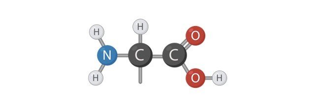
Carbohydrates are one of the organic molecules that are made up of carbon hydrogen and oxygen in the ratio of 1:2:1. These are considered as the major classes of the biomolecules which serves as an important source of an energy. These compounds also play a role in serving as the structural components. Carbohydrates are generally classified into two major groups as simpler carbohydrates and the complex carbohydrates.
Simpler carbohydrates are also known as simple sugar which can be easily digested and it can also be served as the rapid source of energy. Whereas the Complex carbohydrates like cellulose, starch and glycogen takes more time to get digested and metabolized as they are high in fiber unlike simple carbohydrates as they are less likely to cause spikes in the blood sugar.
What are Disaccharide?
The term disaccharide generally refers to the two molecules of monosaccharides. A saccharide generally refers to the structural unit of the carbohydrates. Hence, disaccharide is a compound of carbohydrate which is made up two units of monosaccharides. The term sugar can be referred to as both Monosaccharides and disaccharides.
Monosaccharides are also known as simple sugars as they form the most fundamental type of sugar which is generally known as sucrose. Where as the term table sugar refers to the granulated form of sugar which is referred to as the sucrose. Disaccharide is made up of two monosaccharides namely glucose and fructose.
Characteristic of Disaccharide
Like carbohydrates, disaccharides are also made up of molecules like carbon, hydrogen and oxygen in the ratio of 2:1 which are then referred to as the hydrates of the carbon. Disaccharide are the organic compounds which are linked together by a covalent bond. Disaccharides are the sugar base that comprises of two molecules of monosaccharides that are linked together by a glycosidic bond. Monosaccharides are considered as the most fundamental compound of the carbohydrate.
Glycosidic bonds are considered as the covalent bonds which are formed by two hydroxyl groups having two monosaccharides. Though disaccharides are made up of same chemical formula they are classified into three different kinds however they differ in bond formation and they constitute different properties.
However, disaccharides vary from the other forms of carbohydrates, oligosaccharides and the polysaccharides which all make up the unit of the sugar. Thus, disaccharides are made up of two monosaccharides and the oligosaccharides are made up of three to ten molecules of monosaccharides. Whereas the polysaccharide is made up of several units of monosaccharides.
Disaccharide Synthesis
The chemical process in which the monosaccharides are joined together are known as dehydration synthesis as it results in release of the byproduct, water. Disaccharides are generally formed by displacing a molecule of hydroxyl from the one monosaccharide and the proton taken form the other molecules of monosaccharide.
Thus, formation of two monosaccharides linked together by a covalent bond. Further the proton and the hydroxyl groups are detached and joined to form a water molecule. Thus, disaccharides are formed by the condensation process of two monosaccharide molecules.
Disaccharide is reverted into the monomeric components of monosaccharides by the process of hydrolysis along with the helps of an enzyme disaccharidase. Whereas the condensation process involves the elimination of the water, where it is utilized by the process of hydrolysis.
Classification of Disaccharide and Disaccharide Examples
Disaccharides are generally classified into two types namely reducing and the non-reducing.
A reducing disaccharide is one of the types of disaccharide in which the reducing sugar contains a free hemiacetal unit which serves as the reducing aldehyde group. Examples of one such reducing disaccharide is maltose or cellobiose.
On the other hand, the Non-reducing disaccharides are the ones which do not act as a reducing agent. Both monosaccharides which makes up the molecule of disaccharide does not have hemiacetal unit as they bond through the linkage of the acetal between the anomeric centers. Examples of such non-reducing disaccharides are sucrose and the trehalose.
Importance of Disaccharide
Dietary disaccharides are the other form of carbohydrates which serves as a source of energy. As we intake and digest the disaccharides they are break down into units of monosaccharides which play an important role in metabolizing them for synthesizing the ATP molecules and thus generating the energy. ATPs are thus considered as the biologically synthesized molecules through the process of aerobic and anaerobic respiration.
Glucose is considered as one of the most common form of monosaccharide, where the cellulitis’s the synthesized ATP through the substrate level of phosphorylation. Whereas one of the sources of glucose is disaccharide which contains diet.
However, too much of glucose is considered as hazardous as too much of glucose and leads to diabetes and the obesity risks, and other issues like cardiovascular diseases and formation of tooth decay.
Lactose is one of the disaccharides which is found in the breast milk and it is considered as one of the most important nutrient sources for the infants. Where as the micro-organisms like Lactobacilli has its ability to convert lactose into lactic acid, which is used in the food industry for the production of diary products like yogurt and cheese. The other form of disaccharide named maltose is generally used as a sweetener even it is less in its sugar source.
We all know that our composed is composed of many of the organic compounds, which makes our cells to function efficiently by restoring the energy and its components that synthesis energy rich molecules. One such compounds are monosaccharides which is one of the compounds of the carbohydrate or it may also be referred to sugar A simple sugar which contains the building blocks of a complex forms of the sugar like oligosaccharides and the polysaccharides.
Sugar molecules play an important role in every organism as they help in formation of a glucose which then reduces and undergoes various process to form a molecule of ATP (Adenosine Triphosphate) which are known are considered as the basic energy supplements of our body. However, the term monosaccharide refers to the one saccharide. A saccharide is one of the structures of the carbohydrate. Hence, monosaccharide refers to the carbohydrate that has only one unit of saccharide.
Monosaccharide
Generally, the term sugar refers to both the monosaccharides and the disaccharides. Monosaccharides are thus a simple molecule of sugar as they are considered as one of the significant kinds of sugar. Which does not mean the table sugar or granular sugar, those which are considered as the sucrose’s. Because sucrose’s are the disaccharide molecules, that is it is made up of two monosaccharides named glucose and fructose, which are all come under the group of carbohydrates. Carbohydrates are one of the organic compounds that are made up of carbon, hydrogen and oxygen in the ratio of 1:2:1. These are considered ad one of the major biomolecular compounds, which are the important sources of the energy which also in turn serve as the structural components.
Monosaccharide Features
One of the most fundamental and the common type of sugars known as monosaccharides, which means that they cannot be separated or broken down into any other simpler compounds rather than hydrolysis. Where as monosaccharides has the capability of combining with the other complex types of molecules.
Monosaccharides are usually joined together with the help of a glycosidic bond, which are also known as glycosidic linkages. These glycosidic bonds are considered as the covalent bonds. Thus, the combination of the two simple sugars then refers to as disaccharide, Other molecules which consisting of about three to ten molecules of sugar are called as oligosaccharides.
Apart from them the molecules consisting of about large number of monosaccharide units are referred to as polysaccharides. Where as the chemical process which is involved in combing the molecules of the monosaccharides are known as dehydration synthesis as it results in releasing the water as its by-product. Though this process is irreversible, the complex carbohydrates are broken down into simple sugars, like glycogenolysis where the glycogen is being broken down into the units of glucose which is used further in the energy metabolism processes.
Monosaccharides are usually a colourless and a crystalline substance which are sweet in taste and a solid component. These substances are easily soluble in water. Now a days it occurs in the form of liquid sugars or syrups. However, like other carbohydrates, monosaccharides are also considered as one of the organic compounds and contains carbon which is covalently bonded with the other atoms like hydrogen and oxygen.
Role of Monosaccharide
Monosaccharides play a very important role in performing various kinds of functions. One of the important functions is that it serves as a structural and the multifunctional biological unit. With the help of the glycosidic bonds, they join together and form a complex oligosaccharides and other polymers namely cellular, starch and glycogen. It also serves as a precursor for many other compounds and its formations like galactosamine, glucosamine, sulfoquinovose, mannitol, glucuronic acid and so on.
Monosaccharides are similar to those of other carbohydrate molecules which plays an important role in supplying the nutrition. Monosaccharides are also found in natural sources like fruits, vegetables and other dietary nutritional supplements. Where these are consumed for the purpose of deriving energy in the form of ATP which acts as the biofuel and other sources of minerals. Glucose is one of the most common form of monosaccharide which helps us to synthesize ATP through the levels of phosphorylation reactions.
Classification of Monosaccharide
According to the biochemistry monosaccharides are classified into three types as follows,
Depending on the placement of the carbonyl group.
Number of carbon atoms it contains.
Depending upon its chiral handedness.
Monosaccharide and Metabolic Pathways
Monosaccharides play an important role in many of the metabolic pathways in our body and they are listed below.
Glycolysis: Here the monosaccharide is converted into pyruvate which release a high amount of energy biomolecules.
Pentose Phosphate Pathway: It is one of the alternative pathways for breaking the glucose molecules.
Glyconeogenesis: Here the non-carbohydrate precursors will be converted into monosaccharides.
Glycogenolysis: Here the glycogen is converted into monosaccharide units.
Glycogenesis: Here the glycose is being converted into glycogen.
Lecithinase test can also be called as Naglers reaction. This is considered as one of the important biochemical tests which helps us to identify the organisms that has its ability to liberate the enzyme known as Phospholipases.
this biochemical test is used to identify the different organisms which has its capability to differentiate and specifically found the organisms which has its capability to liberate the enzyme lecithinases, which are also known as phospholipases. The commonly known example of one such species is Clostridium perfringens. Where as these organisms consists of the alpha toxin, which has an ability to activate the phospholipase activity in the medium which helps us to differentiate the species of Clostridium perfringens from the other Clostridium species. Apart from C. perfringes, there are also few other species which has its ability to synthesize the enzyme lecithinase such as C. baratti, C. bifermantans, C. absonum, C. sordelli, and C. novyi.
These species produce an enzyme lecithinase by neutralizing the activity of lecithin C by producing an antitoxin. Phosphatidylcholine or lecithin is a substance which is found abundantly in the animal tissues, egg yolk and few higher plants, which consists of phospholipids linked with the choline. Some species of microorganisms possess a lecithinase (also called as phospholipase C), is one of the enzymes which has the ability of split the Phospholipid lecithin, whose activity is used to characterize the several grams positive and the gram-negative bacteria.
The Lecithinase test is commonly performed in the laboratory by preparing a medium and by following certain procedures, which has been discussed below.
Lecithinase Test Objective
The main of the test is to determine the ability of the micro-organism to synthesize the enzyme named lecithinase.
To detect the species of bacteria which is capable of producing a lecithinase enzyme.
Lecithinase Test Principle
Egg yolk agar is one of the enriched medium that is used commonly in many of the laboratories for isolating the characterising the different species of bacteria on the basis of their lecithinase activity. Lecithin is one of the normal components of the egg yolk. In egg yolk agar, the lipoprotein component lecithovitellin which has a capability to split the enzyme lecithinase into phosphorylcholine and an insoluble diglyceride, which results in the formation of the precipitate in the culture medium. Such micro-organisms which possess an enzyme lecithinase breaks down the lecithin to insoluble diglyceride and the phosphorylcholine, that results in the white opaque precipitation which further spreads beyond the edges of the colony. Thus, such opaque zone of precipitation across the colony when it is grown on the yolk agar medium indicates the positive reaction of the lecithinase activity of the organism present in the medium.
Lecithinase Test Reagents
Egg yolk agar
Composition of Media:
Ingredients
Gram
Pancreatic digest of casein
15.0
Vitamin K1
10.0
Sodium chloride
5.0
Papaic Digest of a soya bean meal
5.0
Yeast Extract
5.0
L-Cystine
0.4
Hermin
5.0
Egg Yolk emulsion
100.0
Agar
20.0
Lecithinase Test Procedure
Initially, a loopful of the test organism is taken and it is streaked as a straight line on the culture plate.
Then the medium is incubated aerobically in the gas pak jar immediately after streaking and it is further transferred into the incubator at a temperature of about 35 to 37ºC. For anaerobes, it is incubated for about 24 to 48 hours, and for aerobes the plate is incubated for 24 to 48 hours.
After incubation, the plate is examined or observed for formation of the opalescent halo surrounding in the inoculum plate.
Lecithinase Test Results
Lecithinase Test Positive Results: If the results of the test are positive, then there will be an appearance of a white opaque and diffused zone which extends along the medium and the surrounding colonies.
Negative Lecithinase Test Results: In case, if the results are negative, there will be absence of an opaque or a white zone that extends along the edges of the colony.
Lecithinase Test Uses
Bacterial lecithinases are considered as one of the special interests as these have their possible role in performing their pathogenicity.
This test also plays a vital role in identification of species like Clostridium perfinges, Pseudomonas aeruginosa, Staphylococcus aureus.
However, the enzymatic activity of the Staphylococcus aureus is greatly used in detecting the coagulase positive strains as they show a high link between the lecithinase activity and the coagulase activity.
This test can also be widely used in differentiating the certain species within the same genus of the Bacillus.
Modified oxidase test is one of the most recommended method for Gram-positive catalase positive cocci. This test is considered as one of the rapid tests, which helps us to differentiate the species of negative Staphylococci’s from the positive strains of Micrococcus by detecting the oxidation of the enzymes present in the medium. To perform this test filter paper disks are being impregnated along with the tetramethyl-p-phenylenediamine dihydrochloride, which is one of the oxidase reagents, added with the dimethyl sulfoxide are used.
However, in the presence of an atmospheric oxygen around the medium, oxidase enzyme that is being present in the medium along with the oxidase reagent and the cytochrome C results in formation of a colored compound, which is referred as indophenol.
Microdase Test Objective
The main aim of the test is to differentiate the gram-positive catalase positive cocci, micrococci from the staphylococci.
Microdase Test Principle
Microdase test is also known as modified oxidase test. This test is considered as one of the rapid tests that helps us to differentiate the species of streptococcus from the micrococcus which are the gram-positive cocci that possess the catalyze enzyme. This differentiation is usually based on process of detecting the enzyme oxidase. For this process of selecting this enzyme a filter paper along with the circular disks are impregnated with a tetramethyl-p-phenylenediamine dihydrochloride which is known as oxidase reagent in the dimethyl sulfoxide DMSO are used.
This modified oxidase reagent is prepared using a1% of tetramethyl-p-phenylenediamine in a certified grade dimethyl sulfoxide. Where as DMSO helps in improving the permeability of the cells to the reagent/ In presence of the atmospheric oxygen, the enzyme oxidase reacts with the oxidase reagent and the cytochrome C which results in the formation of a colored compound. Indophenol is used as an indicator which gives a blue or purple colored formation of a compound on the disc after it is introduced into the disc that contains a bacterial colony.
Microdase Test Reagents
Here, the filter paper disks are impregnated along with the oxidase reagent known as tetramethyl-p=phenylenediamine dihydrochloride in a DMSO (dimethyl sulfoxide).
Microdase Test Procedure
This test is usually performed on the aerobic, catalase-positive and a gram-positive coccus. Initially with the help of a forceps, disk is placed on the empty Petri disk or on a sterile glass slide.
Then a small amount of several colonies is taken from an 18 to 24-hour pure culture which is grown on a blood agar on a small area of the microdase disk, using a wooden applicator.
Then the medium is incubated at a room temperature of about 2 minutes and it is noted that whether there is any change in color. Usually, blue color is noted for positive results.
Microdase Test Results
Positive Microdase Test Results: In case of positive result, the development of blue to purple color can be observed within 2 minutes after incubation.
Negative Microdase Test Results: on the other hand, if the result is negative, Staphylococci should yield a negative result, on exception with the species like Staphylococcus sciuri, S. vitulinus or S. lentus.
Microdase Test Uses
Microdase Test is most commonly used for differentiating the species of Micrococci from the Streptococci. Here most probably Micrococci is considerably yielding a positive result, and the Streptococci yields a negative result, having few exceptions with some of the species of Streptococcus like S. sciuri, S. Vitulinus, S. lentus.
Microdase Test Limitation
Staphylococci yields a negative result for some species of the Streptococci.
Bacteria which are taken from the culture that is grown on a blood agar for 24 to 36 hours only should be used for this test because the cultures that are too young or too old often gives inaccurate results.
Modified oxidase test is usually recommended for the Gram-positive catalase positive cocci only.
Where the species of the Macrococcus and the Kocuria kristinae also often known to possess a cytochrome.
Microdase is not designed for the routine testing of the oxidase activity of the activity instead it is used for testing only the species of Staphylococcus and the Micro coccus.
Malonate test is one of the colorimeter tests which detects the ability of the bacteria to utilize the malonate as the source of carbon, which is the end point for the production of the metabolites of the alkaline that helps in inducing the color change. The medium for the malonate test contains the sodium malonate, here malonate is one the enzyme inhibitor which helps in utilization of the succinic acid by the bacteria, thus shutting the process of the Krebs cycle and the glyoxylic cycles. Let us discuss briefly about this test here in detail.
Malonate Test Objective
The main aim of the test is to test the ability of the organism present in the medium to make use of the malonate as a source of the carbon.
Malonate test was initially found to originally designed to differentiate the several species of the bacteria from the Escherichia, which does not grow in the medium and the Enterobacter.
Malonate test is used as the differential medium has broadened to include the other members of the family of Enterobacteriaceae.
Malonate Test Principle
An organism which has the ability to utilize the sodium malonate in the organisms utilizes malonate as its only as a source of carbon and ammonium sulfate as its only nitrogen source and produces an alkalinity as a result of sodium hydroxide formation, this results in the alkaline reaction which a medium containing a malonate which results in change of the color from original green or light blue color into bromothymol blue. Organism that which does not utilize the compounds of malonate and ammonium sulfate that does not ferment dextrose produce no color change. Organisms which are considered as malonate negative does not ferment dextrose no color change. Organisms which are malonate negate but do not ferment the dextrose and results in the development of a yellow color due to the increase acidity in the medium.
In an inoculum that is collected from a pure culture is transferred aseptically to a sterile tube of malonate broth. The inoculated tube is being incubated at a temperature for 24 hours and the results are being determined. Abundant change and growth in the medium are indicates a positive test for growth using a malonate. However, is a microbe can use malonate for carbon and energy it will grow on the malonate broth. Thus, use of the malonate leads to the rise in the pH of a medium and a pH indicator changes the color of the medium.
Malonate Test Reagents
The medium used in the malonate broth is based on the formula by the Leifson, which is in the liquid form containing an ammonium sulfate as the only source of the nitrogen and malonate as the only source of carbon. It also contains minerals salts, sodium citrate for carbon, and the ammonium phosphate for its source of nitrogen, whereas the malonate as the only source of carbon. It contains minerals salts, ammonium phosphate for its source of nitrogen, and sodium citrate as the source of carbon. Here the pH indicator used is bromothymol blue, which results in the formation of a green color at its neutral pH, and yellow at its acidic range in a pH 6.0 and it turns blue at a basic range in an alkaline medium containing a pH of 7.6.
Media:
Ingredients
Gram/Liter
Ammonium sulphate
2.0
Dipotassium phosphate
0.60
Monopotassium phosphate
0.40
Sodium chloride
2.0
Sodium malonate
3.0
Bromothymol blue
0.025
Malonate Test Procedure
With a help of a light inoculum taken from an 18 to 24-hour pure culture is inoculated into the tube which contains malonate broth.
Then the inoculated broth is incubated at a temperature of about 35ºC for about 24 to 48 hours. It should also be ensured that caps should be loosened before incubating for a good aeratiation.
After incubation, the alkalization of medium is noted which results in formation of a blue color after 24 and 48 hours.
Malonate Test Results
Positive Malonate Test Result: In case of positive malonate test it is indicated by the development of a blue color in the medium.
Negative Malonate Test Result: In case of the negative result, a malonate test is usually indicated by change in color of the medium into green . Sometimes it results due to change in color of the yellow due to the fermentation of a dextrose.
Malonate Test Uses
The malonate test was originally designed to differentiate the species of Escherichia and the Enterobacter.
It is also being used as a differential medium which is now broadened and it is included along with the other members of the Enterobacteriaceae.
Malonate reaction is usually used as a todiffe rentiate among the species of Enterobacteriaceae like Klebsiella pneumonia which is one of the positive strained species.
This test is usually performed to separate the species of Salmonella arizone which is a positive species from the other species of the Salmonella, which is the negative species.
This test also helps in differentiating the Enterobacteriaceae in the food and diary products.
Lipid hydrolysis test one of the such tests which helps us to detect the ability of the bacterium to hydrolyze the lipids that are present in the medium. Lipids are the compounds which are high in the molecular weight compounds that posses the large amount of energy. When these molecules get accumulated into the cell, they start metabolizing through the process of aerobic respiration and thus produces cellular energy known as ATP (Adenosine Triphosphate). These components enters into the other metabolic pathways for synthesing the other protoplasmic cellular components.
However, before the bacteria starts the process of assimilation and gets degraded. The degradation of the lipids which are known as triglycerides is usually accomplished but the extracellular hydrolyzing enzymes that are commonly known as lipases or esterase’s, which plays an important role in cleaving the ester bonds in the molecule by adding the water and thus results in the formation of the building blocks generally called as glycerol and also in fatty acids.
Lipid Hydrolysis Test Objective
The main aim of the Lipid Hydrolysis Test is to determine the ability of the organism that whether it has a capability to hydrolyze a lipid.
To identify the species of the bacteria that are capable producing an exoenzyme lipase.
Lipid Hydrolysis Test Principle
Lipids are generally known as non-polar molecules which to do not dissolve in water. Fats are one type of the lipids which contains a large polymer of fatty acids and glycerol in it which makes the cell to large to absorb those compounds into its membranes. In order to utilize the facts, bacterial cells play an important role in secreting the enzymes known as exoenzymes which are most commonly known as lipases that are present outside the cell and helps in hydrolyzing the lipids that are present into fatty acids and glycerol.
These lipids then result in a formation of an emulsion when they are dispensed with agar and thus produces an opacity when it is further hydrolyzed with the end products such as fatty acids and glycerol which neither forms an emulsion with the agar instead of forming a transparency. In lipid hydrolysis test, the test bacteria that are grown on the agar plate that contains tributyrin as the lipid substrate, which plays a role in forming an opaque suspension in an agar medium.
Incase, if the bacteria have its ability to hydrolyze lipids, the bacterial colonies hydrolysis the tributyrin that is present in the medium in the areas that is surrounded but the, into the soluble glycerol and the butyric acid, where as the rest of the areas on the plate contains the unhydrolyzed tributyrin. Thus, hydrolysis results in demonstration of a transparent clear zone around the colonist and the remaining areas of the plate remain opaque which results as an indicative of the unhydrolyzed tributyrin region.
Lipid Hydrolysis Test Media
Tributyrin agar is very important for performing this test, this medium is prepared using about 5 grams of peptic digest of animal tissue and a yeast extract of about 3 gram and the agar 15.0 gram.
Lipid Hydrolysis Test Procedure
Initially the tributyrin agar medium is inoculated in the medium of the agar using a single line of streaking of organism.
Then the inoculated medium is incubated aerobically in a gas pack jar immediately after streaking.
Then the medium is incubated at a temperature of about 35 to 37ºC for about 24 to 48 hours.
After incubation, if the result is positive then the formation of clear zone around the growth of the colony can be noted.
Lipid Hydrolysis Test Results
Positive Lipid Hydrolysis Test Results: If the results are positive then the clear zone can be seen around the bacterial growth.
Negative Lipid Hydrolysis Test Results: If the results are negative then there will be no formation of clear zone around the growth of the bacteria.
Lipid Hydrolysis Test Uses
This test is helpful in identifying the species of the bacteria which secretes the enzyme lipase. The species mostly include Enterobacteriaceae, fusobacterium, Propionibacterium, Clostridium, Pseudomonas, Corynebacterium, Staphylococcus.
This test also helps us to detect and enumerate the species of the lipolytic organisms which is usually present in the food and other materials.
Lipid Hydrolysis Test Limitations
It is usually suggested that the biochemical, molecular, immunological, or mass spectrometry tests are performed only in colonies that are taken from the pure culture for complete identification of the species.
The movement of water molecules across semi permeable membrane to produce a homeostatic system in cell and its environment is the process of osmosis.
Characteristic of Osmosis
Osmosis was derived from Latin which means impulse or urge; of the solvent to move uphill from lower concentration to higher concentration. The main difference between osmosis in living and non – living cells are the properties of the particular system. Osmosis in non – living cells can be demonstrated by separating the solution and water by a semipermeable membrane.
Osmosis is a type of diffusion between a solution and water when separated by a biological membrane which is permeable for solvents and restrict the entry of solutes.
Fick’s law can be theoretically applied to the diffusion where the rate of diffusion is directly proportional to the concentration gradient and area over the diffusion occurs. Experiments are done to depict the mechanism of osmosis in a living tissue where a solution and pure solvent present in two chambers A and B separated by a semi permeable membrane and observed from the initial time T. After a particular time say T’ to maintain a system equilibrium and restrain by the membrane the pure solvent enters the chamber A of solution to maintain the equilibrium. As the pure solvent enters Chamber A increasing volume of the chamber indicating the movement of the solvent from lower to higher concentration.
Osmosis Examples
Water is transported by vascular tissues xylem where the nonliving tracheid are also taking up the water. In leaves mesophyll cells; the tracheid’s opens to the cells and water enters cell via Osmosis. Semipermeable part of the cells is the plasma membrane. Plasma membrane along with cellular components are protoplast. The liquid part is called as the protoplasm and other cellular components (i.e.) organelles are also separated from each other by membranes. Plant cells are specialized by carrying a vacuole and cell wall both has its specific role in plants structure.
Additionally; plasma membrane is protective and regulative in nature by the mechanism of osmosis and presence of channels and transmembrane which transports particulate compounds for cellular metabolism. Further the plant is surrounded by Rigid Cell Wall which provides a particular shape for the cell.
Vacuoles – storage part of the cells stores nutrients and water in turn maintaining the internal volume of the cell. Externally the cells are connected by rigid cellulose made cell wall mediating communication between each other cells. Inside the cell wall they are separated from other cells by intercellular spaces.
Principle of Osmosis
Osmosis works on a basic principle of chemical potential difference between 2 components separated by a semi permeable membrane. Chemical potential is the free energy available per mole of the substance in a solution. In the osmosis demonstration the pure solvent had high energy compared to the solution which had lower energy (i.e.) lower chemical potential which created a gradient to pure solvent enter the Chamber A. The potential difference arises because of the solute in the cytoplasm which are part of energy metabolic activities constantly replenishes the chemical molecules and are dynamic in nature.
Semi permeable membrane does not allow solute molecules and solute or ion uptake is mediated by ATP utilizing carrier proteins knows as active transport breaking down ATP to produce energy for ion uptake. Active transport of ions constitutes a potential difference between an internal and external environment develops a chemical potential gradient taking up water molecules from xylem. This indicates for every active uptake of ion generates a potential difference involves a passive entry of water into the cell and thereby osmosis is indirectly coupled with energy utilization.
Osmosis is Pressure Dependent
Similar experiments can be quoted to prove the pressure during osmosis. Osmotic pressure is the maximal amount of pressure developed in a system separated by a semipermeable membrane by pure water. Pressure due to osmosis is demonstrated using an osmometer; where a thistle funnel is inverted and covered with a semipermeable membrane is separated by solution A and B; where A is pure solvent and B is solution. At time T’ osmosis happens and the volume of Solution B increases.
On attaching a piston and maintaining the same volume in osmometer by producing a pressure through a piston maintains volume constant for a particular time and when the pressure in piston is increased the water flow reverses from Solution A to Solution B. The pressure of the osmosis is directly dependent on the solute concentration. Dependent on solute concentration makes the process a colligative property of a solution along with 3 other properties.
Osmosis and Turgor Pressure
Turgor pressure or the phenomenon of turgidity is maintained by the process of osmosis; involves vacuoles, protoplast wherein the water uptake in vacuole produces a basic crispness in cell structure and additionally the protoplasm as a whole requires water to maintain rigidity of plant cell thereby making the whole plant stand erect. When a cell loses its water content the leaves and stem wilts and the state is flaccid and prolonged flaccid nature of plants leads to death. To retain the structure water uptake becomes essential which is mediated by osmosis.
Importance of Osmosis
• Partially; the water uptake from soils controlled by osmosis in roots.
• Facilitate water movement from non – living part to the living part of the plant.
• Mechanical support of rigidity is provided by osmosis.
• A special ability of osmosis allows a plant to be turgid and helps in movement of ions in plants.
• Osmotic pressure produces a growth in plants.
• Opening and closing of stomata, flower is done with the help of osmosis.
Minerals and water are transferred from roots to the plant by passive forces of movement namely diffusion and facilitated diffusion; does not constitute to effective transport of the molecules into the cells. Regular metabolic functions require nutrients to enter the metabolic cycle of cells to produce energy for the plant physiological functions. Dynamic molecular movement inside cell and its organelles, between cells and between external environment and plants require other transport mechanism to tag along passive transport.
Active transport is an effective means of transportation of ions and molecule along transcellular pathway. Energy is utilized by the dephosphorylating energy currency of the cells produced by oxidative phosphorylation and Electron Transport Chain in mitochondria of every cell; is effective in transporting voluminous number of ions from the cells. The transport of ions actively involves movement against the gradient of ions and other nutrients constitutes a potential difference electrochemically for the movement of ions and other nutrients.
Movement of ions from cells to the extracellular space and back to cell is contributed by the potential difference between a cell and the external environment constituting a potential difference: Transmembrane potential across these; acts as a driving force for the transport of the ions and charged molecules.
Donan Equilibrium
Donan equilibrium explains the transmembrane potential difference because of the movement of charged ions and the equilibrium of the cell in electrochemical terms. The cell is dynamic and are negatively charged predominantly. Assuming high K+ concentration inside the cell; K+ movement is a simple diffusion or facilitated diffusion pertaining to the protein channels and concentration gradient; to maintain an equilibrium out of the concentration terms. But the mechanism of the cell and its ability not only involves the concentration gradient for the movement but are dependent on both electric and chemical potential difference.
Electrochemically, the K+ ion is coupled with respective cations; are attracted back to the cell by the negative charges. An equilibrium is achieved when the force of concentration gradient pulling the K+ ions outside the cells and electrical gradient pulling K+ inside the cells. Hence a Donan equilibrium is achieved and the mechanism is similar for most of the ions and molecules. Donan’s equilibrium is also termed as Gibbs – Donan equilibrium; as Gibb’s had the equation predicted long back before Donan’s discovery.
Carrier Concept in Active Transport
A general mechanism of movement of ions across the cell membrane mediated by energy dependent protein molecules is clearly explained by the carrier concept. The transmembrane proteins act as a mediator for the movement of ions by binding to the ions and release them to the external or internal membrane based on the requirement.
This mechanism takes place in 3 steps:
Initially, carrier molecules are activated by the dephosphorylating ATP mediated kinases.
Activation of carrier binds to the ion forms a carrier – ion complex
On release of the phosphatase the ions and the carrier molecules are released closing the membrane.
Two theories were put forth based on the carrier concept and are named as Ludegardh’s cytochronme theory and Bennet – Clark’s Hypothesis.
a). Ludegardh's Cytochrome Theory for Active Transport
Ludegardh’s cytochrome theory was formulated by Ludegardh and Burstrom in 1933, assumes 3 postulates. They are: Active transport of anion is present and cation is passively diffused and are independent of anions. Anions are transported through cytochrome system of electron transport chain. Cytochrome acts as a carrier. Oxygen gradient exists across the external and internal surface with oxidation at external surface moves the anion to the internal surface where the reduction takes place. Main disadvantage of the theory is that the carrier molecules present are presumed to transport the anions and cations are transported through passive diffusion.
b). Bennet - Clark's Hypothesis
Bennet – Clark’s Hypothesis proposed in 1956 involves ATP carrier mechanism; associates with phosphatide transport across impermeable membrane. The carrier transports both anion and cation making the protein molecule an “amphoteric” molecule.
Active Transport Types
Active transports are similar inside and across different cell species; mediated by special protein structures in transmembrane walls of the cell directly utilize energy to facilitate the movement of ions across the cell against the concentration gradient are termed as “PUMPS”. Other protein channels utilize energy produced by the potential difference for the movement. Based on this the Active transporters are divided into:
Examples of Active Transporters
Primary Active Transporters
Primary active transport includes movement of ions and nutrients across semi permeable membrane through carrier proteins powered by ATPase. ATPase are protein pumps which transports ions against contraption and electric gradient from inside to outside of the cell and vice versa. The carrier molecules are termed as pumps; facilitating the movement of ions against concentration gradient.
Active transport of ions is further classified as Electrogenic or electroneutral transport based on the potential difference created by the net movement of the ions. Electrogenic transport is well demonstrated by Na/K pump in humans where exit of 3 Na ions 2 K+ ions are taken in; resulting in net outward movement of positive charges making it electro genic.
Similarly, in contrast to Na/K pumps; H /K+ pumps are present where for every exit of H+ ion One K+ ion enters the cell results in a neutral potential difference across the cell membrane. This H+ /K+ ion pump is an example for electroneutral transport. In animals the principal ion in transport to maintain membrane potential is Na+ but in plants H+ ions play important role in movement across the membrane.
The primary transporters implants are predominantly present in Plasma Membrane is H+ ATPase, Vacuolar tonoplast has specialized Hiaasen H+ PP ase in Golgi cisternae also has similar active transport coupled with ATP molecules. Apart from K+ ions Ca+ ions are tyransported and regulated as they act as ligands in many transmembrane proteins. Further primary active transport protein pumps are classified based on their function at different parts of the cell.
Proton Pumps
H+/ ATPase pumps are predominant in plants where the outward transport of H+ ions constitute a potential gradient coupled by ATP utilization in most of the pumps but the H+ pump in chloroplast utilizes light as a source of energy to drive the ATP synthesis. Proton pump allows the transport of ion in single direction hence it is an uniport and are electrogenic.
Further based on their structure the H+ ATP pumps are classified as: F type, V type and P type.
• F type is present in thylakoid membrane utilize the energy created by the potential difference across the membrane to synthesis ATP.
• P Type is the ATP ase present in the Plasma membrane dependent on Mg; drives one H+ ions outside the cell on ATP dephosphorylation.
• V Type is present in the vacuoles of the cells are similar to F type are specialized in making the vacuoles acidic to the plasma membrane pH due to the accumulation of organic and inorganic anions.
Ca Pumps
Ca+ pumps are similar to ATP as present in P type pump in the plasma membrane having a polypeptide chain of about 110kD with extended N – Terminal domain. On Hydrolysis of ATP; 2 Ca+ ions are transported across the membrane regulating the Ca+ ions in cytosolic pool as excess of Ca+ in the cytoplasm may mediate many enzymes activity. Ca binds with calmodulin has inhibitory effects in Ca pump acts as a negative feedback loop to release excess of Ca to external environment.
H+ Pyrophosphatase Pump
ABC Transporters: ATP Binding Cassette Transporters are largest protein molecules found in living organism are similar to P type channels. These are not electrogenic transporters which are involved in nutrient uptake in plasma membrane and takes up molecules for storage in vacuoles. They mainly transport uncharged substances out of cell without any changes in charge and electrochemical gradient.
Secondary Active Transporters
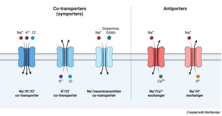
Secondary active transporters are indirect active transporters which utilize the proton motive force created by the H+ gradient to move other minerals and nutrient uptake as the direct active transport moves H+ or Ca+ outside the cell. Secondary transporters involve Symport and Antiport where respective proteins present in the membrane facilitates the transport of the molecules.
Symport involves the movement of molecules and nutrient uptake on same direction involves H+ ions move against the protein gradient. Antiport as the name indicates the H+ ions move against its gradient is coupled with opposite directional movement of mineral or nutrient to be transported. Active Transport includes the transport of ions and nutrients. ATPase in cell membrane is high in number and are varied in action.