Category: Study Materials
-

Pleiotropy: Definition, Mechanism, and Examples
Continue ReadingPleiotropy Definition
Pleiotropy is a phenomenon that occurs when a single gene affects several characteristics of living creatures. Pleiotropy can be caused by a gene mutation. Marfan syndrome, a human genetic disease affecting the connective tissues, is an example of pleiotropy.
The eyes, heart, blood vessels, and bones are all often affected by this illness. Pleiotropy is produced by a mutation in a human gene that causes Marfan Syndrome.
What is Pleiotropy?
Pleiotropy is derived from the Greek pleio, which means “many,” and trepein, which means “influence.” Pleiotropy is the state of having numerous effects.
It is a term used in genetics to describe a single gene that controls or influences several (potentially unrelated) phenotypic characteristics. In pharmacology, it’s a characteristic of a medication that causes it to have extra (positive) effects in addition to the ones it was designed to have.
It is demonstrated in molecular biology by cyclic AMP in a cell, which has a range of effects via controlling a protein kinase, which impacts a variety of other proteins.
Is Cystic Fibrosis an Example of Pleiotropy?
One of the most frequent examples of pleiotropy is cystic fibrosis, which is a hereditary disease. Lung infections are common with this condition. The digestive system and other organs in the body might be affected by this condition. Cystic fibrosis is caused by a mutation in the “cystic fibrosis transmembrane conductance regulator gene,” which prevents the gene from functioning properly.
Is Albinism a Pleiotropy?
Albinism is caused by a pleiotropic gene produced by a tyrosinase (TYR) gene mutation. The afflicted person’s body produces less melanin as a result of the mutation.
Pleiotropic genes are genes that influence the behaviour and functions of several genes with unrelated characteristics. On occasion, such qualities have been seen to be extremely similar in nature, while in other situations, they have been noted to be very unlike.
Pleiotropic characteristics are a term used to describe the issues caused by pleiotropic genes.
Effect of Genes on Traits of Humans
The phenotype refers to the physical characteristics of a person, such as body shape, height, colour, physique, and height. A trait governed or controlled by a single gene is known as a single gene trait. It may be difficult to determine the presence of pleiotropic characteristics until the gene is subjected to mutation.
Mutations are alterations in DNA sequences that occur relative to one another. The point mutation is the most frequent form of gene mutation, which is further divided into silent mutations, nonsense mutations, and missense mutations.
The character of the qualities is generally determined by the two alleles, which are the variation forms of a gene, according to diverse publications. The combinations of particular alleles dictate the synthesis of proteins that derive the process of phenotypic trait development, whilst the DNA sequence of the gene is altered by the mutation that occurs in the gene.
As a result, alterations in the gene segment sequences cause the proteins to stop working. As a result, the progression in the mutation will affect all accessible characteristics that are connected to a single attribute in the pleiotropy process.
Pleiotropy in Genetics
Pleiotropy was first proposed in biology by Gregor Mendel, a well-known geneticist well known for his groundbreaking work on pea plants. He tried purple-flowered plants and white-flowered plants in a series of trials.
Colorful blooms and leaf axils are always visible on plants with coloured seed coats, he observed. An axis is the portion of the plant that connects the stems to the rest of the plant.
He noticed that, although the seed coats were colourless in nature and had no colour in their axis, the pea plants, which were the focus of the study, invariably had white blooms.
After reviewing his findings, it was determined that the colour of the plant’s axil and the seat coat are the most crucial elements in determining whether the plant would produce white or purple flowers. Today, similar findings are attributed to the phenomenon of pleiotropy, in which a single gene contributes to numerous phenotypic characteristics.
Pleiotropy may be caused by a number of different processes, including development pleiotropy, gene pleiotropy, and selectional pleiotropy.
The focus of gene pleiotropy is on the functionality of a specific gene, and this type of pleiotropy is also known as molecular gene pleiotropy. The functions of a characteristic are generally determined by the number of traits and biochemical components influenced by the gene.
The biochemical parameters include the number of enzyme reactions performed by the gene’s protein products. In the evolution of pleiotropy, the major focus is on mutations and their relative influence on a variety of characteristics. It has been shown that single-gene alterations have a broader influence on numerous other potential characteristics.
Furthermore, illnesses involving mutational pleiotropy are defined by deficits in many organs that disrupt the proper functioning of multiple bodily systems. The last process that causes pleiotropy is selectional pleiotropy, which focuses on the impact of gene mutations on the number of distinct fitness components.
The method by which an organism transmits its genes from one generation to the next through sexual reproduction is typically governed by the fitness of the organism. Selectional pleiotropy is frequently concerned with the effects of selection on naturally occurring characteristics.
Polygenic vs Pleiotropy
Many people confuse the meanings of polygenic inheritance with pleiotropy, which is a common finding. The main difference between the two is that pleiotropy occurs when a single gene influences several traits, whereas polygenic inheritance occurs when a single trait is regulated by many distinct genes, such as skin colour.
Pleiotropy vs Epistasis
Understanding the idea and meaning of epistasis, as well as its relationship to pleiotropy, is also critical.
Epistasis refers to the interplay of several genes in influencing phenotypic results.
Pleiotropic gene research is important in biology because it helps researchers understand how specific genes are frequently implicated in genetic diseases. Pleiotropy may be found in abundance throughout nature.
Pleiotropic disorders include fruit flies with vestigial genes, poultry with frizzle characteristics, the process of pigmentation and deafness in cats, pleiotropic sickle cell diseases in humans, and phenylketonuria (commonly known as PKU).
Pleiotropy Examples
Pleiotropy is influenced by both direct and indirect pleiotropy, as evidenced by instances found in numerous literature research.
For example, if a blind mouse is born as a result of changes in a single gene, the odds are quite high that the blind born mouse will struggle with visual learning tests, showing that a single gene is implicated in many pathways.
As a result, there are numerous examples of both direct and indirect pleiotropy, some of which are discussed in further detail in the sections below.
i. The Vestigial Gene and Fruit Flies
Vestigial genes are important in the development of the wing of the fruit fly Drosophila. If these flies are homozygous for the recessive version of the vestigial gene, they have small wings and are unable to fly in the proper manner (VG). As a result, the vestigial gene is pleiotropic, resulting in the fruit fly drosophila’s wings not developing.
Other indirect impacts of pleiotropy in fruit flies include a reduction in the number of eggs present in the ovaries of the flies, a shift in the location of the bristles on the scutellum of the flies, and a shorter life span of the flies. The wings of the first bee are not entirely formed, as opposed to the fully developed wings of the second bee.
ii. Deafness and Pigmentation in Cats
Deafness is present in around 40% of cats with white hair and blue eyes, according to reports. Although this information is intriguing because we have most likely never paid attention to these cats throughout our lives.
It was discovered early on in the research that white cats with one blue eye and one yellow eye were blind in one eye, which was typically the blue eye, but it was subsequently shown that this occurrence of blindness does not necessarily apply to all cat breeds.
Waardenburg syndrome is a human disease that is comparable. Pleiotropic genes are involved in this disease in cats, producing not just deafness but also colouring issues. The goal of the study was to figure out how hearing capacity and the pigmentation process are linked.
Pigmentation has a critical function in regulating fluid flow in the ear canals, according to the findings, which were conducted on mice. Those who lacked pigmentation also lacked the flow of fluids through the ear canals, causing them to rupture and eventually lead to deafness.
iii. Frizzle Traits of Chickens
Pleiotropic genes cause the hens to express a variety of genes. Walter Landauer and Elizabeth Upham discovered in 1963 that hens with the dominant frizzle gene generate feathers that curl all over their bodies rather than laying flat against their skin.
This impact was linked to the genes’ phenotypic effects. Furthermore, it was discovered that these frizzle characteristics induced a variety of alterations in the hens, including aberrant body temperatures, high blood flow rates, high metabolic rates, and increased digestive capacity.
Furthermore, as compared to typical wild eggs, the hens with pleiotropic characteristics produced fewer eggs, affecting their reproduction rates.
iv. Marfan Syndrome
The Marfan syndrome is a hereditary disease that causes problems with tissue connections. The eyes, heart, bones, and blood arteries are the most commonly affected parts of this condition. People who suffer from these disorders generally have long, slender bodies with long legs, arms, fingers, and toes, and the Marfan syndrome can cause mild to severe damage.
The symptoms of the condition differ from one family to the next, and they also differ by age, with some experiencing minor symptoms and others experiencing life-threatening problems. Cardiovascular problems include aortic aneurysms, aortic dissections, and valve abnormalities.
Eye difficulties, such as lens dislocations, retinal issues, and early-onset glaucoma, commonly known as cataracts, are, on the other hand, extremely essential.
v. Sickle Cell Disease
The most frequent kind of pleiotropy that affects humans is sickle cell disease, which is caused by a condition that causes irregularly shaped red blood cells, whereas normal red blood cells have a biconcave, disc-like shape and contain large amounts of haemoglobin.
Red blood cells in the blood are primarily responsible for binding tissues and transporting oxygen to all accessible cells. Sickle cells are most commonly caused by mutations in the beta-globin gene. As a result, irregularly shaped blood cells cluster together, creating a block in the veins and eventually stopping blood flow in the veins.
This obstruction causes a slew of health issues as well as harm to critical human organs, including the heart, brain, and lungs.
vi. Phenylketonuria
Phenylketonuria, or PKU, is another frequent type of pleiotropy, which causes mental impairment, hair loss, and changes in skin colour or pigmentation. The majority of these illnesses are caused by a significant number of mutations in a single gene on a chromosome.
These genes are involved in the synthesis of phenylalanine hydroxylase enzymes. These enzymes are responsible for breaking down the amino acid phenylalanine, which we obtain via protein digestion. Pleiotropy causes the nervous system to be harmed when the amount of amino acids rises owing to pleiotropy.
Other diseases induced by phenylketonuria include intellectual impairments, cardiac issues, developmental delays, and seizures. Classic PKU is the most prevalent kind of PKU, and it usually affects infants. The prevalence of these disorders varies depending on where you live.
In the United States, one out of every ten thousand newborns is affected by this illness. The good news is that doctors can diagnose PKU in newborns based on their early symptoms, allowing them to begin therapy early and save the youngsters from the disease’s devastating consequences.
Antagonistic Pleiotropy
Antagonistic pleiotropy is a theory offered to explain senescence or biological ageing that can be ascribed to natural selection of specific pleiotropic genes.
Natural selection may prefer an allele that has a detrimental influence on the organism if it also provides beneficial aspects of antagonistic pleiotropy. Furthermore, natural selection favours genotypes that improve reproductive fitness early in life but induce biological ageing later in life.
The sickle cell, where the Hb-S allele mutation of the haemoglobin gene gives varied advantages and drawbacks for their survival, is the most typical example of antagonistic pleiotropy.
The homozygous for the Hb-S allele, which has a couple of Hb-S alleles of the haemoglobin allele, has a shorter lifespan due to the negative effects of sickle cell traits, whereas the heterozygous traits, which usually have one normal allele and a single Hb-S allele, are highly resistant to malaria and do not have the same negative symptoms.
Furthermore, it may be deduced that the frequency of the Hb-S allele is higher in geographical areas with higher malaria transmission rates. Lethal alleles are those that result in the death of the individual who has them.
Generally, they are the result of gene mutations that are highly important for an individual’s development and growth. A dominant, recessive, or codominant allele can exist. An individual with the AB blood type possesses both alleles, i.e. allele A and allele B. This is an example of a codominant allele.
Pleiotropy Summary
Pleiotropy is a characteristic that demonstrates that several genes have many phenotypic effects, which may be summarised from the preceding explanation. Pleiotropy may be caused by a number of different processes, including development pleiotropy, gene pleiotropy, and selectional pleiotropy.
The focus of gene pleiotropy is on the gene’s functionality, whereas the focus of development and selectional pleiotropy is on mutations and their relative influence on numerous characteristics, and the effect of gene mutations on the number of distinct fitness components, respectively.
Pleiotropic gene research is important in biology because it helps researchers understand how particular genes are involved in a variety of genetic disorders. Pleiotropy is implicated in a number of genetic diseases that have been reported in the literature.
Deafness and pigmentation in cats, the prevalence of frizzle features in cats, Marfan syndrome in humans, sickle cell disease, phenylketonuria (PKU), albinism, Austin, and schizophrenia are only a few examples of pleiotropic qualities.
Pleiotropy Citations
- Evaluating the potential role of pleiotropy in Mendelian randomization studies. Hum Mol Genet . 2018 Aug 1;27(R2):R195-R208.
- Pleiotropy and Specificity: Insights from the Interleukin 6 Family of Cytokines. Immunity . 2019 Apr 16;50(4):812-831.
- What Is Antagonistic Pleiotropy? Biochemistry (Mosc) . 2019 Dec;84(12):1458-1468.
Share
Similar Post:
-

Arboreal: Definition, Types, and Examples
Continue ReadingArboreal Definition
The word arboreal originates from a Latin word, which means like trees. Thus, it is associated with the trees and the words related to it are arborary, arborous and others.
Arboreal locomotion occurs when the organism residing in the trees show movement such as jumping, swinging and walking and is thus termed arboreal locomotion. As they reside on trees, their body has been adapted in that way, such as their tail, claws or their legs.
Arboreal Animals
Animals which reside on trees are called as arboreal animals, their whole life they lodge on the trees where all their daily activities are carried out such as hunting, mating, sleeping as well as leisure. Their offspring also reside on the trees however as they are very small, these juvenile are most susceptible to tumble down.
Parrots, cat, squirrel, lizards, insects, monkeys, chameleon, koalas, and sloths are the example of arboreal animals, however there are other animals who stay on land but can climb trees such as goats and leopards, where leopards get on the tree so that their prey cannot be taken by other animals.
These arboreal animals form the forest ecosystem and will be seen in such ecosystem, and more specifically in the tropical forests. Arboreal animals reside on trees to protect themselves and their family from scavengers on the land, however there are chances of them tumbling down.
Thus they have acquired certain adaptations such as they make their nests or their habitat at the peak of the trees and those animals which are huge in size will reside in the middle portion of the tress and will hide in the branches and leaves when they feel they could be hunted.
Challenges for Arboreal Animals and Adaptations
There are various problems which could be faced by the arboreal animals such as falling down which results in mishap. Other challenges are looking for food and storing them as well and protection and various daily activities they carry out.
Other problems could be inappropriate weather conditions resulting in loss of habitat as the branches fall of and balancing, walking on tiny branches and other obstacles are some of the issue. Although these problems are overcome as they have stayed on trees for a long duration, thus adapting to it.
i. Gravitational Balance
Center of gravity is responsible for the movement in animals, thus resulting in no friction. When cow walks it does not use both the legs at the same time it uses alternate legs, thus, the center of gravity is from side to side. Other examples of animals with excellent center of gravity are dogs, giraffe, buffalo and elephants.
However, the opposite is that of arboreal animals, i.e., they have low center of gravity which is due to small length of legs. Thus, they can maintain their balance and avoid tumbling down from tress due to center of gravity being low.
ii. Membranes for Gliding
Trees might have spaces between, thus arboreal animals have made gliding adaptations which is due to patagia, which is a membrane allowing sliding found between the legs. As it is flexible, they can jump from one branch to the other and will not fall down but glide in such scenario. These membrane contract and expand but does not possess weight on them.
In animals such as flying frogs, snakes, squirrels, mice and geckos possess this membrane as well, where they glide but not fly. Smallest gliding animal is the flying mouse and they possess the membrane between their knees and their elbows. While gliding it requires its tail and is a rodent.
iii. Body Structure
As arboreal animals spend their whole life on their tree, they have acquired some adaptation such as the gliding membrane, swinging and brachiation. Brachiation is the ability of the arboreal animals to shift from one branch to the other which is seen in monkeys, apes, lemur and other primates.
Other adaptations are long arms which helps them to swing and move from branches to branches and the gaps present between the trees. 35miles/hour is the monkey’s speed. The wrist of arboreal animal can move freely which helps to catch hold off the branches while moving and swinging.
Arboreal animals have prehensile tail and the most classic example are the monkeys, where tail provides support, helps in jumping, swinging, moving, snatching. To prevent from tumbling down and getting bruised, these animals have grip in their feet so that they can hold the branches very firmly and their fingers lack hair thus providing a grip when they hold branches.
Example are squirrel which have easily rotatable ankles, thus can move both the sides very quickly. Other adaptations are shorter feet, thumb, spine, nails and long fingers and forelimb. Some arboreal animal have adhesive feet and its example are tree frogs and salamander.
Arboreal animals are usually small which has various pros like low center of gravity, less weight and more stability. The exception are orangutans which are around 300 pounds in weight and reside in tropical forest.
Arboreal Locomotion
Arboreal locomotion occurs when the organism residing in the trees show movement such as jumping, swinging and walking and is thus termed arboreal locomotion. As they reside on trees, their body has been adapted in that way, such as their tail, claws or their legs.
The adaptations are long arms which helps them to swing and move from branches to branches and the gaps present between the trees. Many times, when they fight and play, they don’t tumble because of the adaptations made by the body and their locomotion which prevents from falling when they have skipped a branch and are about to fall.
The type of locomotion varies from animals to animals such as concertina locomotion is seen in snakes.
Arboreal Examples
The examples of Arboreal animals residing in the tropical, subtropical area are:
a) Orangutan: They are found at the peak of trees and don’t come down very often. Their feet and hands are not considered separate as they both can perform the same functions like swinging, climbing, snatching and etc.
b) Tree Kangaroo: They reside in the New Guinea tropical rainforest and are actual arboreal staying on trees. Tree kangaroo also reside on the peak and have human speed when on the land. The features of kangaroo are strong forelimbs and legs. Their jump is quite excellent as they possess the ability to jump 30 feet down from one tree to the other.
c) Sunda Flying Lemur: Lemur are agile during night, thus sleep in day at the top or in the holes of the tree and requires all its four leg to obtain a hold on the tree’s branches. To climb they using hoping where they spread their legs and jump.
Sunda colugo and Malayan colugo are the other names of the lemur. It does not stay on the ground and has gliding membrane which is present from the neck to the toes, thus making gliding very smooth.
Arboreal Citations
- Positional behavior and body size of arboreal primates: a theoretical framework for field studies and an illustration of its application. Am J Phys Anthropol . 1992 Jul;88(3):273-83.
- Hindlimb suspension and hind foot reversal in Varecia variegata and other arboreal mammals. Am J Phys Anthropol . 1997 May;103(1):85-102.
- Fomitopsis officinalis: a Species of Arboreal Mushroom with Promising Biological and Medicinal Properties. Chem Biodivers . 2020 Jun;17(6):e2000213.
Share
Similar Post:
-

Plasmolysis: Definition, Mechanism, and Examples
Continue ReadingPlasmolysis Definition
Plasmolysis is the process in which cells lose water in a hypertonic solution. The reverse process, deplasmolysis or cytolysis, can occur if the cell is in a hypotonic solution resulting in a lower external osmotic pressure and a net flow of water into the cell.
What is Plasmolysis?
Plasmolysis can be divided into two words, where plasma means matrix and lysis means loosening. The desiccation of the cell’s protoplast due to loss of water, which occurs at a distance from the plant, is called as Plasmolysis.
Thus, voids are seen between the plasma membrane and the cell wall. Convex and concave are the two plasmolysis types.
Shrinking of protoplasm whereas, concave pockets are formed by plasma membrane in concave plasmolysis, however there exist common places between the protoplasm and cell wall. With hypotonic solution, the situation can be reversed for concave plasmolysis.
From the cell wall, protoplast has separated itself and the cell is now of spherical shape and this convex protoplast, which cannot be reversed.
Crenation is seen in animals, which is nothing but plasmolysis, and the cells are contracted, however plants does not contract because of the cell wall, thus possess concave pockets or are circular.
Plasmolysis vs Cytolysis
Plasmolysis and cytolysis are separate as the cell ruptures due to the amount of water present in the cell, more than the cell can hold and this happens when the cell comes in contact with a hypotonic solution and the water keeps on entering the cell more than the limit of the cell, resulting in cytolysis. This is seen in red blood cell, which explodes but does not happen in plants as they turgor pressure and cell wall.
Due to variation in osmotic pressure and movement plasmolysis and cytolysis occurs. In plasmolysis water exits from the cell due to hypertonic environment, whereas in cytolysis water moves inside due to hypotonic conditions. Thus, they are the opposite of each other.
Plasmolysis vs Turgidity
Due to variation in osmotic concentration of solutes in the solution and movement plasmolysis and turgidity occurs. In plasmolysis, the water is thrown out of the cell, whereas in turgidity, there is influx of water. Thus, contraction of cell takes place due to plasma membrane and protoplasm gets isolated from cell wall and the opposite of this occurs in turgidity.
Turgidity takes place when cell is placed in hypotonic solution and plasmolysis occurs when there is hypertonic condition. Thus, turgor pressure elevates in turgidity and drops in plasmolysis. Thus, the plants in turgidity stands straight and bend down in plasmolysis.
Plasmolysis vs Flaccidity
In plasmolysis, the water is thrown out of the cell, due to hypertonic environment. Thus, contraction of cell takes place where, plasma membrane and protoplasm gets isolated from cell wall. Due to absence of water between the plant and the surrounding, turgor is lost, thus flaccidity occurs. Such a cell is neither contracted nor expanded.
However, these processes quite resemble each other as water is lost and they start to bend down which is the plant. Both these conditions can be normalized once the cell is near the hypotonic environment.
Plasmolysis Process
Appropriate solute concentration and pressure in the plant is maintained by the vacuole, whose work is osmoregulation. Water diffuses into the cell when there is variation in solute or water concentration. The movement of water from a region of high water to a region of low water is called as Osmosis. Water will always move to a region which has a greater number of solutes, when referring to solute.
Turgor pressure can be kept under control if salt and water amount is maintained. To maintain the structure of plants, water molecules possess pressure which will move them towards the plant cell, thus maintain structure.
Thus, plant turgidity is very vital, as it limits the water amount and takes up only the required amount of water and if this turgor pressure is imbalanced or damaged then the plant will not be able to stand through in isotonic environment, which means when the concentration of solute within the environment and the cell is similar.
Such a cell is called plasmolyzed and is no more turgid and can be called as a flaccid cell. Hypertonic is those that has more salt than water, and in such a hypertonic condition, cell will release water and this process is plasmolysis and the cell is in plasmolyzed condition.
However, this process can be over turned by de-plasmolysis and if the water is effluxed continuously it would lead to cytorrhysis, where the wall of the cell is disintegrated. Plasmolysis is performed in lab when cells ae exposed to sugar or salt in high concentration. However, this process does not happen in the environment.
Concave vs Cconvex Plasmolysis
The desiccation of the cell’s protoplast due to loss of water, which occurs at a distance from the plant, is called as Plasmolysis. Thus, voids are seen between the plasma membrane and the cell wall. Convex and concave are the two plasmolysis types.
Shrinking of protoplasm whereas, concave pockets are formed by plasma membrane in concave plasmolysis, however there exist common places between the protoplasm and cell wall. With hypotonic solution, the situation can be reversed for concave plasmolysis, by de-plasmolysis.
From the cell wall, protoplast has separated itself and the cell is now of spherical shape and this convex protoplast, which cannot be reversed. Plants produce wax and control stomata so that water is not released.
Plasmolysis Examples
Plasmolysis is performed in lab where cells are exposed to sugar or salt in high concentration. However, this process does not happen in the environment. However, some examples are flooding of coastal areas with elevated salt content and when they are left unprotected to chemicals such as weedicides.
Plasmolysis Importance
In plasmolysis, the water is thrown out of the cell, due to hypertonic environment. Thus, contraction of cell takes place where, plasma membrane and protoplasm gets isolated from cell wall. Voids are seen between the plasma membrane and the cell wall.
From the cell wall, protoplast has separated itself and will signal the plant to start absorbing water and stop further loss of water, which is the backup plan of plasmolysis until cytorrhysis has arrived, which will eventually disintegrate the cell wall and finally apoptosis.
Plasmolysis Citations
- The biophysics of the gram-negative periplasmic space. Crit Rev Microbiol . 1998;24(1):23-59.
- Salt stress or salt shock: which genes are we studying? J Exp Bot . 2013 Jan;64(1):119-27.
- Significance of plasmolysis spaces as markers for periseptal annuli and adhesion sites. Mol Microbiol . 1994 Nov;14(4):597-607.
Share
Similar Post:
-

Diploid: Definition, Structure, and Examples
Continue ReadingDiploid Definition
The word diploid originates from a Greek word, which can be broken into two word “di” meaning two and “ploidy” means the chromosome set present within the cell. thus, diploid means there are two set of similar chromosome, each coming from the parent cell.
The chromosome are similar as they possess traits which are obtained from the parent and similar case is seen in humans, where they obtain genes from parents. As diploid means two set of chromosomes, haploid comprises of one set of chromosome.
Example of haploid cell are sex cells which come together and form a zygote which is diploid. Example of diploid cell are somatic cell. thus, humans have 2 cell types and they are; somatic and sex cell. the chromosome number is “2n” in diploid and “n” in haploid.
Ploidy
Depending on the number of set of chromosome present, they can be categorized. This is called as ploidy. Those are termed as polyploidy which contain either three set or more than that.
In silkworms, there are 10,48,576 ploidy, whereas in human it is deadly and can result in complication in the pregnancy and has to be terminated. Visibility of an extra chromosome can result in a condition which is aneuploidy observed in humans, resulting in disorder such as trisomy 21 or Down syndrome, trisomy 18 or Edwards syndrome and trisomy 13 which is Patau syndrome.
In monosomy, chromosomes are absent, example is Turner syndrome, where female lack chromosome or is dysfunctional. Polyploidy occurs in plants and not in higher animals. Example are African frog, potato, rat in which polyploidy is observed.
In heptaploid, seven chromosome sets are observed. In hexaploidy six sets of chromosomes, five set of chromosomes in pentaploidy, four sets in tetraploidy, three sets in triploidy, in diploid two sets of chromosome and in haploid one set of chromosome.
Humans have a pair of 23 chromosomes, thus 2n = 46. These 23 can be further divided into 22 somatic cell and one sex cell. all the cells in the human body are diploid except the sex cell which consist of egg and sperm and are haploid, with 23 chromosomes. However, these sex cells when fuses they form a zygote which is diploid. Thus, the chromosome number remains stable.
For the formation of sex which are haploid, meiosis occurs and when these sex will tur to diploid after which they will undergo mitosis. Meiosis takes place to form four daughter cell, with each daughter having half chromosome from the parent cell. formation of two daughter cell, where each one contain similar chromosome number. Example are the bees, ants which are developed from meiosis and are known as haploid organism.
Haploid cell possess one chromosome set, whereas the diploid has two sets. Diploid cells further undergo mitosis and haploid cells are formed by meiosis. Example of diploid are somatic cell and that of haploid are sex cells. The daughter cells formed in meiosis are not similar to the parent, whereas in mitosis daughter cells are identical. Eggs and sperm are haploid and skin cells are diploid.
Diploid Examples
There are 23 pair of chromosomes in human which means 46 chromosome in total. Earthworm possess 18 chromosome pair and dogs have 39 chromosome pair, thus 78 chromosomes. There is just a single chromosome in E. coli, animals cannot change from haploid through diploid but plants ae capable to do so and is called as alternation of generation. Example is during gamete formation, plants are haploid and during spore production, diploid. Viruses are also diploid as they possess two RNA.
Biological Importance of Diploid
It is said that there are high mutations probability in diploid than in haploid which is because of more chromosome, thus doubling it in diploids. Although these mutations will affect those diploid cells which are surviving through difficulty, but diploids have better thriving rate than the haploid, thus if a haploid cell gets mutated it comes into action at that possible time and in diploid, they are effective when they are heterozygous.
Mutations can be hold on to in haploids than in diploids, thus diploids are quite adapted when changes are occurring due to mutation, however, haploids are better adapted when changes are due to natural selection.
Diploid Citations
- Diploid sperm and the origin of triploidy. Hum Reprod . 2002 Jan;17(1):5-7.
- Physiological polyspermy: Selection of a sperm nucleus for the development of diploid genomes in amphibians. Mol Reprod Dev . 2020 Mar;87(3):358-369.
- Unzipping haplotypes in diploid and polyploid genomes. Comput Struct Biotechnol J . 2019 Dec 9;18:66-72.
Share
Similar Post:
-

Humans are Omnivores? Diet and Evidence
Continue ReadingHumans are Omnivores
There are various misbelieves about the diet of a human, some say they are herbivores, consume plant-based diet and some say they are complete carnivores, eating animal’s flesh. Thus, to prove this various scientist have undergone research and have found that humans are actually omnivores, feeding on both. This fact was proved by Dr. McArdle, a primatologist and anatomist.
Taxonomy and Diet
The myth that humans are complete vegetarians have been arisen due to lack of certainty about the diet and the taxonomy. Carnivores can be specific to a particular diet or can eat various flesh, thus belonging to carnivores.
The diet cannot be just classified into herbivores or carnivores but insectivores are those which eat insects, frugivores which consume fruits, gramnivores eat seed and nuts, whereas folivores consume leaves.
He concluded by saying that every organism has a particular function and those functions are also found in other species.
Omnivores
Omnivores are said to be those organism, that are both herbivores as well as carnivores, which is a type of adaptation accomplished by them. Thus, to thrive in the environment they have acquired this type of adaptation and feed on whatever is available.
This adaptation is seen only in animals and humans have the freedom to choose what they want to consume. However, in animals a few characteristic have been seen such as modified teeth which can also be an adaptation.
Great Apes and Their Diet
Species differ from each other in their food characteristic, is said by Dr. McArdle. Animals that consume fruits are the apes, however there are other types of apes which also eat fruit but differ in the habits and the habitat, example gibbons and siamangs.
There are other animals that are on plant-based diet but does not eat much fruit are the gorilla and orangutans which have not been viewed eating non-vegetarian food. A comparison was done to identify the link between the primate’s diet and their size. It was found out that the tiniest species fees on insect, called the insectivores and the one which is the hugest is an herbivore.
Thus, it determines the amount of food consumed according to the size and the food available on the basis of the location. Organism that quite resemble the humans in their habits, physiology, characteristic and the genetics are the chimpanzee, which hunt the prey to feed themselves.
Evidence that Humans are Omnivores
a) Archeological Records: From the records it has been very evident that since the ancient times humans have been killing other animals to obtain food and they are potential carnivores. Thus, humans are omnivores.
b) Anatomical Features: There are features that humans have similar to omnivores on the basis of their body. Example are mice, pigs, rodents and others. Although omnivores do not have vats where food is broken down by microflora, but is present in deer and cattle and there are sacs found in animals such as monkey, rhinos and horses, and sharp teeth, but humans do not possess such features. However, humans have other features.
c) Jaws and Teeth: One of the most important feature of omnivores that are humans are the teeth which have canines which are quite small due to cranium enlargement and small size of the jaw. However, in animals the canines are huge as they help to prove their dominance as well as to eat their food. In humans the canine and premolar and molar are enough to break the food.
d) Intestines: The most tedious digestive system is of herbivores due to the presence of various compartments, then the carnivores and omnivores. As the plant-based diet is quite rigid, thus breaking it is more complex in nature. Thus, they possess more organs for digestion to occur. The easier to broken one are the carnivores, whereas the omnivores are in between the herbivore and carnivore, not complex as well as not easy. Those plant substances that cannot be broken by humans are thrown out of the body.
Thus, humans are omnivores and have the freedom to choose the type of diet, however in animal it is an adaptation made by them for survival.
Humans are Omnivores Citations
- Vegetarian diets : nutritional considerations for athletes. Sports Med . 2006;36(4):293-305.
- Nutritional considerations for vegetarian athletes. Nutrition . Jul-Aug 2004;20(7-8):696-703.
- Vegetarian and Omnivorous Nutrition – Comparing Physical Performance. Int J Sport Nutr Exerc Metab . 2016 Jun;26(3):212-20.
Share
Similar Post:
-

Polysaccharide: Definition, Structure, and Examples
Continue ReadingWhat is Polysaccharide?
Polysaccharide are chains of carbohydrate that are connected to each other by glycosidic bond and consist of repeating carbohydrate to form a long chain. The term polysaccharide can be broken into words, where poly means many and saccharide means various sugars.
Thus, it means a group of various sugars consisting around 10 moieties. These carbohydrate moieties, are the biomolecules which are made up carbon, hydrogen and oxygen. Carbohydrates can be categorized in two; simple and complex.

Carbohydrates forms the structure and are the energy source. Carbohydrates that can be easily broken down, to provide energy are termed as simple carbohydrate, whereas those that require time to be broken down, however does not interfere with the increment of sugar level and are fibrous in nature are called as complex carbohydrates.
Example of simple carbohydrate are glucose and that of complex carbohydrate are chitin, glycogen and cellulose.
Polysaccharide Characteristics
a) The formula for polysaccharide are Cx (H2O) y.
b) The ration of carbon to hydrogen to oxygen is 1:2:1.
c) Polysaccharide do not dissolute in water.
d) They are less active and are present in condensed form.
e) In taste they aren’t pleasing.
f) They cannot form crystals
g) They are white in color on isolation.
The polysaccharide can be heterogenous and its structure will be branched or linear depending on the polysaccharide it forms. On the basis of sugar molecules, oligosaccharide and disaccharide differ from polysaccharide, where 2 sugar molecules are the disaccharide and more than that sugar moieties are called as oligosaccharide, however they aren’t huge like the polysaccharides.
Polysaccharide Dehydration Synthesis
Dehydration means water is been worn out of a molecule, in polysaccharide it happens when to a sugar molecule another molecule gets attached and results in the expulsion of water molecule. Another way for synthesis to occur is when the sugar molecule attach to the other molecule, they are in a condensed form and water is expelled.
Polysaccharide Hydrolysis
Hydrolysis is the exact reversion of condensation, where a water molecule is lost. In hydrolysis water molecule gets used. To form a monosaccharide from polysaccharide it is known as saccharification. Enzymes such as maltase, pancreatic and salivary amylase break down the sugar molecule, such as the enzyme acting on starch is salivary amylase, resulting in the formation of maltose.
In the small intestine, further the digestion of carbohydrate will take place. In the small intestine, when the partly digested sugar molecule reach, pancreatic juice is released by the pancreas, which will further degrade them into small sugars.
There are enzymes present on the borders of intestine, which will take up the simple sugars with the use of transporters and through the passive transport will reach the capillaries and will be moved to organs such as liver, where it will serve as reserves for glycogen or it could also synthesize ATP.
The enzymes are found on the border of intestine are sucrase, lactase, maltase and isomaltase. These polysaccharide will be attacked at the 1-6 linkage and will form maltose, which will further be cleaved by the enzyme maltase forming glucose, explicitly two molecules.
If instead sucrose or lactose would be present then the respective enzyme sucrase and lactase would act on it. Only those sugar molecules which reach the large intestine, which cannot be absorbed and will be colonized by the microflora in the intestine anaerobically and release gases and fatty acid which are utilized by the body and the gases are released when we fart.
Glycogenesis
From glucose the formation of glycogen is known as the glycogenesis, which would occur when there is huge amount of glucose in the liver and the muscles. Large glucose chains are formed from small glucose molecule, and in the glycogenesis process from those sugar molecules which are present in the cell, glycogen is formed. These molecules when have to be utilized are again broken into glucose by the glycogenesis process.
Glycogenolysis
The metabolization of glycogen is known as glycogenolysis. From the glycogenolysis process, glucose is formed, where from the glycogen a glucose molecule is cleaved and forms glucose-1-phosphate and then again forms glucose-6-phosphate to proceed with the glycolysis. Glycogenolysis takes place in the liver.
Glycosylation
The attachment or linkage of a protein, organic molecule or a biomolecule to the glycan is known as glycosylation. For example, in O linked glycosylation, the O glycan is linked to the oxygen of amino acid such as tyrosine, threonine and etc.
Another example is N linked glycosylation where the N glycan is linked to nitrogen atom of another amino acid which is asparagine. There are various example such as Sulphur linked glycan, Phosphorous linked glycan, Carbon linked glycan and others.
Polysaccharide Classification
When the polysaccharide is formed from a single type of sugar it is called as Homopolysaccharide. Heteropolysaccharide is made up of various sugar molecules. These are two types of polysaccharide classification on the basis of the type of sugar present.
There are two types of polysaccharide and they are Storage polysaccharide and Structural polysaccharide. On the basis of their name is their function.
Structural polysaccharide are like chitin, cellulose which forms the structure of a certain animal. Example is to make the exoskeleton of animals, chitin is required.
Storage polysaccharide are those stress various sugar molecules serving as the reserves. For example, the storage of glycogen in animals in its simple form.
Polysaccharide Example
There are various polysaccharide such as cellulose which is made up of glucose molecules chains in a linear array. Another polysaccharide is glycogen which is formed in liver and muscle and comprises of glucose in a branched chain and is a storage polysaccharide in animals.
Another polysaccharide is Starch which connects glucose moiety to each other by glycosidic bond. A polysaccharide with nitrogen is the chitin, which is a structural polysaccharide that forms structure in various organism. Other example are Zylan, fucoidan, arabinoxylan, galactomannan and others.
Biological Importance of Polysaccharide
The major energy source is the carbohydrates, which are taken up by animals to produce ATP. Example using the substrate level phosphorylation, from glucose ATP is produced. However excess amount of glucose can result in diabetes and similarly excess of fructose can could result improper absorption from small intestine.
Thus, to prevent that fructose is relocated to large intestine where it will be colonized by the micro-flora. In plants, they function as storage polysaccharide, such as storage of glucose in the starch form, which will used by plants to prepare food.
Glycogen is stored in animals, where it can be broken into glucose to meet the energy requirements. In animals, they also form the skeleton and covering of various organism and have industrial applications as well.
Polysaccharide Citations
- Biological activities and pharmaceutical applications of polysaccharide from natural resources: A review. Carbohydr Polym . 2018 Mar 1;183:91-101.
- Polysaccharide of Ganoderma and Its Bioactivities. Adv Exp Med Biol . 2019;1181:107-134.
- Natural Polysaccharide Nanomaterials: An Overview of Their Immunological Properties. Int J Mol Sci . 2019 Oct 14;20(20):5092.
Share
Similar Post:
-

Smooth Endoplasmic Reticulum: Structure and Function
Continue ReadingWhat is Smooth Endoplasmic Reticulum?
In eukaryotes, smooth endoplasmic reticulum is found. smooth endoplasmic reticulum is a type of endoplasmic reticulum, in which ribosomes are absent and in structure is tubular.
Regulation of calcium level, removal of toxins from drug, metabolism of sugars are the function of smooth endoplasmic reticulum. It is a vital organ with various functions and comprises of a chain of tubules which are the cisternae.
Within the cell membrane and the nuclear envelope, the endoplasmic reticulum extends. There are two types of ER; smooth and rough ER. rER is located in protein generating cells.
Smooth endoplasmic reticulum is found in fatty acid cells and steroid hormones. Example of rER are plasma cells, liver cells, pancreatic cells and goblet cells.
Smooth endoplasmic reticulum is found in leucocytes, interstitial cells, heart’s conducting fibers, adipose cells and others.
Smooth Endoplasmic Reticulum Discovery
In 1902, smooth endoplasmic reticulum was first time seen in light microscope by a group of scientist and one of them was Charles Garnier, before that an important organelle like ER was not even discovered. Although it had to be proved whether ER actually subsisted, took around 50 years of wait, after the arrival of electron microscopy.
Inside the cytoplasm it appeared like netted structure and was thus called ER and its function, structure, working was all identified by 1960. It played a role in the protein synthesis was realized by Gunter Blobel around 1971.
Smooth Endoplasmic Reticulum Structure
As it does not possess ribosomes its surface is smooth, hence the name smooth endoplasmic reticulum. It is present in the cytoplasm and is linked to the nuclear envelope. Inside the reticulum is the net like structure in which tubules are present along with vesicles in the cytoplasm. As there are folding present in the ER, which increases the surface area so that the enzymes can act.
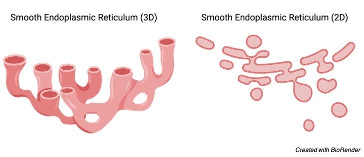
Lumen is present inside the smooth endoplasmic reticulum, which is encapsulated by a membrane of phospholipid. The tubules branches with the other tubules resulting in the formation of a 3D reticulum, however they may also exist in spiral shape in cells generating hormones.
Types of Smooth Endoplasmic Reticulum
Sarcoplasmic Reticulum: A type of smooth endoplasmic reticulum is the sarcoplasmic reticulum which is found in cells of muscle, whose function is to regulate the homeostasis of calcium. In structure they are tubular in shape surrounding the muscle and gather around the myofibril, in which is the cuff like structure of SR.
The functions are it controls the calcium concentrations, while the muscles are contracting. In structure there exist a net like structure in which are tubules spreading throughout the muscle, surrounds the myofibrils. Longitudinal and junctional sarcoplasmic reticulum are its domains.
The similarity in both the domains is that there is an arrangement of myofibrils which orient themselves with the sarcomeres. Tubules are in linear fashion and are connected to each other, surrounding the myofibril is called as longitudinal SR and the area where these tubules end are sac like structures called the terminal cisterna.
Smooth endoplasmic reticulum is the point of entry to the SR and the exit points are the ryanodine receptors along with cisterna. During the contraction of muscle, calcium is taken out from the SR and moves to the ryanodine reception, thus allowing the contraction to take place.
Then again, the calcium is reached to SR with the help of smooth endoplasmic reticulum during relaxation. Thus, acting like a pump and transporting calcium from cytoplasm to the SR and vice-versa.
Smooth Endoplasmic Reticulum Location
All types of eukaryotic cell possess smooth endoplasmic reticulum except mature RBC, embryo cells and ova. Hepatocytes, striated muscle cell, gonadal and sebaceous cell contain huge amount of smooth endoplasmic reticulum.
Smooth Endoplasmic Reticulum Function
Production of lipid, intracellular calcium regulation, drug detoxification, metabolism of sugar are some of the functions. Sebaceous cell, gonadal cell, hepatocytes contain huge amount of smooth endoplasmic reticulum. Although the amount of cholesterol produced by ER is quite minimal but homeostasis maintenance is the function of smooth endoplasmic reticulum. Movement of molecule to Golgi complex from ER is also another function of smooth endoplasmic reticulum.
i. Lipid Synthesis
The sites where membrane comes in close proximity to other organelles such as plasma membrane, lysosomes, chloroplast, Golgi apparatus and lysosomes and others are the MCS also called the membrane contact sites. Thus, transfer of molecules happen through them. For the production of lipid, in huge amount enzyme are present within the ER to carry out the synthesis.
On receiving a signal, these enzyme start to act and maintain homeostasis, allows the growth and differentiation to occur. Phospholipid’s synthesis safeguards the cell and its content, allows the growth, provides immunity and transfers signal along with transportation of lipid. Between the mitochondria and the ER is the MCS which produces phospholipids.
Another type of lipid are ceramides, produced in the smooth endoplasmic reticulum and are moved to Golgi apparatus. Its functions are cell adhesion, migration, signaling, cell death and cell cycle.
In the smooth endoplasmic reticulum the steroid hormones are formed, however as they cannot be stored in vesicles, hence depending on the requirement they are produced from precursors. smooth endoplasmic reticulum is abundant in testis, ova, hepatocytes and other cells.
ii. Carbohydrate Metabolism
In eukaryotes the source of energy is the carbohydrate. In varying conditions glucose is synthesized from precursors such as pyruvate, lactate, succinate which are non- carbohydrate and the process is known as gluconeogenesis. Although this is a multi-step process.
Glucose-6-Phosphate is the final compound synthesized and cannot pass through the cell and is therefore stored in the cell, thus converting glucose-6-phosphate to glucose by the enzyme present in the smooth endoplasmic reticulum which is glucose-6-phosphatase. Its function are maintaining homeostasis and glucose level and is present in liver and kidney, where the conversion takes place.
iii. Calcium Concentration Regulation
The calcium concentration is regulated by sarcoplasmic reticulum, which is a type of smooth ER. This calcium regulation takes place in the muscle cell. smooth endoplasmic reticulum is the point of entry to the SR and the exit points are the ryanodine receptors along with cisterna.
During the contraction of muscle, calcium is taken out from the SR and moves to the ryanodine reception, thus allowing the contraction to take place. Then again, the calcium is reached to SR with the help of smooth endoplasmic reticulum during relaxation. Thus, acting like a pump and transporting calcium from cytoplasm to the SR and vice-versa.
iv. Drug Detoxification
There are various enzymes present in smooth ER and they are cytochrome P450, which helps in removals of toxins from the liver. The mechanism behind it is to solubilize the drug by attaching a 0H- group so that it can be removed out of the body.
Examples of drug working on the same mechanism are barbiturates and phenobarbital. The detoxification rate can be increased by barbiturates and alcohol; however, this is not possible with small volume of doses, due to wide action of smooth endoplasmic reticulum.
Disorders Caused by Dysfunctional Smooth Endoplasmic Reticulum
i. Cytochrome P450 Oxidoreductase Deficiency
The enzyme present in the smooth endoplasmic reticulum is Cytochrome P450 and its enzyme is Cytochrome P450 reductase and its absence can result in a disorder impacting the production of steroid hormone. It would lead to improper growth, effect on the reproductive system and other issues.
Moderate symptom patient would be infertile, whereas those with severe symptom are impacted with the Antley Bixler syndrome where bones have pre-fused, ears are in lower position, and face is also flattened.
ii. Von Gierke Disease
The disorder is named so due to the person who discovered it in 1929.The disease which has an impact on the metabolism of glycogen is called as Glycogen storage disease (GSD-1). Its symptom are improper growth and blood sugar levels quite low and can be deadly too. Glucose-6-phosphatase is the enzyme which converts glucose-6-phosphate to glucose and the process is gluconeogenesis. This enzyme is found in the smooth endoplasmic reticulum.
The absence of this enzyme can lead to this disorder. In this disorder, due to absence of the enzyme, glycogen cannot be broken thus gets deposited in kidneys and liver and thus increasing the liver size. Out of 100,000 people 1 is affected by this disorder and is an autosomal recessive disorder.
Smooth Endoplasmic Reticulum Citations
- Smooth endoplasmic reticulum and axonal transport. J Neurochem . 1980 Jul;35(1):16-25.
- Is it time to reconsider how to manage oocytes affected by smooth endoplasmic reticulum aggregates? Hum Reprod . 2019 Apr 1;34(4):591-600.
- Endoplasmic Reticulum Stress Induced by Toxic Elements-a Review of Recent Developments. Biol Trace Elem Res . 2020 Jul;196(1):10-19.
Share
Similar Post:
-

Ionic Bond: Definition, Types, and Examples
Continue ReadingIonic Bond Definition
A bond which links atoms, molecules together is known as chemical bond. There exists 3 types of chemical bond and they are Ionic bond, covalent bond and hydrogen bonds and these are bound by electrostatic attraction.
In ionic bond there is shift of electron from the donor to the acceptor. The shift happens usually from a metal which is the donor and will be a cation after donating (positive charge) to the non-metal which is the acceptor and will be anion after receival of electron.
To the name of the ion or molecule “ide” is attached at the end which obtains an electron. Example is conversion of chlorine to chloride and sulfur to sulfide.
What is Ionic Bond?
There could be two types of forces seen between the atoms and ion. When two oppositely charged ions attract each other it is called as electrostatic attraction, whereas ions of same charge will oppose each other, thus electrostatic repulsion. There is electrostatic attraction in the ionic bond, where anions get drawn towards the cation which depends on the gap between the two atoms, their size and the forces on them.
Until the octet configuration is accomplished, electrons will get shift from the donor to the receiver, resulting in zero charge on the donor existing in the form of crystals.
Ionic Bond Examples
A bond which links atoms, molecules together is known as chemical bond. A chemical compound consist of various forms of atom, resulting from a chemical bond. Chemical element are those which contain one type of atom. The bond which clutches the chemical compound is called as ionic compound.
Sodium chloride is the ionic compound in which sodium and chloride are bound by ionic bond.
The overall charge is zero as one ion is positive and the other is negative, thus balancing and resulting in zero charge. Those compounds are said to be acidic which has hydrogen ions and those are basic which has hydroxide ion, together forming a salt.
Ionic Bond vs Covalent Bond
Shift of electrons take place between the donor electron to receiver in ionic bond, whereas in covalent bond there is electron sharing taking place in between the two atoms. After the ionic bond, an ionic compound is formed.
Electronegativity is same in covalent bond, whereas in ionic bond the one that receives the electron is more electronegative. When the cations and anions come together in an electrostatic attraction, they form a salt, whereas no salt formation is seen in covalent bond.
Covalent compound are in liquid or gaseous state at room temperature, whereas ionic compound are in the crystalline form. Covalent compound can have single, double and triple bond and are less polar. Ionic bond has high polarity.
Ionic Bond vs Hydrogen Bond
Shift of electrons take place between the donor electron to receiver in ionic bond, whereas in hydrogen bond, between the two atom a bridge is formed.
Example of ionic bond is sodium chloride and of hydrogen bond is water molecule. For the formation of hydrogen bond, a minute positive hydrogen atom with covalent bond that is polar interacts with polar negative atom with covalent bond results in a hydrogen bond.
Although hydrogen bond is the weakest, the secondary and tertiary protein structures are formed by them.
Ionic Bond Citations
Share
Similar Post:
-

Turgidity: Definition, Types, and Examples
Continue ReadingTurgidity Definition
The state of being turgid or swollen, especially due to high fluid content, is referred to as turgidity. Turgidity refers to the feeling of being bloated, distended, or swollen in general.
Turgidity, in a biological context, explains how plant cells may remain upright despite the absence of a skeletal structural structure like mammals. Plants gain stiffness as a result of it.
As a result, cell distention is a common occurrence in plants. In fact, if you don’t provide it to the plant, it will appear withered and sick. The existence of the cell wall and the osmoregulatory function of the vacuole in plants allow for turgidity.
The cell wall protects the cell against lysis caused by excessive water inflow, whereas the vacuole controls solute concentration and promotes osmotic water transport into and out of the cell.
Turgidity Etymology
Turgidity is derived from the Latin turgidus, which derives from the Greek turgēre , which means “to swell.”
Turgidity in Plants
Plant turgidity is accounted for by the cell wall, which is one of the most important characteristics of a plant cell. Aside from the plasma membrane, the plant cell wall is another layer that surrounds the cell. It might be made up of one or two layers. The main cell wall is in charge of secreting the secondary cell wall, which is located atop the plasma membrane.
Plant turgidity refers to a situation in which the cells of a plant become turgid owing to turgor pressure, which is the pressure applied by water inside the cell against the cell wall. The cell wall of a plant organism is one of its most essential characteristics. A cell wall is an additional layer that surrounds a cell. They are absent in the animals, leaving just the cell membrane.
Plants possess both of these qualities. The plant’s cells have an extra protective covering called the cell wall. It is made up mostly of cellulose, pectin, and hemicellulose and is robust and stiff. Plant cell walls are made up of one or two layers. The main cell wall is the initial layer. This layer has the potential to generate a layer underneath it. The secondary cell wall is the new layer.
The second layer is a thick lignin-depositing layer. Lignin contributes to the cell’s waterproofing. These characteristics of the cell wall aid the plant cell’s resistance to osmotic pressure, which is caused by a difference in solute concentrations between solutions separated by a semipermeable barrier, such as the cell membrane, during osmosis.
Turgid Cell
The cell wall and the cell membrane of a plasmolyzed plant cell have gaps between them. When a plant cell is put in a hypotonic solution, this happens. The decrease in turgor pressure is caused by water molecules moving out of the cell. The cell membrane of a flaccid plant cell is not inflated and does not push strongly against the cell wall.
When a plant cell is put in an isotonic solution, this happens. Between the cell and the surrounding fluid, there would be no net flow of water molecules. A cell with turgor pressure is referred to as a turgid cell. When a plant cell is submerged in a hypotonic solution, osmosis allows water to enter the cell, resulting in high turgor pressure on the plant cell wall.
A cell with turgor pressure is referred to as a turgid cell. The plant that seems to be healthy (i.e. not wilted) contains turgid cells. Solutes (such as ions and carbohydrates) are stored in the plant cell (particularly, inside its vacuole). Water prefers to flow in because the inside of the cell has a greater solute concentration (and hence fewer water molecules) than the exterior.
Hypotonic refers to a solution (surrounding the cell) with a lower solute concentration than the solution inside the cell. When a plant cell is submerged in a hypotonic solution, osmosis allows water to enter the cell. A high turgor pressure is applied against the plant cell wall as a result of the inflow of water.
The cell becomes turgid as a result of this. Plants have a cell wall, which protects the cell from bursting (osmotic lysis), which occurs when there is no cell wall. In a hypotonic solution, an animal cell, for example, would swell.
If osmosis continues, the pipe will ultimately explode. As a result, the plant cell’s cell wall is required to maintain cell integrity and prevent the cell from bursting. The osmotic pressure exerted by the cell wall prevents excessive osmosis in the plant cells.
The cell wall, on the other hand, is unable to protect a plant cell that has been exposed to an isotonic or hypertonic solution. These solutions might cause the plant to get wilted and lose its vitality.
Flaccid Cell
An isotonic solution is one in which the concentration of solutes in the solution is the same as the concentration of solutes inside the cell. There would be no net movement of water molecules between the two, implying that there would be no net movement of water molecules between the two. If you put a plant cell in an isotonic solution, it will become flaccid. Flaccidity is the medical term for this disorder.
The cell membrane of a flaccid plant cell is not firmly pressed against the cell wall and is not bulging. Thus, the turgor pressure is the difference between turgidity and flaccidity. Because of the turgor pressure applied to the cell wall, a plant cell appears swollen or distended in turgidity, but in flaccidity, the plant cell loses its turgidity and appears limp or flaccid.
Plasmolyzed Cell
A hypotonic solution is one in which the concentration of solutes is higher than the concentration of solutes inside the cell. The turgor pressure of a plant cell in a hypotonic solution decreases as water molecules migrate out of the cell. Plasmolyzed refers to a cell that has lost its turgor pressure. Plant cells that have been plasmolyzed have holes between the cell wall and the cell membrane.
In addition, the cells looked to be shrinking. Plasmolysis is the process or situation in which protoplasm shrinks as a result of water loss through osmosis. Plasmolysis, on the other hand, is an uncommon occurrence in nature. Rather, plant cells are submerged in powerful saline or sugar solutions in the lab to induce them.
Turgidity and Rigidity
As previously stated, turgidity refers to the state of being turgid or bloated as a result of the fluid present. Rigidity, on the other hand, refers to the state of being stiff and unbending. Turgidity and stiffness are key characteristics of plants because they help them stay erect. Both of these characteristics are due to the turgor pressure exerted on the cell wall.
As previously stated, the cell wall protects the cell from osmotic pressure, which, if too high, might cause osmotic lysis in cells without it. By creating a thicker secondary layer containing lignin, the cell wall also offers structural support. Aside from that, cellulose is present in the cell wall, which makes it stiff and durable.
Another layer of pectin-rich intercellular substance lies between the cell walls. The middle lamella is the name given to this stratum. Its main purpose is to hold neighbouring cells together. Overall, the plant’s cellular characteristics allow it to resist gravitational force and remain erect towards the source of light.
Importance of Turgidity in Plants
Plants require turgidity because it offers structural support and strength. Without it, the plant would be unable to maintain its upright position, which is the optimal position for collecting light energy for photosynthesis. Aside from that, it gives plants rigidity.
The plant cells will not be completely dilated if there is not enough water absorbed to generate turgor. If this situation is not corrected, the plant will become wilted and sickly. The drooping caused by turgor loss can be restored by providing enough water for the vacuole to process through osmoregulation.
Turgidity Citations
- The Nanoscale Organization of the Plasma Membrane and Its Importance in Signaling: A Proteolipid Perspective. Plant Physiol . 2020 Apr;182(4):1682-1696.
- Muscle Articulations: Flexible Jaw Joints Made of Soft Tissues. Integr Comp Biol . 2015 Aug;55(2):193-204.
- Nuclear envelope: a new frontier in plant mechanosensing? Biophys Rev . 2017 Aug;9(4):389-403.
Share
Similar Post:
-

Allele: Definition, Characteristic, and Examples
Continue ReadingAllele Definition
Allele is a variation of a gene that controls the same characteristic and is located in a certain chromosomal region (called the locus). Allele is derived from the Greek ἄλλoς (állos), which means “other.” Allelomorph is a synonym for allele.
What is Allele?
An allele is a word used to define a gene’s unique copy. In eukaryotic genomes, genes, or DNA sequences that govern human characteristics, are generally present in two copies; each copy (allele) is inherited from one parent. Each allele is assigned to a unique gene locus on the chromosome. The two alleles of the gene are found in the same area of two homologous chromosomes, one from each parent. It’s possible that the alleles are dominant or recessive. The influence of the recessive allele is masked by the dominant allele (definition: the allele that is expressed) (definition: the allele that is not expressed).
An observable trait or attribute is referred to as a “phenotype.” If the trait is regulated by only one gene, the genotype is made up of both alleles. As a result, the term genotype refers to an organism’s set of alleles that code for each characteristic. The genotype of a gene can be characterised as homozygous or heterozygous depending on the DNA sequence of each allele.
The term homozygous genotype refers to an organism’s genome having two identical alleles for the same gene. Both alleles contribute equally to the trait’s manifestation. In contrast, a heterozygous genotype has two distinct versions of the same gene. However, some genes contain more than two different allelic forms, which are referred to as many alleles.
Allele Examples
Alleles and how they are expressed are shown in the examples below. As in the case of full dominance, allelic expression may follow a Mendelian pattern of inheritance. The alleles may be manifested by codominance, partial dominance, or polygenic inheritance in nonMendelian inheritance.
i. Complete Dominance
The alleles responsible for blossom colour in garden peas, one of the seven characteristics examined by Gregor Mendel in his studies that subsequently established the laws of genetics, are a classic example of how alleles function.
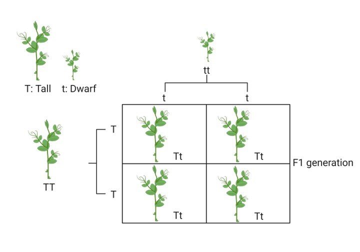
The blooms of garden peas can be purple or white in hue. The allele for purple colour and the allele for white colour are present in the gene coding for this characteristic, which is responsible for generating a purple pigment.
To characterise genotypes, alleles are assigned letters. The allele for tall (referred to as T) can naturally express tall phenotype, but the allele for dwarf phenotype (referred to as t) can’t. As a result, the tall genotype is abbreviated as TT, Tt (Tall phenotype) whereas tt for dwarf phenotype. In this case, however, one T allele is capable of producing enough genes to turn the height tall. Plants with either the TT or Tt genotypes will produce tall phenotype, whereas only the tt genotype will have dwarf phenotype.
The dominant allele is the one that exhibits the characteristics above the other one in this scenario (the T allele in this case). The other allele (t) is recessive, meaning it is only expressed when two copies are present.
This isn’t always the case, though. Codominance is a situation in which two distinct alleles (heterozygous genotype) are equally expressed in some genes. ABO blood grouping alleles are one of the most common instances of this sort of inheritance.
Many ABO blood types are found in humans. Antigens unique to blood types A, B, AB, and O can be seen in the red blood cells. The A antigen is found in blood type A. The B antigen is found in blood type B. A and B antigens are found in blood type AB. On the cell surface of red blood cells, blood type O lacks antigens A and B.
ii. ABO Codominance
There are three allelic forms of the ABO locus: A, B, and O. The blood group phenotypic is determined by the genotype of this gene, which determines the antigens on red blood cells. The A and B alleles both code for glycosyltransferase, which catalyses the last step in the production of A and B antigens, but the O allele codes for an inactive form of the enzyme. Due to numerous single nucleotide polymorphisms (SNPs) in the ABO gene, the enzymes transcribed by the A and B alleles differ in only four amino acids.
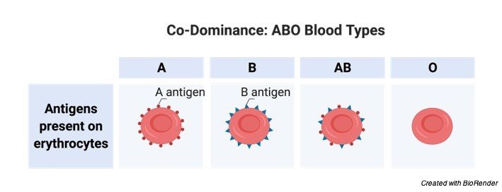
The ABO gene’s inheritance pattern is shown in the table below. Because the locus has three different alleles, mating can result in six genotypes. A, B, AB, and O are the only four phenotypes (blood groupings) that exist. In blood groups, there are two inheritance patterns. When the O allele is present, both the A and B alleles become dominant. If a person has the AB genotype, however, both alleles are expressed equally (codominance), resulting in a phenotype in which both A and B antigens are present on the surface of red blood cells.
iii. Multiple Alleles
It’s possible that certain genes have more than two allelic variants. Many people are aware that human genes have many alleles. Each gene may only have two alleles per person. Within a population, however, certain genes may contain hundreds of alleles. Any allele is the consequence of a change in the DNA sequence of a gene, even if the change is only one nucleotide long.
As a result, having many alleles of a gene does not always imply phenotypic diversity. Many allelic variants are caused by variations in the gene’s sequence that have no effect on the protein’s sequence or function. In comparison to mutant alleles that alter protein structure or function, all of these “normal” alleles are referred to as “wild-type” alleles.
All mutations discovered in a gene in a population are sometimes referred to as multiple alleles. Multiple loss-of-function mutations, for example, can be found in a group of individuals with a genetic illness. Although various mutations result in the same consequence, loss of protein function, each mutation is treated as a separate mutant allele if it occurs in a distinct gene location.
iv. Beta-Thalassemia
The decrease or lack of beta-globin chains in haemoglobin is known as beta-thalassemia. So far, more than 300-thalassemia alleles have been identified, with the majority inheriting in a Mendelian recessive manner. The majority of reported mutations in the-globin gene or its surrounding areas included a single or a few nucleotides.
There have also been reports of deletion mutations that result in the entire lack of chain manufacturing. The severity of the illness is mostly determined by the chain’s quantitative decline level. Beta-thalassemias are a category of hereditary blood diseases characterised by a wide range of mutations in the globin gene and a wide range of symptoms.
v. Short Tandem Repeats (STR)
Short tandem repeats (STRs) are short DNA sequences (2-6 nucleotides) found throughout the genome. Although they are non-coding DNA sequences, they have been related to particular genetic disorders. However, forensic investigation is the most common and effective application of STR markers. The amount of repetitions in each STR locus varies a lot from person to person.
Using these differences within a population, scientists devised a forensic identification technique based on several STR markers. The combination of several markers in the same test results in a high level of discrimination in the form of a unique “DNA fingerprint” that may be used to identify people in instances like unidentified bodies or criminal suspects.
Allele Variation
Returning to the ABO blood groups, genetic differences between alleles might result in protein differences. Even a modest change in protein levels, in this example, four amino acids, can have a significant impact on phenotypes. Because the recipient’s immune system reacts to the ABO antigens present in the donor’s RBCs, a blood transfusion to an incompatible patient might result in death. As a result, allelic variation between genes can be considered one of the main mechanisms contributing to the genetic diversity observed between people.
i. Discontinuous Variation
Discontinuous variation, which occurs when a characteristic exists in two or more distinct alternative forms, is shown in blood groups and flower colour phenotypes. Different phenotypes can be clearly identified in this sort of variation. Polymorphism is a word used by geneticists to describe characteristics that have two or more common phenotypes in a population, while morphs is a term used by geneticists to describe individual phenotypes.
Rare, extraordinary phenotypes exist in some situations; they are referred to as mutants, whereas the more frequent normal phenotype is referred to as wild-type. Although both polymorphisms and mutations are caused by changes in DNA sequence, polymorphism alterations have become increasingly prevalent.
ii. Continuous Variation
Continuous variation is the second form of genetic variation. As opposed to discontinuous variation, this form of variation exhibits a continuous range of phenotypes that can not be identified as discrete phenotypes. Weight, height, eye colour, and other quantifiable traits are examples of characters that demonstrate continual fluctuation. These phenotypes are more difficult to analyse than those with discontinuous variation since they are generally encoded by several genes.
Molecular Basis of Allelic Variation
Phenotypes can be caused by the activity of a single gene, as in most discontinuous phenotypes, or by the action of multiple genes, as in continuous phenotypes. The majority of genes code for proteins that express the characteristics directly. Proteins are the key factors presenting the phenotype through executing their biological activities, as they are the direct product of gene expression. Proteins might be antigen receptors, pigments, hormones, antibodies, or enzymes that deliver antigens to immune cells.
Variation in the genes that code for these proteins can have a variety of effects, ranging from no impact to altering the function of these genes. A single nucleotide, the building block of DNA, can alter some genetic variants. The most prevalent and simplest kind of genetic variation is nucleotide substitution. Amino acid substitution will occur if a substitution occurs in the coding region of the gene.
The impact of amino acid substitution on protein function and phenotype is largely determined by the altered amino acid location within the three-dimensional structure of the protein. An enzyme’s job, for example, is to catalyse chemical reactions by attaching to its substrates via an active site, which is an area inside the enzyme’s 3D structure that is lock-and-key bonded to a substrate. Because biological activity is based on structure, any minor alteration in the 3D structure of the active site will have a direct impact on enzyme function.
As a result, a base-pair substitution that changes an amino acid in the active site or in a different area of the active site, causing the active site’s 3D structure to alter, will stop the enzyme from working. Other nucleotide changes can result in a stop codon, causing translation to end prematurely and the production of truncated protein products.
Mutations can also take the form of one or more nucleotide additions or deletions. Because the mRNA molecule is read in three “codons,” the effect of these mutations is typically more harmful to the protein; thus, unless the deletion or insertion contains a multiple of three nucleotides, a shift in the reading frame will occur, altering all the subsequence codons. “Frameshift mutations” is the name given to such mutations.
Allele Citations
Share
Similar Post: