Data analysis tools are software and programs that collect and analyze data about a business, its customers, and its competition in order to improve processes and help uncover insights to make data-driven decisions.
20 Best Data Analysis Software Tools for 2022
(1) Sequentum Enterprise
Sequentum provides complete control for web data extraction, document management and intelligent process automation (IPA). Our end-to-end platform provides the flexibility to be used in-house or you can outsource your web data extraction needs to our experienced Managed Data Services group. Our tools create software configuration files that define exactly what data to extract, quality control monitors, and output specifications to any format or endpoint.
If you have multiple databases, data saved in external applications or an indefinite number of spreadsheets, you’ve come to the right place. Our goal is to make it as convenient as possible for you to combine and understand all your data in one single source of truth.
Looker is a business intelligence software and big data analytics platform that helps you explore, analyze and share real-time business analytics easily.
KNIME Analytics Platform is the open source software for creating data science. Intuitive, open, and continuously integrating new developments, KNIME makes understanding data and designing data science workflows and reusable components accessible to everyone.
The Lexalytics Intelligence Platform is a modular business intelligence solution focused on solving text data’s specific challenges. Data analytics companies and data analyst teams use our platform to gain the richest possible insights from complex text documents.
Manage your organizational planning challenges by generating forecasts on an enterprise scale – quickly, automatically and as accurately as you can reasonably expect, given the nature of the behavior being forecast. The software delivers results for millions of forecasts at breakthrough speeds, enabling you to plan more efficiently and effectively for the future.
RapidMiner’s data science platform delivers transformational business impact for over 40,000+ organizations in every industry to drive revenue, reduce costs, and avoid risks.
OpenRefine always keeps your data private on your own computer until YOU want to share or collaborate. Your private data never leaves your computer unless you want it to. (It works by running a small server on your computer and you use your web browser to interact with it)
Maintain healthy data across all your on-premises and cloud systems. Talend brings it all together with support for virtually any cloud data warehouse and all major public cloud infrastructure providers.
NodeXL Pro gives data-driven marketers access to powerful social media analysis features including influencer identification, brand evaluation, social listening, content analysis, ideation, lead harvesting, competitor social and campaign analysis, automation, white-labeling, and more!
(11) Alteryx: Self-Service Analytics, Data Science
The Alteryx Analytic Process Automation (APA) Platform™ accelerates data-driven business outcomes across lines of business in any industry. Develop faster results, deeper insights, better decisions, and ultimately, a cultural shift where anyone can drive business outcomes through self-service analytics and data science.
Automate routine analysis with RocketBots, specialized applications that leverage AI and machine learning to execute analytics workflows, produce decks, and compose data stories. RocketBots can run on demand, on a set schedule, or whenever there’s new data available.
Arcadia Data is the first AI-driven analytics and BI platform built for the cloud and data lakes. Our in-data-lake BI architecture gives you faster, deepest insights from modern data platforms like cloud object stores, Apache Kafka, and Apache Hadoop.
Infor industry analytics deliver relevant and meaningful insights for everybody from the boardroom to the shop floor. Infor Birst® makes business intelligence and analytics easy to consume with pre-built industry and role-specific content and metrics embedded wherever business users need information. Improve decision-making with Birst.
Self-service Analytics, Planning, Forecasting, and Predictive Analytics. All of Board’s functionality is completely unified and shares the same metrics, data, and view of customers, products, and markets. It is a unique Decision-Making Platform that helps you achieve your goals and make a difference to your business.
For over a decade, we have loved serving tens of millions of charts on millions of dashboards for thousands of incredible companies. All of you have helped us create a product that truly enables anyone in a company, not just data teams, to explore and understand their data.
Build a modern business, driven by data. Connect to any data source to bring your data together into one unified view, then make analytics available to drive insight-based actions—all while maintaining security and control.
We know what it means to be data-driven. We were born industrial and we breathe digital, so data is in our DNA. That’s how we help mission-critical organizations use data to get from Now to Next – going from data-rich to data-driven.
This integrated data and AI platform unifies data management, DataOps, business analytics and automates AI across multicloud environments such as AWS, Azure, IBM Cloud and private cloud.
Create a data-driven culture with business intelligence for all Enable everyone at every level of your organization to make confident decisions using up-to-the-minute analytics.
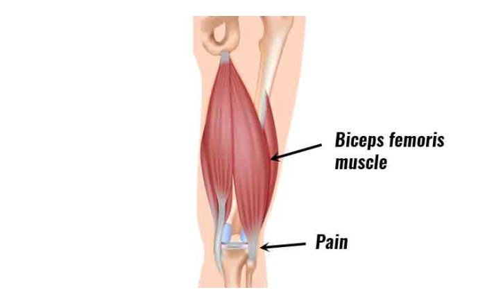
The muscle found at the back of the thigh is called the biceps which belongs to the hamstrings muscle group. The biceps muscle is found in two parts and is a spindle-shaped muscle. It consists of two heads, the long head, and the short head.
The sites of origins of both heads are different which are innervated by different nerves. But, the heads are inserted at the same location and joined together.
The biceps femoris helps in the movement of hip joints and controlling the knee. Commonly, it gets injured in high-intensity sports.
Anatomy of Biceps Femoris
At the posterior to the short head, the long head of the muscle lies thus, we can only see the long head on looking at the thigh from behind. Although, a short head can be observed peeking out from underneath.
On the outer side of the leg, the muscle is the closest and it is the most lateral of the muscles in the back of the thigh.
Image Credit: https://www.sportsinjuryclinic.net/
There are two different divisions of the sciatic nerve that innervate in muscle. The divisions are the common fibular division or the common peroneal nerve, and the tibial division.
The common fibular division innervates the long head of the biceps femoris, whereas the tibial division innervates the short head.
1. The Long Head
The long head is originated from the upper and medial part of the back of the ischial tuberosity of the pelvis. The ‘butt bone’ is the origin of the long head. The parts of the hamstring muscles are formed by the long head.
Along with the hamstring muscle, the long head also forms parts of two other muscles, named the semimembranosus and semimembranosus muscles. The long head of the biceps muscle is the strongest muscle of the hamstring group.
2. The Short Head
The short head of the biceps femoris has originated from the linea aspera and the supracondylar ridge of the femur. The ridge is located around the middle of the femur towards the knee.
Therefore, as compared to the long head the short head starts significantly lower down the leg. Unlike the long head, the ischial tuberosity is not the origin of the short head, thus it is not considered part of the hamstring.
As criteria are made that a muscle arising from the ischial tuberosity is classified as one of the hamstring muscles. Interestingly, the short head is altogether absent in humans also is show some anatomical variations.
3. Insertion
A tendon called the biceps femoris tendon is formed by the long head and short head by coming together at the distal thigh. This tendon inserts onto the lateral side of the head of the fibula and the lateral condyle of the tibia. Thus, on the outer side of the leg and the upper and outer surface of the shin bone, the two heads connect to the calf bone near where it meets the kneecap.
Functions of the Biceps Femoris
The biceps femoris provide movement and stability of the joints by acting on both the knee and hip. The long head acts on both head and knee but the short head only acts on the knee.
The actions are influenced by the short head and the long head, which the muscles take in different ways. The long head of the biceps femoris influence some actions such as the bending or the flexion of the knee, lateral rotation of the tibia, and extension of the hip joint such as stretching the leg backward.
The short head influences actions including lateral rotation of the tibia, flexion of the knee (bending of the knee).
Biceps Femoris- Associated Pain and Injury
The muscles of the biceps femoris are most commonly injured in the posterior thigh. This is due to the innervation of the long and short heads by different nerve branches. It is believed that sometimes, the muscles behave ‘out of sync’ and make them more vulnerable to injury.
Typically, during high-intensity exercise, the muscles get injured. Particularly during hip extension, such as when the leg is stretched out behind the body. The activities such as sprinting, American football, rugby, and hockey are associated with these injuries.
Usually, the force on the muscle causes these strains. The fibers will tear if the muscle is not strong enough to withstand the forces acting upon it and can cause intense, sharp pain at the back of the thigh.
The movement can also be impacted by these strains and cause pain in the knee and hip. These injuries can be prevented by carrying strength conditioning and stretching exercises.
The flexibility and range of movement of the joint will be improved by these exercises and it also allows the muscles to withstand increased force during exertion.
Treatment of Biceps Femoris Strains
The treatment of the biceps femoris includes reducing the inflammation and resting the muscle to allow it to heal and the thigh should be iced and rested. The use of anti-inflammatories helps in pain and swelling. Surgery may be required in some cases when the tendons have snapped or are severely torn.
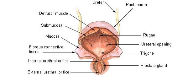
An organ present in the urinary tracts of several animal species is called the urinary bladder. The urine is stored in the urinary bladder before urination. The urine is produced and delivered by the kidneys through two ureters. The urinary bladder in humans is capable to hold up to four cups of urine in its hollow, muscular structure.
Overview of Urinary Bladder
The structure of the bladder is composed of a broad fundus, body, apex, and neck. There are three openings in the human bladder, each of them is covered by a mucosal flap.
The flab prevents urine from flowing back into the ureters. The bladder differs its anatomical position between men and women.
Image Credit: https://training.seer.cancer.gov/
The urinary bladder in men is found in front of the rectum, whereas the position of the bladder in women is in front of the uterus. The bladder wall can stretch from 5 mm to 3 mm due to rugae or folds covering the inner walls.
The kidneys can continue filtering the blood due to the holding capacity of the bladder. When an organism cannot urine, the bladders store the urine. For example, during sleep, extra urine is stored in the bladder, which allows an organism to rest without urinating.
Urinary Bladder Function
The major function of the urinary bladder includes storing and collecting urine from the kidneys. They store urine, until its excretion via urination. An average of 300mL to 500mL of urine can be stored in a typical human bladder.
Due to the flattening of the rugae folds, the urinary bladder has high elasticity and can accommodate an increased volume of liquid. However, urination is not controlled by the bladder, but the pontine micturition center in the brain controls it.
Diseases of the Urinary Bladder
There are several diseases of the urinary bladder. Frequent urination, pain, irritation, and incomplete emptying are some common symptoms of bladder diseases. Occasionally, the urinary bladder can be affected by diseases of other tissues or organs.
For example, frequent urination can be caused by an enlarged prostate. Some most common pathologies of the urinary bladder are as follows:
1. Bladder Cancer
Bladder cancer occurs commonly in the epithelial lining of the bladder. Approximately 90% of all bladder cancers are carcinomas. The common causes of bladder cancer include infection, cigarette smoke exposure, and medications.
2. Urinary Tract Infection
A major public health concern can be posed by urinary tract infections, which can be severe. Some bacterial infections in the bladder are the major cause of these infections. The bacteria traveling up the urethra and into the bladder may cause infections.
UTIs can be very dangerous when left untreated because the infection can increase and infect the bladder and kidneys.
Burning sensation during urination, frequent urge to urinate, despite little urine being released, foul-smelling urine are some common symptoms of UTIs.
3. Bladder Stones
Hard deposits found in the bladder are called bladder stones. The stones are composed of minerals and caused by highly concentrated urine resides in the bladder, dehydration.
The size of the stone can vary and typically it is asymptomatic. Pain, blood in the urine, and irritation are the common symptoms of bladder stones. Typically, the bladder stone can be identified by using ultrasound, X-rays, and CT scans.
4. Neurogenic Bladder
It is a type of brain disorder, due to which, an organism’s ability of urination impacts. Urination is typically aided with a catheter because the peripheral nerves involved in urination are affected. Patients suffering from neurogenic disorder use intermittent catheterization to empty their bladder several times a day.
5. Bladder Exstrophy
Bladder exstrophy is a congenital abnormality. It involves the protrusion of the bladder, which is a rare condition. Generally, the condition arises due to abnormal development of the pelvic floor and other muscles. In women, it causes the development of genitals.
6. Bladder Sphincter Dyssynergia
The condition in which the urethral sphincter cannot be relaxed in coordination with bladder contraction is referred to as bladder sphincter dyssynergia. The common causes for this disease include injury or disorder in the central nervous system. In this disorder, patients have a history of bladder infections and they also frequently feel urine retention.
7. Paruresis
A phobia in which a person cannot urinate due to the presence or perceived presence of others is called paruresis. A sympathetic nervous system response causes this disease by tightening the sphincters in the bladder in response to adrenaline and finally preventing urination. Psychological therapy is required to treat this disease.
8. Trigonitis
The condition that involves inflammation of the trigone region of the bladder is called trigonitis. In response to stretching, the trigone, which is a smooth triangular-shaped region, signals the need for urination. When the region is inflamed, it causes an urgent need for urination, pain in the pelvic region, and pain during urinating. The most common cause of trigonitis is bladder infections, but it can be caused due to several causes.
9. Interstitial Cystitis
Chronic bladder pain is defined as a condition of interstitial cystitis. However, damage in the epithelial lining of the bladder is seen in patients, but the specific causes of interstitial cystitis are unknown. The symptoms include an urgent need to urinate, pain, and frequent urination. Typically, a negative urine culture is used for diagnosis because the symptoms are highly similar to urinary tract infection.
10. Urinary Retention
A condition in which an individual becomes unable to completely empty the bladder is called urinary retention. Poor pressure when urinating, straining to urinate, the sensation of a full bladder are some common symptoms of this disease. If the disease cannot be treated, it causes a ruptured bladder, thus it is considered to be an emergency. The cause is several and it can be treated by catheterization, surgery is required in some cases.
The area of tissue from which new growths are formed in plants is called meristem. The cells at meristem continually proliferate and do not differentiate. The new leaves arise from the meristem tissue, which depends upon the signals received by it. It also gives rise to flowers or roots of the plant. A plant cannot produce new cells without the meristem.
Apical Meristem Function
The apical meristem is responsible for the growth of the plant and its length and height. The location of apical meristem is at the ends of roots, known as root apical meristem, or at the tops of shoots, which are known as shoot apical meristem.
This meristem is responsible for the primary growth of the plant. The vertical growth is promoted by the axillary buds, which are exerted by the presence of an apical bud.
Shoot Apical Meristem Function
The shoot apical meristem is developed to become one of three primary meristems that are the protoderm, ground meristem, and procambium. The shoot apical meristem is mainly composed of undifferentiated cells found above the ground.
The epidermal tissues of the plant are formed by the protoderm while the ground develops into the cortex and pith of the plant. And the xylem and phloem, collectively known as vascular bundles are formed by the procambium.
The leaves of the plant are also included in the shoot that grows from the sides of the apical meristem. A bump or an axillary bud is formed by the beginning growth of the leaf at the node.
The axillary bud remains dormant if the terminal bud is in close proximity to it. However, the increased distance or removal of the terminal bud results in the disappeared or diminished exerting apical dominance.
It allows the growth of leaves at the lateral buds of the apical meristem. The shoot apical meristem converts into the inflorescence meristem when the angiosperm plant becomes ready to bloom and emerge various flower parts such as petals, sepals, stamens, carpels, etc.
Root Apical Meristem
It is found below the ground. The root apical meristem is responsible for the growth and development of a plant’s root. The root apical meristems have the ability to yield two types of tissues at the same time and can produce cells in a bilateral direction.
Among these two types of tissues, one typically comprises the main roots of the plant and the other type consists of a root cap, the main roots are responsible for continuous growth and supply proliferative, undifferentiated cells for continued growth while the root cap protects the apical meristem.
As the root cap grows deeper into the soil, the cells of the root cap are continuously being shed and replaced by new cells, which are provided by the main root. This is typical of the taproot.
The lateral growth of the root is conducted by the lateral root meristem, which helps in efficient water supply and nutrient absorption in the plant. It also helps in nutrient storage and stability for aerial growth.
Basal Meristem Function
Basal meristem is also called intercalary meristem. It is located between mature, differentiated tissues. The intercalary meristem is distinctly different from the apical meristem, which is located relatively near an apical meristem.
The vertical growth of the plant is promoted by the apical meristem and the intercalary meristem works independently of the apical meristem. However, it works in the base of the plant, not at the tip of the plant tip. The grasses and other plants can grow continuously after being cut due to the intercalary meristem.
Lateral Meristem Function
The vertical growth of the plant is controlled by the apical meristem, similarly, the lateral meristem is responsible for the lateral growth of the plant. Lateral growth is also known as secondary growth.
The lateral growth is conducted around an already established stem thus it is known as secondary growth. There are two types of lateral meristem in woody plants, named the vascular cambium and the cork cambium.
The vascular cambium is similar to the procambium of the apical meristem, which is responsible for the development of wood and increased width of the plant. The periderm is developed from the cork cambium, similar to the protoderm.
The primary epidermis growth is produced from the protoderm. And the epidermis is replaced by periderm to produce bark, which acts as a shield and protects the plant from physical damage and water loss by a waxy layer of suberin.
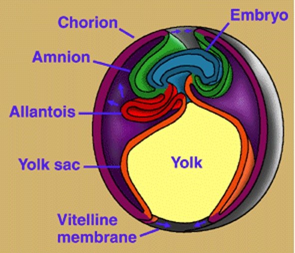
The vertebrates which have a fetal tissue known as the amnion are called amniotes. A membrane that surrounds the fetus and is derived from the fetal tissue is called the amnion. It protects the fetus and can be found within the egg, or can simply enclose the fetus. For instance, lizards and birds, it found within the egg or enclose the fetus within the uterus.
Amniotes Description
Most of the vertebrates, excluding fish and amphibians are amniotes. Amphibians and fishes are known as amniotes, which means “without an amnion”. The species often lay their eggs in water to protect them from any damage.
Image Credit: https://www2.gwu.edu/
Most amniotes are terrestrial animals thus they have to protect their fetus under the weight of gravity. The amnion protects the developing fetus. Whales are the only exceptions of amniotes because they are aquatic animals.
It may be due to the development of amnion in whales before their move to the sea. Some marine turtles also live in the sea but return to land to lay their eggs.
Characteristic of Amniotes
The fetus of amniotes is surrounded by three membranes, which are the amnion, the top chorion layer, and the waste-absorbing allantois. In general, the amniotes share several characteristics such as they all are developed from a common ancestor and the ancestor has developed the amnion character.
Most of the egg-laying species consist of the amnion. For example, the birds and reptiles, and some mammals consist of this protective layer. However, the shell is not present in the human egg but in many ways, the development of the human egg is similar to the chicken eggs.
Animals Considered Amniotes
Sauropsid Amniotes
Amniotes are divided into two divisions, the sauropsid amniotes, and the synapsid amniotes. The reptiles and birds are included in the sauropsid amniotes. Formally, many different groups are included in this group, but they are separated from the synapsids because they share many derived characteristics.
However, both the groups are evolved around the same time from a common ancestor. The ancestor was likely not terrestrial. After the evolution of both sauropsids and synapsids, both had to make some adaptations to sustain themselves in terrestrial environments.
The heart, lungs, and kidneys reflect the differences mainly. The lungs of synapsids differ from the lungs of sauropsids that have foveolar lungs. The small chambers present in foveolar lungs open to a common space.
The heart of sauropsids lacks the ventricle and some sauropsids have almost 4 chambered hearts. The process for waste excretion is also different in the sauropsid amniotes than the synapsids.
Sauropsids excrete uric acid. Much of the water can be absorbed in the cloaca in which the substance precipitates out of the urine. Thus, uric acid become a more water-efficient way of excreting nitrogenous wastes than the synapsid method. The turtles, lizards, crocodiles, and birds, which lay eggs are included in sauropsids.
Synapsid amniotes
The sauropsids are separated from the synapsid amniotes since millions of years ago, but they have slight differences. They excrete all urea as nitrogenous waste. Urea is concentrated in the synapsid kidney and excreted with a small amount of water.
The strategy is much more efficient than the excretion of straight ammonia but it is not water-efficient as uric acid. Ammonia is usually excreted by fish and amphibians. Synapsids consist of 4 chambered hearts, which have a partition between ventricles.
Thus, the efficiency of oxygenating the blood is improved due to separate paths for blood. The structure of the lungs is also different in synapsids than the sauropsids. They consist of an alveolar lung which has many branches of the trachea instead of small pockets from a central chamber.
The branches end at an alveolar sac. Synapsids consist of only three extant groups, all of them are mammals. However, the amniotic sacs are present in all of these amniotes but they reproduce by different methods.
For instance, the monotremes lay eggs in nests and after the eggs hatch, they feed them milk like other mammals. The marsupials are considered as a median between the monotremes and the placental mammals.
In the marsupials, the young are developed in the uterus but born at a very early age. They must climb along with the mother into the marsupial pouch, where the mother feeds them milk for their proper development.
The rest of the synapsid amniotes are represented by the placental mammals. In these animals, a placenta or oxygen and nutrients passing maternal membrane are used to feed the offspring within the womb.
They produce the largest offspring among all amniotes but they have fewer offspring compared to sauropsid amniotes.
Evolution of Amniotes
The evolution of amniotes may occur when first terrestrial animals are venturing onto land. The two main groups of amniotes were divided due to the much different terrestrial environment.
The division may occur around 400 million years ago, in the Devonian period. Later, various considerable differences have evolved between these groups. As discussed above, they differ in their anatomy and physiology.
At the time, they also diversified due to the new terrestrial environment which provided them several new niches for the animals to fill. An organism named parieasaur, a cow-sized organism could have been a common ancestor of modern amniotes.
It is a large reptile-looking organism from the Devonian period that may have primitive hearts, lungs, and kidneys and likely had an amnion. Thus it is likely one of the first amniotes.
The set of glands between the bladder and the penis is called the prostate gland. A component of seminal fluid, called prostate fluid is secreted by this gland, which helps sperm cells swim. Skene’s gland is the female homolog of the prostate gland, but in this article, we only focus on the prostate gland.
Prostate Gland Description
The size of the prostate is about the size of a walnut (1.5 in). It is penetrated by the urethra that directs urine out of the body. An alkaline seminal fluid is expelled into the urethra by the prostate gland during ejaculation.
The alkaline nature of this fluid helps to counter the acidic environment of the vagina and protects sperm in the vaginal tract. The prostate gland also helps in sperm ejection.
Image Credit: https://www.cancer.org/
The function of the prostate gland is simple but when it becomes swollen, infected, or damaged by disease, it can cause pain and discomfort. The gland blocks the urethra and holds urine in the bladder when the gland swells.
The swollen prostate gland causes several problems such as pain during urination and an inability to empty the bladder at old age, which can lead to further complications if not corrected.
Prostate Gland Function
The primary function of the prostate gland is the secretion of prostate fluid. The prostate fluid is a seminal fluid, which ejects during ejaculation. The seminal fluid is ejected into the urethra with the help of prostate muscle.
The weight of the muscle is about twenty grams and surrounds the urethra just below the bladder. The drainage tubes are known as vas deferens shuttle sperms into the site of the prostate gland.
The proteolytic enzymes exit the prostate through ducts. Before ejaculating, the ducts open into the urethra. The prostate squeezes with the help of its surrounding, it closes the opening found between the urethra and bladder.
The fluid is directed into the urethra and pushes the semen out. Enzymes, zinc, and citric acid are present in the seminal fluid. The semen becomes slightly basic due to the fluid produced by seminal vesicles while the prostate fluid is slightly basic.
The basic nature of semen is to protect it and to prolong the lifespan of each sperm. An enzyme, called PSA or prostate-specific antigen is another element of prostate fluid. The function of this enzyme is to liquefy seminal fluid and to allow sperm to swim easily.
The levels of PSA in the patient’s blood can be measured by a blood test. A high level of prostate fluid increases the risk of prostate cancer. However, the swollen prostate gland can cause many difficulties but its site makes it a great location for delivering fluid.
Prostate Gland Location
In males, the prostate gland is the largest accessory gland. The location of the prostate gland is above the urethral sphincter and inferior to the neck of the bladder.
The prostate is located in front of the rectum, which makes it easy for physicians to conduct DRE. Digital rectal exams are conducted to inspect the health of the gland. The prostate is monitored by doctors for any signs of disease, such as swelling.
Prostate Gland Structure
There are four anatomical lobes in the prostate gland. But a histological parsing into zones is a more significant delineation.
• The central zone surrounds the ejaculation dust. This zone is derived from the Wolffian duct located in human embryos.
• The transitional zone is the area near the center, which surrounds the urethra. It is originated from the urogenital sinus.
• The peripheral zone forms the body of the prostate gland. The area originated from the urogenital sinus and is located toward the back.
The gland is well-innervated and notably vascular. The prostate gets the supply of oxygenated blood from the prostatic arteries and the oxygen-depleted blood drains into the prostatic venous plexus and enters the iliac veins of the pelvis.
The prostate gland innervates in a little simpler manner and is innervated by the inferior hypogastric nerve plexus. A bundle of nerves, which innervates the smooth muscle of the prostate is called a plexus.
Prostate Gland Pathology
The urination could be blocked by a swollen prostate by pressing against the urethra. This will irritate the bladder and the surrounding area. In older gentlemen, gland swelling or urinary discomfort occurs more frequently.
Benign prostatic hyperplasia (BPH) or the enlargement of the prostate gland is very common in old men and up to half of the men over sixty suffer from this. Between the ages of seventy and eighty, this statistic jumps to ninety.
Frequent urination and leaking are the symptoms of this affliction, which can be treated by alpha-blockers to relax the muscles around the urethra. 5-alpha reductase inhibitors also help in reducing the levels of DHT testosterone and shrink the prostate.
Another condition where the prostate tissue is inflamed by an infection is called prostatitis, which can be treated by antibiotics. All prostate growth is not benign. According to the National Cancer Institute, over 200,000 men are diagnosed with prostate cancer each year.
The five-year survival rate for prostate patients is only 29 percent, which means that under 30,000 deaths are caused by this disease each year. In America, prostate cancer is the third leading cause of death.
Prostate cancer can be treated by a combination of surgery, radiation, and chemotherapy. Sometimes, it becomes necessary to remove the prostate gland because it can lead to additional tissues.
Aix galericulata or the Mandarin duck is also known as yuan-yang in Chinese. The bird is native to China, Japan, and Siberia and closely related to the North American wood duck. Mandarin duck is a beautiful bird where the male is more colorful than the female and has quite striking plumage like most other duck species.
The male mandarin duck has a red/ orange face with a white crescent above his eyes. It consists red bill and the rest of his plumage is red, orange, brown, purple, and green colored.
The male also consists of a sail of feathers in his back during the breeding season. While female ducks have brown and grey feathers and a light bill, which has a tinge of pink. Female also consist a white eye-ring.
Mandarin Duck Habitat and Nesting
The mandarin ducks live in pools, rivers, lakes, marshes, fast-flowing streams, and swamps surrounded by dense forests. The courtship rituals in mandarin ducks are much elaborated. At a beginning of the mating season, these ducks form pairs and look for a nest.
The female is accompanied by the male on this nest hunt. The nest of the ducks can be up to 32.8 feet above ground in a hole in a tree. Nine and twelve eggs laid by the female at a time. The eggs are incubated for up to four weeks.
The eggs hatch within a few hours of each other. The mother will coax them out of the nest by calling to them from the ground. Before the move to feeding grounds, the chicks will free fall to the ground from the nest.
The chicks are ready to fledge after 45 days after hatching and will fly off to join another flock. Habitat destruction is the main threat to the Mandarin duck populations. The predators include raccoons, minks, eagles, snakes, and otters.
Fun Facts About Mandarin Duck!
The fascinating plumage of Mandarin ducks makes them the most beautiful duck species. In China, Japan, and Korea, it is presented as a symbol of love and fidelity because they often form long bonds with their partner.
Mandarin ducks sometimes form pairs for life and usually, they pair up for several breeding seasons. These ducks also show several interesting biological concepts. Let’s understand and take a closer look at these concepts.
i. ZW Sex-determination System
Sex is determined by the presence of specific chromosomes in humans and other animal species. For instance, humans have an XY system of chromosomes in which the sex is determined by a pair of chromosomes in an individual.
The females have two homologous chromosomes (XX) while the males have two different types of chromosomes (XY). The Mandarin ducks have a ZW sex-determination system like most other birds. This system is different from the XY system in most mammals.
Besides birds, the ZW system is also observed in several species of fish, crustaceans, and some insects such as moths and butterflies. The ZW system of sex- determination is relatively widespread in nature.
During the incubation period, the male duck stays around the female while once the eggs hatch, he leaves the female to rear the chicks and he molts and sheds his colorful plumage. During this time, the male is unable to fly because he also sheds his primary flight feathers.
During this time, the male also resembles a female because his bright plumage is gone and only brown and grey color remains. Thus he blends with his environment and also hides from its predators.
iii. Dabbling Ducks
Dabbling ducks include eight genera and 50-60 species, which also includes Mandarin ducks. However, there are many differences between these ducks and other duck species but they are named after the way they feed.
These ducks usually live in shallow waters such as flooded fields and marshes. Instead of diving, they feed by tipping up in the water and their rear end out of the water with their head down.
Their feeding manner is defined as dabbling in which they move their bill around in the water. They have flat, broadbills and float high on the surface of the water. This way of feeding helps them to feed on small insects living on the surface of the water and to forage through floating algae and plants.
The legs of dabbling ducks are more central than the diving ducks that helping them to walk well on land thus they can also graze. The dabbling ducks are strong fliers and able to take a flight straight from the water, unlikea diving ducks.
Lovebirds are chunky, short-tailed birds belonging to the genus Agapornis. These birds are native to savannas of sub-Saharan Africa and Madagascar and live in forests. There is a total of nine species of lovebirds among which eight species are distributed across Africa.
And one species is called the grey-headed lovebird or Madagascar lovebird found in Madagascar. The species is the only lovebird, which is endemic to this island.
Lovebirds mostly occur in green colors like small parrots but some members also have orange, yellow, grey, black, or red colors on their heads and neck.
They have a prominent ring around their eyes and their beaks are relatively large and sharp. The rosy-faced lovebird is the largest species of lovebird, distributed from Angola to South Africa.
Lovebird Habitat
Lovebirds are social birds and always found in flocks, and forage together sometimes. They obtain their nutrition from seeds, fruits, and berries and are herbivores. Some species of lovebirds are specialists that only feed upon particular plant material whereas some other species are generalists and feed on whatever they can find.
The homes of lovebirds are generally made on tree holes, rocks, and shrubs. In some species, the members build their nests together while in other species they pair off and build a nest away from the flock.
The birds make pairs for their whole life and are monogamous thus they are named “lovebirds”. Courtship rituals are also seen in these birds in which males feed small pieces of food to the female, in some species males dance and sing to impress the female.
Around 4 to 6 eggs are laid by the female during the breeding season. The incubation period is of approximately 20 days after which the eggs hatch and both the parents take care of the chick for one month of age.
Lovebird Predators
Lanner falcons are one of the predators of lovebirds, who often seek refuge in thorny bushes to avoid capture. Besides this, habitat destruction and capture for the pet trade are some other threats to lovebirds.
Their bright colors, small size, and high energy makes them popular pets. If they are kept alone and bond with humans, they are known to be extremely affectionate.
Lovebirds are facing threats due to habitat destruction but besides this six species are listed as least concerned species and three species are listed as vulnerable and near threatened among which the black-cheeked lovebird is listed vulnerable and the Nyasa and Fischer’s lovebirds are listed as near threatened.
Fun Facts About Lovebird
There are several biological adaptations made by the lovebirds, which help them to live in their environment. They are the smallest species of parrots and can perform acrobatics in the air. Let’s understand some biological concepts about lovebirds!
i. Head movements during flying
Lovebirds have great flying skills and they are also able to maneuver quickly in dense areas. Researchers study their swift and agile flying skills by using high-speed cameras to film the birds.
With the help of the captured footage, they discovered that these birds can move their heads very quickly. Lovebirds can move their heads up to 270 degrees at a speed of 2700 degrees per second.
The speed is similar to insects and this swift movement is one of the fastest animal movements. Thus, their ability helps them to see in all the directions while flying in the air, which means that they get more time to see and react to the environment and helps to avoid collisions, enabling them to keep a clear line of sight during twisting and turning.
ii. Beak adaptations
The robust, hooked bill is the characteristic feature of lovebirds, which is also present in lovebirds. The bill looks very similar to the bill of owls and hawks from distance but actually, the bill of lovebird have the upper and lower mandibles sharper, more uniform curve.
The upper mandible of the lovebird’s beak has hooked and fits into the lower mandible. The bills of lovebirds are broad and powerful and thus enable them to crack seeds and tear them into fruits.
The beak has continuous growth because it is made of keratin. These birds nibble their beak on hard objects like branches to ensure that their beak has a perfect length and is sharp.
iii. Monogamy
In some parts of the animal kingdom, monogamy is a fascinating behavior. In some animals, when the offspring had much better chances of survival in presence of both the parents, monogamy was likely evolved.
Lovebirds can often be observed feeding and pruning each other which resembles their strong bond with each other. They form mating pairs for their whole life. Unlike other bird species, who make pairs for only one or more breeding seasons, lovebirds make bonds for up to 15 years or form mating pairs for life.
They are monogamous, which has various advantages. For instance, the birds can share parental responsibilities. After laying eggs, the female sits on her nest to incubate the eggs for approximately three weeks and the male brings her food.
After the eggs hatched, both the parents take care of their brood where usually males gather food while females stay in the nest. Thus in monogamous pair, one parent is always able to protect the chicks from predators.
Another advantage of monogamy is that they won’t have to waste their energy trying to find a mate after bonding. There is not any requirement and advantage to waste energy searching for a new mate at each breeding season if the bond between the birds is right
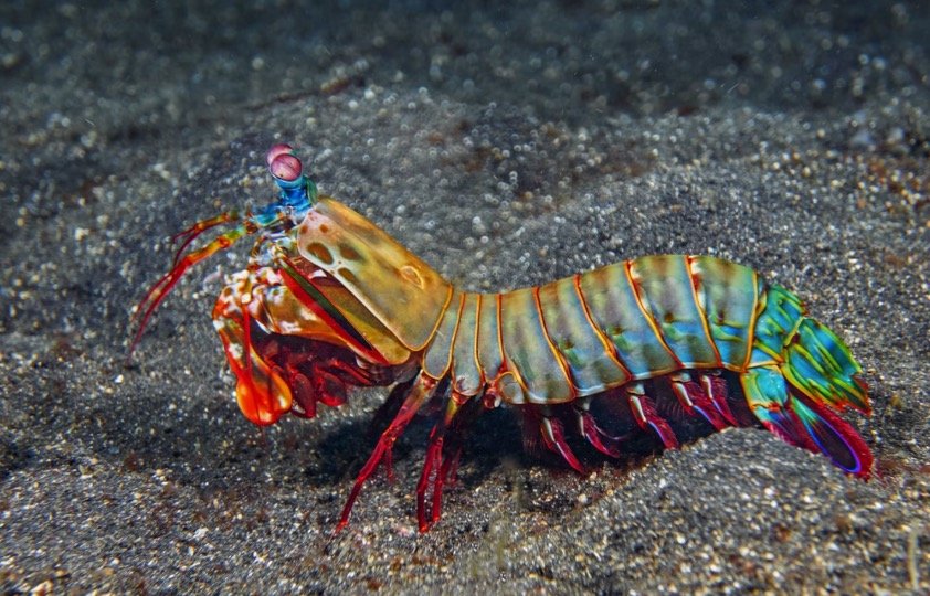
Mantis shrimp are an extremely fascinating group of shrimp species. They are true predators, and take nutrition from tiny organisms or scavenge on dead organisms. The mantis shrimps have modified their forelimbs to shaft or stun their prey. Based on their features, mantis shrimps are divided into subgroups.
The forelimbs are used as clubs by “smashers”. They can create forces unequaled in the animal world due to the massive power behind their modified forelimbs. The club can reach speeds of nearly 50 mph in a fraction of a second and over 1,500 newtons force created by this, which is similar to a 340-pound object falling directly onto the prey.
But, a sonic wave is also created by the acceleration of the club that smashes into the prey after the actual club and can cripple much larger prey. Comparatively, the sharp points on their limbs are used by “Spearers” that help them to impale small fish and other prey.
The speed and precision of the strike of Spearers are amazing. Unlike Smashers, the habitat of Spearers includes soft substrates such as sand. They wait for a fish to swim by and impale it at lightning speed.
The coloration of most mantis shrimp is very beautiful and striking, which helps them in communication and camouflage. The color of some species is very dark that helps them in blending with the seafloor.
The peacock mantis shrimp is very bright colored that helps it to scare off any predator. If the camouflage doesn’t work, many potential predators get injured or scared by their powerful strike.
Fun Facts About Mantis Shrimp
The mantis shrimp is not cool enough, but there are many interesting biological concepts related to these organisms. Let’s take a closer look.
i. Hunting Adaptations
We can see the adaptations made by crustaceans by the two different types of mantis shrimps- Smashers and Spearers. However, both types were evolved from a common ancestor nearly 200 million years ago, but the use of front limbs is quite different in both.
The direct force is used by the Smashers by their mantis-like front legs to strike their prey, on the other hand, Spearers modified their front legs into sharp barbs to impale and capture prey.
The prey is also different in both groups. Spearers usually prey upon soft, fleshy fish, while Smashers eat other crustaceans, snails, and oysters. The shells of these animals can be broken apart from their club-like limbs so they can get the nutrients inside.
Maybe the process of speciation in this group started by the difference in usage and prey type, which may have been the driving factor. Today, mantis shrimp represents 450 different species found in coastal regions all over the world.
ii. Compound Eyes
To become an incredible predator, mantis shrimps made another adaptation besides their weapon-like appendages. Like many arthropods, mantis shrimps consist of compound eyes. The function of these eyes is similar to human eyes, though the construction is very different.
The receptor cells of these eyes are right at the surface, instead of a single lens that funnels light onto a retina. The eyes of mantis shrimp are considered the most complex and functional eyes of any animal.
In humans, different colors and wavelengths of light can be detected by 3 types of cells, mantis shrimp have up to 16 different types of cells in their eyes. Due to this, several wavelengths that are far beyond human perception can be seen by mantis shrimp.
It can also detect both infrared and ultraviolet light, which cannot be seen by humans. It also provides a clue about the bright colors of mantis shrimp.
iii. Coloration as Communication
The coloration of many organisms helps them in communication. For instance, many insects including bees display “warning coloration”, which is recognized as dangerous by other animals. Similarly, peacocks and other animals use colorful coloration to attract mates.
Over time, females select more colorful mates, which leads to the birth of very colorful males and females. Mantis shrimp seem to be using a little of both. They use bright coloration to warn their potential predators.
However, according to some researches, mantis shrimps use bright coloration to attract mates and communicate with other members. Some species signal to their mates and other shrimp invading their space by using fluorescence.
The mantis shrimp can see patterns and colors of light, which is not visible to the human eye due to their amazing compound eyes. However, the exact mechanism and process of communication via colorful signals is not fully understood.
The reason behind this is the need for complex scientific instruments to measure what light is displayed by the mantis shrimp and how they process light signals.
The slowing down of enzyme-catalyzed chemical reactions is called allosteric inhibition. The proper functioning and maintenance of our bodies’ equilibrium are done by these metabolic processes and the process is regulated by allosteric inhibition.
All the important molecules are broken and built up by these metabolic processes. From the digestion of food to the repairing of our muscles, the metabolic processes are fundamental for everything.
Lock and Key: Substrate Binds to Enzyme at the Active Site
A series of chemical reactions consist of metabolic processes, which produce end products. Enzymes are the key drivers of metabolic processes. The reactions are catalyzed by these specific proteins. These enzymes reduce the amount of required energy and speed up important chemical reactions.
First, an enzyme bind to a substrate. A product is then created by this reaction. At the next metabolic step, the product serves as a subsequent substrate for a different enzyme. Finally, until a final product is created at the end, the chain of reactions occurs.
The interesting fact is that an enzyme binds to a specific substrate. Thus the enzyme can be compared to a lock and the substrate is compared with the key. A specific enzyme only binds with a specific substrate. The location, where an enzyme binds to a substrate is called the “active site”.
The wrong key will not fit the specific lock on an enzyme. The enzyme cannot catalyze a reaction if the substrate cannot fit into an active site. Even if the substrate is correct for the specific enzyme, an allosteric inhibitor can prevent the enzyme from having the correct conformation.
Allosteric Inhibition and Enzymatic Activity
Allosteric inhibition is used to control the speed of metabolic reactions. They deactivate the enzyme and thus slow down the enzymatic activity. A molecule that binds to the enzyme at an allosteric site is called an allosteric inhibitor. This site is located at a different location from the active site.
The enzyme changes its 3D shape after binding with the inhibitor. Allosteric inhibition is a form of noncompetitive inhibition. It means that at the active site, the inhibitor does not compete directly with the substrate but indirectly changes the composition of the enzyme.
The enzyme becomes inactive after changing its shape and cannot bind with its corresponding substrate. The formation of subsequent products will be slowed down by this.
The allosteric inhibitor can be compared with a locksmith. The lock (enzyme) is changed by the locksmith (allosteric inhibitor) so that the key (substrate) will no longer be able to open the lock (enzyme).
Allosteric Inhibition Prevents the Over-accumulation of Products
Allosteric inhibitors prevent the body from creating unnecessary products and wasting energy. If a metabolic pathway is compared with an assembly line at a factory, a machine alters the products at each station in the assembly line before passing it to the nest station.
Then the intermediate products are moved from station to station by the assembly line until they get the final product at the end. For example, pants are produced in this factory. At the first station, a pair of pants are cut from raw material by a machine, then the hems of the pants are stitched together by a machine at the second station.
At the third station, the zippers are attached by a machine, and finally, at the fourth station, the products (pants) are tagged and shipped in the shipment pile. The assembly line is smooth and there is no hold-up when the machines are working properly and stations will produce products at similar rates.
However, if the machine at the third station breaks down that attaches the zippers, there is now a hold-up. We must stop the first two stations of the supply to prevent products from piling up.
In this way, the supply and demand for each intermediate product are controlled and it is ensured that they are equal at each station. Similarly, the chain of reactions can be slowed down by allosteric inhibition. In this way, the over-accumulation of unnecessary products is prevented.
Examples of Allosteric inhibition
ATP in cellular respiration is an example of an allosteric inhibitor. This metabolic process is operated as a feedback loop. The speed of upstream reactions is controlled by downstream products. Phosphofructokinase is an enzyme involved in glycolysis. It converts ADP into ATP.
The ATP serves as an allosteric inhibitor when there is too much ATP in the system. The ATP slows down the conversion of ADP by binding with phosphofructokinase. Thus, ATP prevents its unnecessary production by itself because when there are already adequate amounts of ATP, it is no need to produce more ATP.
The antibiotic penicillin, which is an important drug acts as an allosteric inhibitor. Penicillin has saved millions of lives by helping the body to kill harmful bacteria. The enzyme DD-transpeptidase helps the harmful bacteria to create strong cell walls.
Penicillin binds to this enzyme and counteracts this process. Penicillin inhibits the formation of bacterial cell walls by acting as an inhibitor. The surrounding fluids of the bacterial cell can then push itself in through osmosis with a weak wall and the cell burst and die.