Author: Admin

Author: Admin

  • Fungi: Definition, Characteristics, Types, and Facts

    What are Fungi?

    o Fungi represent a distinct kingdom of organisms with tremendous diversity and are eukaryotes but have characteristics of both prokaryotes and eukaryotes (such as a cell wall).

    o Three divisions exist within this kingdom: Zygomycota, Ascomycota and Basidiomycota.

    o Fungi, like plants, are separated into divisions not phyla.

    o If something contains “mycota” in its division then it’s a fungus.

    o All cells of Zygomycota are haploid, except for the zygospore and don’t have cell walls except in their sexual structures.

    o Oomycota, which are slime molds and water molds, are not true fungus but are part of the Protista kingdom.

    o All fungi are eukaryotic heterotrophs that obtain their food by absorption rather than by ingestion: they secrete their digestive enzymes outside their bodies and then absorb the products of digestion.

    o Although most fungi are considered saprophytic (live off dead matter), many fungi do not distinguish between living and dead matter, and thus can be potential pathogens.

    o Fungi are larger than bacteria.

    Characteristics of Fungi

    o With the exception of yeasts, fungi are multicellular.

    o Yeasts are unicellular fungi.

    o Most fungi possess cell walls, called septa, made up of the polysaccharide, chitin.

    o Chitin is more resistant to microbial attack than is cellulose.

    o Arthopods’s (insects and crustaceans) exoskeleton is made up of chitin as well.

    o Septa is usually perforated to allow exchange of cytoplasm between cells, called cytoplasmic streaming.

    o This allows for rapid growth.

    o A fungal cell can contain multiple nucle.

    o The nuclei, in a single cell, may or may not be identical.

    o Fungi lack centrioles (as well as prokaryotes), mitosis takes place entirely within the nucleus, and their nuclear membranes don’t disappear during mitosis.

    o In the growth state, fungi consist of a tangle mass (called mycelium) of multiply branched thread-like structures called hyphae (haploid).

    Fungi Structure

    Fungi, Fungi Definition, Fungi Characteristics, Fungi Types, Fungi Facts 1

    Fungi Reproduction and Life Cycle

    o Like most organisms, fungi alternate between haploid and diploid stages in their life cycle; however, the haploid stage predominates, and is their growth stage.

    o Hyphae are haploid and some hyphae may form reproductive structures called sporangiophores.

    o By far the most important type of asexual reproduction is that of spore formation.

    o Asexual reproduction is extremely important to fungi.

    o It is responsible for the production of large numbers of spores throughout the year.

    o These structures release haploid spores that give rise to new mycelia in asexual reproduction.

    o Spore formation is NOT always via asexual reproduction.

    o Haploid spores can form and spread faster and more efficiently than diploid zygotes because they don’t undergo meiosis.

    o Yeasts rarely reproduce sexually by producing spores.

    o More often in yeasts, asexual reproduction occurs via budding.

    o Also called cell fission, in which a smaller cell pinches off from the single parent cell.

    "Sporangiophores release haploid spores that give rise to new mycelia in asexual reproduction"

    o When sexual reproduction occurs it is between hyphae from two mycelia of different mating types.

    o These two hyphae (haploid) grow towards one another eventually touching and forming a conjugation bridge, and becomes a gamete producing cell, called a gametangium.

    o In Zygomycota, the gametangia remain attached to the parent hyphae and the nuclei fuse with one another to produce a diploid zygote, called a zygospore.

    o After its formed it usually goes dormant, but when it is activated, the zygospore undergoes meiosis to produce haploid cells, one of which immediately grows a short sporangiophore to asexually reproduce many spores.

    o Sporangiophores release haploid spores that give rise to new mycelia in asexual reproduction.

    o Except for the zygospore all cell in Zygomycota are haploid, so they undergo mitosis.

    "When sexual reproduction occurs it is between hyphae from two mycelia of different mating"

    o The important thing to understand about fungal reproduction is that asexual reproduction.

    o Ex. Budding or mitosis normally occur when conditions are good.

    o Sexual reproduction (meiosis) occur when conditions are tough.

    Haploid state ⇒ asexual reproduction (mitsosis/budding (cell fission)) ⇒ conditions are good

    o Diploid state (zygospore) ⇒ sexual reproduction (meiosis) ⇒ conditions are bad.

    o Homologous chromosomes are chromosomes in a biological cell that pair (synapse) during meiosis.

    o The pair are non-identical chromosomes that both contain information for the same biological features and contain the same genes at the same loci but possibly each have different alleles (that is, different genetic information) at those genes.

    o Any cell that contains homologous chromosomes are called diploid.

    o Any cell that doesn’t contain homologous chromosomes are called haploid.

    o Lichens are composite organisms consisting of a symbiotic association of a fungus with a photosynthetic partner usually either a green alga or cyanobacterium.

    o The mixture of organisms regularly found at any anatomical site is referred to as the normal flora.

    Fungi Citations

    Share

    Similar Post:

    Continue Reading
  • Endospores: Definition, Function, and Diagram

    What is Endospore Formation?

    o An endospore is a dormant, tough, and non-reproductive structure produced by Gram-positive bacteria which forms when a bacterium produces a thick internal wall that encloses its DNA and part of its cytoplasm.

    o Remember that Gram + bacteria produce endospores.

    o The primary function of most endospores is to ensure the survival of a bacterium through periods of environmental stress.

    o They are therefore resistant to ultraviolet and gamma radiation, desiccation, lysozyme, temperature, starvation, and chemical disinfectants.

    Endospore Formation

    Endospore, Endospore Definition, Endospore Function, Endospore Diagram 1

    Endospore Formation Steps

    o In endospore formation, the bacterium divides within its cell wall.

    o One side then engulfs the other side.

    o The chemistry of the cell wall of the engulfed bacterium changes slightly to form the cortex of the endospore.

    o Several protein layers lie over the cortex to form the resistant structure called the spore coat.

    o A delicate covering called the exosporium, sometimes surrounds the spore coat.

    oThe outer cell then lyses, releasing the dormant endospore.

    o The endospore must be activated before it can be germinated and grow.

    o Activation usually involves heat.

    o Germination is triggered by nutrients.

    Endospore Formation Citations

    Share

    Similar Post:

    Continue Reading
  • Bacterial Growth Curve: Definition, Stages, and Graph

    Bacterial Growth Curve

    Bacterial growth in batch culture can be modeled with four different phases: lag phase, exponential or log phase, stationary phase, and death phase.

    Bacterial Growth Curve

    Bacterial Growth, Bacterial Growth Curve, Bacterial Growth Definition, Bacterial Growth Stages, Bacterial Growth Graph 1

    I. Lag Phase

    o During lag phase, bacteria adapt themselves to growth conditions.

    o It is the period where the individual bacteria are maturing and not yet able to divide.

    o During the lag phase of the bacterial growth cycle, synthesis of RNA, enzymes and other molecules occurs.

    II. Exponential Phase

    o Exponential phase (sometimes called the log phase) is a period characterized by cell doubling.

    o The number of new bacteria appearing per unit time is proportional to the present population.

    o If growth is not limited, doubling will continue at a constant rate so both the number of cells and the rate of population increase doubles with each consecutive time period.

    o For this type of exponential growth, plotting the natural logarithm of cell number against time produces a straight line.

    o The slope of this line is the specific growth rate of the organism, which is a measure of the number of divisions per cell per unit time.

    o The actual rate of this growth (i.e. the slope of the line in the figure) depends upon the growth conditions, which affect the frequency of cell division events and the probability of both daughter cells surviving.

    o Exponential growth cannot continue indefinitely, however, because the medium is soon depleted of nutrients and enriched with wastes.

    III. Stationary Phase

    o During stationary phase, the growth rate slows as a result of nutrient depletion and accumulation of toxic products.

    o This phase is reached as the bacteria begin to exhaust the resources that are available to them.

    o This phase is a constant value as the rate of bacterial growth is equal to the rate of bacterial death.

    IV. Death Phase

    At death phase, bacteria run out of nutrients and die.

    Bacterial Growth Curve Citations

    Share

    Similar Post:

    Continue Reading
  • Bacterial Conjugation: Steps, Definition, and Diagram

    Bacterial Reproduction: Bacterial Conjugation

    o Bacteria (prokaryotes) CAN’T undergo meiosis or mitosis, don’t have centrioles, and they can ONLY undergo Asexual Reproduction.

    o The type of cell division they are capable of undergoing is called binary fission, which exhibits exponential growth.

    o In binary fission, the circular DNA is replicated in a process similar to replication in eukaryotes.

    o Two DNA polymerases begin at the same point on the circle (ORI) and move in opposite directions making complementary single strands that combine with their template strands to form two complete DNA double stranded circles.

    o The cell then divides, leaving one circular chromosome in each daughter cell.

    o The daughter cells are genetically identical.

    Genetic Recombination

    There are three types of genetic recombination that can occur in bacterium:

    I. Bacterial Conjugation

    o Bacterial Conjugation requires that one of the bacterium have a plasmid with the gene that codes for the sex pilus.

    o If the plasmid can integrate into the chromosome of the host it’s called an episome.

    o In order for a bacterium to initiate conjugation, it must contain a conjugative plasmid.

    o Conjugative plasmids possess the gene for the sex pilus.

    o The sex pilus a hollow protein tube that connects the two bacteria to allow the passage of DNA.

    o The plasmid replicates differently than the circular chromosome.

    o One strand is nicked, and one end of this strand begins to separate from its complement as its replacement is replicated.

    o The loose strand is then replicated and fed through the pilus.

    o Two plasmids of interest are: the F plasmid and the R plasmid.

    o The F plasmid is called the fertility factor or F factor.

    o It allows the production of the sex pilus.

    o A bacterium with an F factor is designated as F+, and one without it is designated F-.

    o The F plasmid can be in the form of an episome and if the sex pilus is made while the F factor is integrated into the chromosome, then some or the entire chromosome may be replicated and transferred.

    Bacterial Conjugation Diagram

    Bacterial Conjugation, Bacterial Conjugation Steps, Bacterial Conjugation Definition, Bacterial Conjugation Diagram 1

    o R-plasmids often contain resistance genes coding for multiple antibiotic resistance.

    o As well as resistance transfer genes, they also code for the production of a conjugation (sex) pilus.

    o The conjugation pilus enables the donor bacterium to transfer a copy of the R-plasmid to a recipient bacterium, making it also multiple antibiotic resistant and able to produce a conjugation pilus.

    o Conjugation is conservative because the donor retains a complete original copy of the plasmid after the transfer is complete.

    II. Bacterial Transformation

    Transformation is the process by which bacteria may incorporate DNA from the external environment into their genome.

    III. Bacterial Transduction

    o Sometimes the capsid of a bacteriophage will mistakenly encapsulate a DNA fragment of the host cell.

    o When these new virions infect a new bacterium, they inject harmless bacterial DNA fragments instead of virulent viral DNA fragments.

    o This type of genetic recombination is called transduction.

    o The virus mediates that mediates transduction is called the vector.

    o This can be done artificially in a lab.

    o Mnemonic for transduction: the bacteriophage induces a change.

    Bacterial Conjugation Citations

    Share

    Similar Post:

    Continue Reading
  • Bacterial Cell Wall Structure: Gram Positive vs...

    Bacterial Cell Wall

    o The bacterial plasma membrane and everything inside of it is called the protoplast.

    o A protoplast isn’t a complete bacterium.

    o Protoplast: Plant, bacterial or fungal cell with the cell wall removed using either mechanical or enzymatic means.

    o Surrounding the protoplast is the bacterial envelope.

    o The component of the envelope, adjacent to the plasma membrane is the cell wall.

    o One of the functions of the cell wall is to prevent the protoplast from bursting.

    o Most bacteria (prokaryote) are hypertonic (hypotonic solution) to their environment (this means that the aqueous solution of their cytosol contains more particles than the aqueous solution surrounding them.

    o The resulting osmotic pressure causes a net movement of water into of the cell).

    o Compare isotonic where the cytosol contains the same amount of particles and hypotonic (hypertonic solution) where the cytosol contains less particles.

    o The cell wall is strong and able to withstand high pressure.

    o As the cell fills with water and the hydrostatic pressure builds, it eventually equals the osmotic pressure and the filling stops.

    o If the cell wall is removed, the plasma membrane cannot withstand the pressure.

    o Osmosis describes the movement of water.

    Peptidoglycan

    o Peptidoglycan, also known as murein, is a polymer consisting of sugars and amino acids that forms a mesh-like layer outside the plasma membrane of bacteria, forming the cell wall.

    o The polymers are crosslinked by an interbridge of more amino acids.

    o It is porous, so it allows large molecules to pass through.

    Structure and Synthesis of Peptidoglycan

    Peptidoglycan Cell wall, Peptidoglycan Structure, Peptidoglycan Synthesis, Gram Positive vs Gram Negative

    o Archaea don’t have peptidoglycan cell walls.

    o Peptidoglycan is more elastic than cellulose, which as we recall is carbohydrate formed by plants and contains Beta linkages (only bacteria eat Beta linkages and is part of the plants cell wall).

    o Some antibacterial drugs such as penicillin interfere with the production of peptidoglycan by binding to bacterial enzymes which make the crosslinks.

    "One of the functions of the cell wall is to prevent the protoplast from bursting"

    o Lysozyme, an enzyme produced naturally by humans, attacks the crosslinks as well.

    o In both cases the cell wall is disrupted and the cell lyses killing the bacterium.

    o Don’t confuse this with a lysosome which are organelles containing digestive enzymes

    o Capsules: This type of surface layer is composed primary of polysaccharides.

    o If the layer is strongly adhered to the cell wall, it is called a capsule; if not, it is called a slime layer.

    o Not all bacteria have this.

    o These layers provide resistance to phagocytosis and serve as antigenic determinants.

    o One method of classification of bacteria is according to the type of cell wall that they possess.

    o A staining technique, called gram staining, used to prepare bacteria for viewing under the light microscope, stains two major cell walls differently

    Gram Positive Bacteria

    o The first type is called gram-positive bacteria.

    o Gram-positive bacteria are those that are stained dark blue or violet by Gram staining.

    o This is in contrast to Gram- negative bacteria, which cannot retain the crystal violet stain, instead taking up the counterstain and appearing red or pink.

    o Gram-positive organisms are able to retain the crystal violet stain because of the high amount of peptidoglycan in the cell wall.

    o Thick peptidoglycan layer.

    Gram Positive Cell Wall
    Gram Positive Bacteria - Gram Positive Bacteria Cell wall - Gram Positive Bacteria Diagram - Gram Positive Bacteria Definition

    o Stain blue/violet.

    o The space between the peptidoglycan layer and the plasma membrane is known as the periplasmic space and it contains proteins that help the bacteria acquire nutrition.

    Gram Negative Bacteria

    o Gram-negative bacteria appear red or pink in color when gram stained.

    o Many species of Gram-negative bacteria are pathogenic, meaning that they can cause disease in a host organism.

    o This pathogenic capability is usually associated with certain components of Gram-negative cell walls, in particular the lipopolysaccharide (also known as LPS or endotoxin) layer.

    Gram Negative Cell Wall
    Gram Negative Bacteria - Gram Negative Bacteria Cell wall - Gram Negative Bacteria Diagram - Gram Negative Bacteria Definition

    o The following characteristics are displayed by Gram-negative bacteria:

    1. Thin peptidoglycan layer (which is present in much higher levels in Gram-positive bacteria)

    2. Outer membrane containing lipopolysaccharide (LPS) (can form a protective barrier from antibodies and many antibiotics) outside the peptidoglycan layer, this outer membrane is also more permeable than the inner, even allowing molecules the size of glucose to pass through

    3. Porins exist in the outer membrane, which act like pores for particular molecules

    4. A lipoprotein in the outer membrane called Braun’s lipoprotein points inward toward the cell wall and attaches covalently to the peptidoglycan

    5. There are two spaces between the layer of peptidoglycan and the two membranes

    6. Stain red/pink

    7. The periplasmic space is the space between the two membranes.

    o Some gram-negative bacteria possess fimbrie or pili (not to be confused with the sex pilus discussed below).

    o Fimbriae are short tentacles that can attach a bacterium to a solid surface.

    o They are NOT involved in cell motility.

    "Outer membrane is also more permeable than the inner, even allowing molecules the size of glucose to pass through"

    o Bacterial flagella are long, hollow, rigid, helical cylinders made from a globular protein called flagellin; these shouldn’t be confused with eukaryotic flagella which are made up of microtubules.

    o They rotate counterclockwise.

    o When they are rotated clockwise, the bacterium tumbles.

    o This tumbling acts to change the orientation of the bacterium allowing it to move forward in a new direction.

    o The movement of a bacterium toward or away from a particular stimulus is called taxis.

    o Such stimuli include chemicals (chemotaxis) and light (phototaxis).

    o The flagellum is propelled using the energy from a proton gradient rather than by ATP.

    Bacterial Cell Wall Citations

    Share

    Similar Post:

    Continue Reading
  • Membrane Transport: Protein, Examples, and Types

    Membrane Transport

    o Concentration of ions inside and outside a cell are very different:

    o Na+ is more plentiful outside the cell

    o Cl- is more plentiful outside the cell

    o Ca+2 and Mg+2 are more plentiful outside the cell

    o K+ is more plentiful inside the cell

    o A couple rules dictate the permeability of solutes and ions:

    1) The smaller the molecule the more permeable it is.

    2) the lower the polarity, the more permeable it is.

    3) Lipid bilayers are impermeable to all ions and charged molecules, no matter how small they are.

    o Cell membranes allow water and small nonpolar molecules (including steroids) to permeate by simple diffusion (dictated by the chemical concentration gradient).

    o For molecules with a charge there is also an electrical gradient pointing in the direction that a positively charged particle will tend to move.

    o The two gradients can be added to form a single electrochemical gradient.

    o The electrochemical gradient for compound X points in the direction that particle X will tend to move.

    o Heat and pressure also affect the way particles diffuse.

    o In strict terms, diffusion occurs in the direction of decreasing free energy and in increasing universal entropy.

    o The ion flow changes the voltage across the membrane.

    o The membrane potential, thus altering the electrochemical driving forces for the transmembrane movement of all the other ions.

    Examples of Membrane Transport

    Membrane Transport, Membrane Transport Protein, Active Transport, Diffusion, Facilitated Diffusion, Membrane Transport Examples, 2

    o If compounds X and Y are separated by an impermeable membrane, diffusion is stopped.

    o However, if the molecules of X can wiggle their way across the membrane then diffusion is only slowed.

    o Since the membrane slows the diffusion of X, but doesn’t stop it, the membrane is said to be semipermeable to compound X.

    o Facilitated diffusion is also said to make the membrane semipermeable.

    o Most of the diffusion of polar and charged molecules across a natural membrane takes place through incidental holes (sometimes called leakage channels) created by the irregular shapes of integral proteins.

    o This is passive diffusion (down its electrochemical gradient).

    Types of Membrane Transport Proteins

    1) Carrier Proteins:

    a) allows passage only to solute molecules that fit into the binding site on the protein. Bind their solutes with great specificity much like how enzymes do

    2) Channel Proteins:

    a) discriminates mainly on the basis of size and electric charge

    b) Most of the channel proteins in the plasma membrane of animal and plant cells are quite different and have narrow, highly selective pores.

    o Almost all of these proteins are ion channels.

    o Concerned exclusively with the transport of inorganic ions, mainly Na+ , K+, Cl-, and Ca+2

    o There are three types of ion channels:

    i. Voltage gated ion channels: probability of being open is determined by the membrane potential.

    o All living cells have gated sodium-potassium ATPases and/or other pumps that are voltage dependent

    ii. Ligand gated ion channel: controlled by the binding of some molecule (the ligand) to the channel protein.

    iii. Stress-activated channel: opening is controlled by a mechanical force applied to the channel.

    o Ex. In your ear

    o Ion channels are either fully open or fully closed.

    Facilitated Diffusion

    o Facilitated diffusion is diffusion in which the molecule cant naturally diffuse (because of its size, polarity or both) across the membrane so one of these membrane transport proteins assists it.

    o It is still down the electro-chemical gradient however.

    o Most, but not all, human cells rely on facilitated diffusion for their glucose supply.

    o Only certain epithelial cells in the digestive tract and the proximal tube of the kidney are capable of absorbing glucose against a concentration gradient.

    o This is done via a secondary active transport mechanism down the concentration gradient of sodium.

    o In the absence of insulin, only neural and hepatic cells are capable of absorbing sufficient amounts of glucose via the facilitated transport system.

    Passive Transport
    Passive Transport, What is Passive Transport, Passive Transport Definition, Passive Transport vs Active Transport,

    o Facilitated diffusion is said to make the membrane selectively permeable because it is able to select between molecules of similar size and charge.

    o To move a solute against its concentration gradient, a transport protein must do work: it has to drive the “uphill” flow by coupling it to some other process that provides energy (usually via ATP hydrolysis or through coupling it with a “downhill” movement). This is called active transport.

    o It can also be accomplished indirectly by using ATP to create an electrochemical gradient, and then using the energy of the electrochemical gradient to acquire or expel a molecule.

    o The latter method is called secondary active transport.

    o Whenever we are moving against an electrochemical gradient it MUST be active transport

    "Ion channels are either fully open or fully closed"

    o For coupling transport of two molecules:

    o If both solutes move in the same direction its called symport.

    o If they move in opposite directions its called antiport.

    o If a carrier protein ferries only one type of molecule across the membrane (and is therefore not a couple transporter) its called a uniport.

    Ion Transporters
    Membrane Transport, Membrane Transport Protein, Active Transport, Diffusion, Facilitated Diffusion, Membrane Transport Examples, 1

    o Under normal conditions, the interior of most cells is at a negative electrical potential compared to the exterior, so that positive ions tend to be pulled into the cell.

    o Thus the inward electrochemical driving force for Na+ is large, as it includes the driving force due to the concentration gradient and a driving force in the same direction to the voltage gradient.

    o Therefore to maintain this balance we need a pump which pumps out Na+.

    o This is achieved through the Na+-K+ pump.

    o It couples the export of Na+ to the import of K+ and hydrolyzes ATP to do so.

    o The Na+-K+ pump helps to maintain osmotic balance of pressure in a cell so it wont flood with H2O and burst.

    Membrane Transport Citations

    Share

    Similar Post:

    Continue Reading
  • Cell Membrane / Plasma Membrane: Structure and...

    Plasma Membrane / Cell Membrane

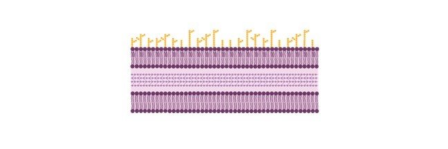
    o The cytosol of nearly all prokaryotes is surrounded by a phospholipid bilayer called the plasma membrane (the membranes of archaea differ in their lipid structure).

    o It gives the cell its basic structure and serves as a permeability barrier.

    o The phospholipid is composed of a phosphate group, two fatty acid chains, and a glycerol backbone.

    o The phospholipid group is polar, while the fatty acid chains are nonpolar making the molecule amphipathic.

    o When placed in aqueous solution, amphipathic molecules spontaneously aggregate, turning their polar ends toward the solution, and their nonpolar ends toward each other.

    o The resulting spherical structure is called a micelle.

    o If enough phospholipids exist, and the solution is subjected to ultrasonic vibrations, liposomes may form.

    o A liposome is a vesicle surrounded and filled by aqueous solution.

    o It contains a lipid bilayer like that of a plasma membrane.

    o The inner and outer layers of a membrane are referred to as leaflets.

    o The level of saturation in the fatty acids of the phospholipids also determines the membranes fluidity; an increase in the unsaturation of these fatty acids increases the fluidity of the membrane.

    o If we increase the temperature as well the membrane fluidity increases.

    o The plasma membrane contains other types of lipids such as glycolipids.

    o Different lipid types are arranged asymmetrically between the leaflets.

    o For instance, glycolipids are found on the outer leaflet only.

    o Unlike eukaryotic membranes, prokaryotic membranes usually DON’T contain steroids such as cholesterol.

    Instead, some bacterial membranes contain steroid like molecules called hopanoids.

    o Cholesterol tends to stiffen the bilayer, making it more rigid and less permeable.

    o Hoponoids probably reduce the fluidity of the membrane in Prokaryotes.

    o In eukaryotes nearly all new membrane synthesis occurs in the ER.

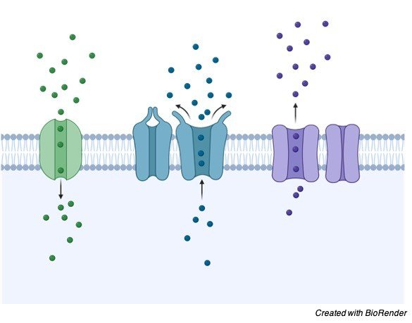
    o Also embedded within the plasma membrane are proteins.

    o Most of the functional aspects of membranes are due to their proteins.

    o Membrane proteins act as transporters, receptors, attachment sites, and enzymes.

    Organization of Plasma Membrane

    Cell Membrane, Cell Membrane Function, Cell Membrane Structure, What is Cell Membrane,

    Types of Protein in Plasma Membrane

    o Two types of proteins are involved with the plasma membrane:

    1) Integral Proteins: Ampipathic proteins that transverse the membrane from inside the cell to the outside.

    o Are usually ion channels.

    2) Peripheral or Extrinsic Proteins: are situated entirely on the surfaces of the membrane.

    o They are ionically bonded to integral proteins or the polar group of a lipid.

    o Both integral and peripheral proteins may contain carbohydrate chains making them glycoproteins and the carbohydrate portion always protrudes toward the outside of the cell.

    o Proteoglycans also exist on the membrane.

    o It can thus form a carbohydrate layer (glycocalyx) which protects the cell surface from chemical and mechanical damage.

    o Proteoglycans are also a mixture of proteins and carbohydrates, but they generally consist of more than 50% carbohydrates.

    o Proteoglycans are a major component of extracellular matrix.

    o Glycoproteins are proteins with a carbohydrate group attached and they are a component of cellular plasma membranes.

    o Also serve as markers for cellular recognition.

    o Lipoproteins also exist in some plasma membranes with their lipid portions embedded in the membrane and their protein portions at the surfaces.

    o Membrane proteins are distributed asymmetrically throughout the membrane and between the leaflets.

    o Neither proteins nor lipids easily flip from one leaflet to the other.

    o The shape of the cell and the mechanical properties of the plasma membrane are determined by a meshwork of fibrous proteins, called the cell cortex, that is attached to the cytosolic surface of the membrane.

    o It is made up primarily of spectrin.

    o Since the forces holding the entire membrane are intermolecular, the membrane is fluid; its parts can move laterally but can’t separate.

    o The model of the membrane as just described is known as the fluid mosaic model.

    Plasma Membrane Citations

    Share

    Similar Post:

    Continue Reading
  • Prokaryotes: Prokaryotic Cells, Structure, Organization, and Facts

    What are Prokaryotes

    o Prokaryotes don’t have a membrane bound nucleus.

    o They are split into two distinct domains called Bacteria and Archaea.

    o Archaea have as much in common with eukaryotes as they do with bacteria.

    o They are typically found in the extreme environments such as salty lakes and boiling hot springs.

    o Unlike bacteria, the cell walls of archaea are not made from peptidoglycan.

    o Most known prokaryotes are members of the domain Bacteria.

    o The introduction of the two domains makes the kingdom Monera obsolete.

    o The kingdom Monera was the kingdom containing all prokaryotes.

    o In order to grow; all organisms require the ability to acquire carbon, energy and electrons (usually from hydrogen).

    o Organisms can be classified according to the sources from which they gather these commodities.

    o A carbon source can be organic or inorganic.

    o Most carbon sources also contribute oxygen and hydrogen.

    o CO2 is a unique inorganic carbon source because it has no hydrogens.

    o To some degree, all microorganisms are capable of fixing CO2 (reducing it and using the carbon to create organic molecules usually through a process called the Calvin Cycle).

    "Autotrophs are organisms that are capable of using CO2, as their sole source of carbon"
    "Heterotrophs use preformed, organic molecules as their source of carbon"
    Classifications

    o Organisms can be classified as well according to how they get their energy:

    1) Phototrophs use light as their energy source.

    o Ex. Cyanobacteria: blue/green algae that uses photosynthesis.

    o Only prokaryotes can acquire energy from an inorganic source other than light.

    2) Chemotrophs use the oxidation of organic and inorganic matter as their energy source.

    o Electrons or hydrogens can be acquired from inorganic matter by lithotrophs, or organic matter by organotrophs.

    o You can recall this one because lithium is an inorganic molecule and lithotrophs get their hydrogens from inorganic molecules.

    o Humans are chemoheterotrophs.

    All organisms can be classified as one of each of the three types.

    o Bacteria are found in all classifications.

    o Some bacteria are capable of fixing nitrogen.

    o Atmospheric nitrogen is abundant, but in a strongly bound form that is useless to plants.

    o Nitrogen fixation is the process by which N2 is converted to ammonia.

    o Most plants are unable to use ammonia however and must wait for other bacteria to further process the nitrogen in a process called nitrification.

    o Nitrification is a two step process that creates NITRATES, which are useful to plants, from ammonia.

    o Nitrification requires two genera (genus’s) of chemoautotrophic prokaryotes.

    o Nitrification is the biological oxidation of ammonia with oxygen into nitrite followed by the oxidation of these nitrites into nitrates.

    o Degradation of ammonia to nitrite is usually the rate limiting step of nitrification.

    o Genus – (plural: genera) taxonomic group containing one or more species.

    o Chemoautotrophy is an inefficient mechanism for acquiring energy, so chemoautotrophs require large amounts of substrate.

    o All known chemoautotrophs are prokaryotes.

    o Only prokaryotes can acquire energy from an inorganic source other than light.

    Structure of Prokaryotes

    o Prokaryotes have a single, circular double stranded molecule of DNA.

    o This molecule is twisted into supercoils and is associated with histones in Archaea and with proteins that are different from histones in bacteria.

    o The DNA, RNA and protein complex in prokaryotes forms a structure visible under the light microscope called a nucleoid (also called the chromatin body, nuclear region, or nuclear body).

    o The nucleoid is not enclosed by a membrane.

    Structure of Bacteria

    o There are three major shapes of bacteria:

    1) cocci (round)

    2) bacilli (rod shaped)

    3) spiral

    o There are other shapes, including helical.

    o Helically shaped bacteria are called spirilla, if they are rigid. Otherwise they are called spirochetes.

    o Certain species of spirochetes may have given rise to eukaryotic flagella through a symbiotic relationship.

    o The name of the bacteria often reveals the shape.

    o Prokaryotes have no complex, membrane-bound organelles.

    o The key words are ‘complex’ and ‘membrane bound’.

    o They have organelles: ribosomes, nucleoids, and mesosomes etc….; just not complex membrane-bound organelles like mitochondria, ER, Golgi, etc.

    o Prokaryotic ribosomes are smaller than eukaryotic ribosomes.

    o They are made from a 30S subunit and a 50S subunit to form a 70S subunit.

    o A prokaryote may or may not contain a mesosome.

    o Mesosomes are invaginations of the plasma membrane and can be seen under the light microscope.

    o Their exact function is unknown, but may be involved in cell wall formation during cell division.

    o Prokaryotes also have inclusion bodies.

    o Inclusion bodies are granules of organic or inorganic matter that may be visible under a light microscope.

    o Inclusion bodies may or may not have a membrane.

    Prokaryotes Citations

    Share

    Similar Post:

    Continue Reading
  • Viruses: Structure, Function, and Facts

    What are Viruses

    o Viruses are tiny infectious agents, much smaller than bacteria.

    o They are comparable in size to large proteins.

    o In its most basic form, a virus consists of a protein coat, called a capsid, and from one to several hundred genes in the form of DNA or RNA inside the capsid.

    o No virus contains both DNA and RNA.

    o Most animal viruses, some plant viruses, and very few bacterial viruses surround themselves with a lipid-rich envelope either borrowed from the membrane of their host cell or synthesized in the host cell cytoplasm.

    o The envelope typically contains some virus-specific proteins.

    o The capsid usually accounts for most of the weight of the virus.

    o A mature virus outside the host cell is called a virion.

    Viruses Types and Structures

    Viruses, Viruses Structure, Viruses Function, Viruses Types, Viruses Facts 2

    o Viruses are not currently classified as living organisms; they do not belong to any of the taxonomical kingdoms of organisms.

    o Viruses differ from living organisms in the following ways.

    o They ALWAYS require the host cell’s reproductive machinery in order to replicate.

    o Viruses don’t metabolize organic nutrients.

    o Instead they use the ATP made available by the host cell.

    o Viruses possess either RNA or DNA, but never both.

    o Thus, there are viruses with the familiar double-stranded DNA, with single-stranded DNA, with double-stranded RNA, and with single-stranded RNA.

    o Also the nucleic acid could be linear or circular.

    o Viruses can be crystallized without losing their ability to infect.

    o A viral infection begins when a virus adsorbs to a specific chemical receptor site on the host.

    o The host is the cell being infected.

    o The chemical receptor is usually a specific glycoprotein on the host cell membrane.

    Life Cycle of Viruses

    Viruses, Viruses Structure, Viruses Function, Viruses Types, Viruses Facts 1

    o The virus can’t infect the cell if the specific receptor isn’t available/there.

    o Next, the nucleic acid of the virus penetrates into the cell.

    o In a bacteriophage, a virus that infects bacteria, the nucleic acid is normally injected through the tail after viral enzymes have digested a hole in the cell wall.

    o Most viruses that infect eukaryotes are engulfed by an endocytotic process.

    o They could also be engulfed by a process called membrane fusion, in which the envelope of the virus is actually incorporated into the plasma membrane of the host cell releasing the capsid into the cytoplasm.

    Types of Viruses Infection

    Once inside the cell, there are two possible paths;

    I. Lytic infection

    o The virus commandeers the cell’s reproductive machinery and begins reproducing new viruses.

    o There is a brief period before the first fully formed virion appears.

    o This period is called the eclipse period.

    o The cell may fill with viruses until it lyses or burst, or it may release the new viruses one at a time in a reverse endocytotic process.

    o The period of infection to the lysis is called the latent period.

    o The latent period encompasses the eclipse period.

    o A virus following the lytic cycle is called a virulent virus.

    Lytic and Lysogenic Cycle- Bacteriophage- Definition, Structure, Diagram, and Function - research tweet
    II. Lysogenic infection

    o The viral DNA is incorporated into the host genome, or, if the virus is an RNA virus and it possesses the enzyme reverse transcriptase.

    o DNA is actually reverse-transcribed from RNA and then incorporated into the host cell genome.

    o When a host replicates its DNA, the viral DNA is replicated as well.

    o A virus in a lysogenic cycle is called a temperate virus.

    "The chemical receptor is usually a specific glycoprotein on the host cell membrane"

    o While the viral DNA remains incorporated in the host cell, the virus is said to be dormant or latent, and is called a provirus (a prophage if the host cell is a bacterium).

    o There are 2 important results from the lysogenic cycle:

    1) The cell infected is immune to reinfection by the same phage.

    2) The host cell may exhibit new properties, this is known as phage conversion.

    o The dormant virus may become active when the host cell is under some type of stress.

    o Examples of stress include UV light or carcinogens.

    o When the virus becomes active, it becomes virulent.

    o Exhibits exponential growth b/c each new cell will create more viruses.

    o It is a longer cycle than the lytic cycle.

    Type of Viruses

    o There are many types of viruses.

    o One way to classify them is by the type of nucleic acid that they posses.

    o A virus with unenveloped plus-strand RNA is responsible for the common cold.

    o The “plus-strand” indicates the proteins can be directly translated from the RNA.

    o Enveloped plus-strand RNA viruses include retroviruses such as the virus that causes AIDS.

    o A retrovirus carries the enzyme reverse transcriptase in order to create DNA from its RNA.

    o The DNA is then incorporated into the genome of the host cell.

    o Minus-strand RNA viruses include measles, rabies, and the flu.

    o Minus- strand RNA is the complement to mRNA and must be transcribed to plus-RNA before being translated.

    o There are even double stranded RNA viruses, and single and double stranded DNA viruses.

    o Reassortment – is a method with which virus’ can alter their genetic makeup it occurs If a virus has a segmented genome and if two variants of that virus infect a single cell, progeny virions can result with some segments from one parent, some from the other.

    o Viroids are a related form of infectious agent.

    o Viroids are small rings of naked RNA without capsids.

    o Viroids only infect plants.

    o There also exist naked proteins called prions that cause infections in animals.

    o Prions are capable of reproducing themselves apparently without DNA or RNA.

    Defense Against Viral Infection

    o Although the lipid rich envelope is borrowed from the host cell, spike proteins encoded from the viral nucleic acids protrude from the envelope.

    o These proteins bind to receptors on a new host cell causing the virus to be infectious.

    o However, it is also the spike proteins that human antibodies recognize when fighting the infection.

    o Since RNA polymerase doesn’t contain a proofreading mechanism, changes in the spike proteins are common in RNA viruses.

    o When the spike proteins change, the antibodies fail to recognize them, and the virus may avoid detection until new antibodies are formed.

    o A vaccine can be either an injection of antibodies or an injection of a nonpathogenic virus with the same capsid or envelope.

    o The later allows the host immune system to create its own antibodies.

    o Vaccines against rapidly mutating viruses are generally not very effective.

    o Another difficulty of fighting viral infections is that more than one animal may act as a carrier population.

    o Even if all viral infections of a certain type were eliminated in humans, the virus may continue to thrive in another animal, thus maintaining the ability to reinfect the human population.

    o For instance, ducks carry the flu virus, apparently without any adverse symptoms.

    o One of the reasons that the fight against smallpox was so successful was because the virus can only infect humans.

    "The structure of a virus: capsid, nucleic acid, and lipid-rich protein envelope for some viruses: tail, base plate, and tail fibers for most bacteriophages"
    Viruses Citations

    Share

    Similar Post:

    Continue Reading
  • Threshold Frequency: Definition, Equation, and Examples

    Threshold Frequency Definition

    The threshold frequency is defined as the minimum frequency of the incident radiation below which photoelectric emission or emission of electrons is not possible.

    The threshold frequency refers to the frequency of light that will cause an electron to dislodge emit from the surface of the metal.

    If γ signifies the frequency of incident photon and γth signifies threshold frequency, then;

    • If γ < γTh, then this denotes that no ejection of photoelectron will occur.

    • If γ = γTh, then this denotes that photoelectrons are just ejected from the surface of the metal, however, the kinetic energy of the electron is equal to zero.

    • If γ > γTh, then this denotes that the photoelectrons are ejected from the metal surface. Photoelectrons ejected have some kinetic energy.

    These trends are thus termed as the photoelectric effect.

    Kinetic energy (K.E) is equal to half times the mass (or abbreviated as m) multiplied by the square of the velocity (or abbreviated as v) of the electrons as shown below;

    K.E = 1/2 (mv2)

    Photoelectric Effect

    The photoelectric effect is referred to a phenomenon in which electrons are expelled or ejected from the surface of a metal when light is incident on it. Electrons thus emitted are also termed as photoelectrons.

    Therefore, the threshold frequency is referred to as the frequency of the light which carries sufficient energy to extricate an electron from an atom.

    According to Albert Einstein, the photoelectric effect is described as follows:

    Thus,

    hν = W + E

    Where

    • h signifies Planck’s constant.

    • ν signifies the frequency of the incident photon.

    • W signifies a work function.

    • E signifies the maximum kinetic energy of ejected electrons: 1/2 mv².

    The Work Function

    The work function of a metal is referred to as the minimum amount of energy which is required to start the emission of electrons from the surface of the metal. The work function is expressed in electron volts. One electron volt is referred to as the energy required to move an electron across a potential difference of one volt. Different metals have characteristic work functions, and also distinctive threshold frequencies.

    For instance, aluminum has a work function equal to 4.08 eV, however, potassium has a work function equal to 2.3 eV.

    1eV = 1.6 x 10⁻¹⁹ Joule

    Photons

    A photon can be defined as a quantum of light that has zero rest mass and moves at the speed of light in the vacuum. The phenomena of the photoelectric effect cannot be defined by considering light as a wave. Though, this effect can be described by considering the particle nature of light, which further states that light can be imagined as a stream of particles of electromagnetic energy. Hence, these particles of light are termed as photons.

    The energy held by a photon is as follows;

    E = h𝜈 = hc/λ

    Where,

    • E signifies the energy of the photon

    • h signifies Planck’s constant

    • 𝜈 signifies the frequency of the light

    • c signifies the speed of light (in a vacuum)

    λ signifies the wavelength of the light

    Work Function and Threshold Frequency Formula

    The theory of the photoelectric effect was proposed by Einstein by using Max Planck’s theory of light energy. It was thus considered that each packet of light energy (or commonly called as photons) carried energy equal to hv where h represents a proportionality constant known as the Planck constant and v represents the frequency of the electromagnetic waves of light.

    Kmax represents the maximum amount of kinetic energy carried by the atoms before leaving their atomic bonding.

    To describe the threshold frequency the equation for the photoelectric effect can be written as follows:

    Kmax = hv-W

    Where,

    W represents the work function of the metal. It is defined as the minimum energy that needs to be supplied to the metal body for the discharge of photoelectrons.

    Now W can be written as follows:

    W = hvo

    Here

    vo represents the photoelectric threshold frequency of the electromagnetic radiation.

    Threshold Frequency Applications

    The concepts of threshold energy in photoelectric effect and threshold frequency find their application in several devices and processes. Some of which are as follows;

    • Photoelectron Spectroscopy: Photoelectron spectroscopy measurements are often done in a high vacuum environment to avert the electrons from being dispersed by gas molecules that are present in the air. In this process, we use monochromatic X-rays or UV rays of known frequency and kinetic energy (K.E) to determine experimentally the composition of given area samples.

    • Night Vision Devices: When Photons strike alkali metal or semiconductor material (such as gallium arsenide) in an image intensifier tube, then this causes the expulsion of photoelectrons because of the phenomena known as the photoelectric effect. This is further accelerated by an electrostatic field where electrons strike a phosphor-coated screen hence converting electrons back into photons. Signals are thus produced and intensified due to the acceleration of electrons. This concept which is mentioned here is used in night vision devices.

    • Image Sensors: Television in the early days contained video camera tubes that made use of the photoelectric effect to convert an electronic signal into an optical image. Though, presently, the mechanism of television working has been reformed.

    The concept of photoelectric emission, work function, and photoelectric threshold frequency are essential to understand quantum physical sciences. This is also required for constructing various devices and to study various other phenomena.

    Threshold Frequency Examples

    Q. Calculate the threshold frequency for a metal with a work function of 5 electron volt or eV?

    Solution: The equation for work function is given as-

    W = hvo

    vo = W/h

    Where h represents Planck’s constant

    vo represents the threshold frequency of metal

    Converting 5eV into Joules as we know

    1eV = 1.6 x 10⁻¹⁹ Joule

    So, 5 eV = 5 x 1.6 x 10-19 Joule

    vo = (5 x 1.6 x 10-19) / (6.63 x 10⁻³⁴)

    Thus,

    vo = 1.20 x 10¹⁵ Hertz or Hz

    Threshold Frequency Citations

    Share

    Similar Post:

    Continue Reading