Author: Admin

Author: Admin

  • Drosophila melanogaster (Fruit Flies): Overview and Life...

    Drosophila melanogaster

    Drosophila melanogaster, which is commonly known as fruit fly and comes under the family Diptera.

    It is mostly seen in all regions, where it flies above the fruits and undergoes its life cycle mostly in ripened fruits such as banana.

    This is mostly used as a specimen in biology to undergo a study or research in its lifecycle.

    It is also used in the field of genetics, pathology and microbiology for research purposes.

    Drosophila melanogaster is most commonly used in the field of research as it has a shorter life span and also consisting of only four pairs of chromosomes, and it produces a greater number of off springs in a quitter period of time.

    It is known as the African species having all the non-African lineages.

    More than a research specimen it is generally a common pest or insect in restaurant, home and in road side food stalls.

    Scientific Classification of Drosophila melanogaster

    Kingdom : Animalia

    Phylum : Arthropoda

    Class : Insecta

    Order : Diptera

    Family : Drosophilidae

    Genus : Drosophila

    Species : D. melanogaster

    Physical Characteristics of Drosophila melanogaster

    Drosophila melanogaster is considered as a wild type of flies, and they are usually colored with yellow and brown along with black transverse rings in the region of their abdomen.

    The eyes are brick red in color. And this color is due to the presence of pigments xanthommatin.

    drosophila melanogaster - research tweet 2

    These flies have their sexual dimorphism such that females have their backs longer than males.

    Males have dark rounded short backs. However, they are easily distinguished; as males have their abdomen darker than females and they also contain spiky hair around their reproductive regions.

    These flies are also capable of sensing air currents which makes their hair move along the air currents and as well their eyes are also sensitive to intense light, so that they are away from predators when a shadow falls on them.

    Life Cycle of Drosophila melanogaster

    Generally, these flies have a growth range of about 50 days from the stage of egg to death.

    However, the development depends upon the temperature, humidity and other notable factors.

    Considering the shortest growth rate i.e., from egg to adult it is of 7 days, and the temperature noted here is of 28ºC.

    Under increased temperature and at crowded areas the development of these flies increases but it results in smaller flies.

    Life cycle of Drosophila melanogaster with stages is shown below

    drosophila melanogaster - research tweet 1

    Where as Drosophila melanogaster is an holometabolous insect, which means that it has the capability to undergo full metamorphosis.

    The life cycle of Drosophila melanogaster is divided into four stages as embryo/ egg, larva, pupa and adult.

    The eggs hatch after half a day that is from 12 to 15 hours of laying which are about 0.5 mm in its size.

    The larva which comes out of the embryo has its duration of about 4 days, during this larval stage mounting undergoes twice which results in instars as second and third instar.

    During these stages they feed on decomposed matter such as decayed mushrooms, microorganisms and in fruits and enters through the pupal stage where it undergoes metamorphosis for 4 days at 25º C and after this stage come the adult fly.

    Reproduction in Drosophila melanogaster

    Males Drosophila melanogaster perform a behavioral pattern to attract female Drosophila melanogaster, where males place themselves extending their wings horizontally and starts singing a courtship song by vibrating their wings, then male make their position beyond the female’s abdomen and undergoes copulation.

    If females are not interested, they kick the males out.

    During copulation males transfer their hundreds of long sperm cells into females, then females store their sperm cells in the seminal vesicle which is present between spermathecae.

    Sperm cells compete and undergoes fertilization, then it is being followed by fertilization after several copulation processes.

    Female lays about five eggs at a time and almost 400 eggs in its lifetime.

    The female mates with first mater throughout its life as it has its sensory neurons in their uterus; which responds to it, by sending signals to the hypothalamus.

    Why Drosophila melanogaster is a Model Organism in Genetics?

    Drosophila melanogaster is the most commonly used specimen in the life study of genetics and has its own importance in the field of developmental biology too.

    It is used to study the environmental mutagenesis.

    These were considered as the first organism to use in research field as fruit flies help in the better understanding of transcription and replication in all living eukaryotes inclusive of humans.

    Thomas Hunt Morgan, who was the first researcher to use fruit fly in his hereditary experiments, at Columbia University in a Fly room, where the scholars used milk bottle to capture the flies and rear them and as well, they started observing their traits in a microscope which results in better observations.

    Only through this study Morgan and his colleagues found out the basic theories and principles of heredity such as gene mapping, epistasis, multiple alleles and sex-linked inheritance.

    drosophila melanogaster - research tweet 4

    They are being used since the historical period to study the patterns of inheritance and to know about mutagenesis particularly about environmental mutagenesis.

    There are several valuable reasons why we use these flies in laboratory and few of them are:

    There is no need for any large culture mediums or equipment’s to carry out experiments with them.

    It can also be anesthetized just by giving ether or carbon dioxide as it does not require any medications or vaccines.

    Its morphology can be read easily in anaesthetized flies.

    Since it has a short life span it helps us to read the mutations and hereditary variations that it undergoes genetically for generations.

    It has also had the capability to produce more individuals at a short period of time.

    It can also be cultured easily without spending any funds against it.

    Females can also be identified easily as, virgin or not and thus helps in experimenting cross genetic techniques.

    Where as the mature larva contains a giant chromosome in their salivary glands which is also known as polytene chromosomes.

    It also has a puff like structure in the center which helps us to identify the region where transcription takes place.

    These flies show its developmental period in a shorter sequence of time. So, it is very helpful in reading developmental biology.

    Citations

    Share

    Similar Post:

    Continue Reading
  • Trypan Blue Exclusion Test of Cell Viability:...

    About Trypan Blue Cell Counting

    Majority of experimental procedure in cell cultures, such as transfections, cryopreservation, cell fusion techniques and subculture routines, it is necessary to count the cell number prior to use.

    Using a consistent number of cells will help to maintain optimum growth of cells and also help researchers to optimize experimental procedures using cell cultures.

    Accurate cell number in various experiments in turn gives results with better reproducibility.

    The most common method of cell quantitation is by using hemocytometer.

    The hemocytometer, one of the most important device was invented by Louis-Charles Malassez. Hemocytometer is a thick glass microscope slide with a rectangular indentation that creates a chamber.

    Hemocytometer chamber is engraved with a laser etched grid of perpendicular lines.

    The ruled area of the hemocytometer consists of several, large, 1 x 1 mm (1 mm2) squares.

    These are subdivided in 3 areas; 0.25 x 0.25 mm (0.0625 mm2), 0.25 x 0.20 mm (0.05 mm2) and 0.20 x 0.20 mm (0.04 mm2).

    The central, 0.20 x 0.20 mm marked, 1 x 1 mm square is further subdivided into 0.05 x 0.05 mm (0.0025 mm2) squares.

    This is used in RBC counting while 1 x 1 quadrant is used in WBC counting.

    The raised edges of the hemocytometer glass slide hold the coverslip 0.1 mm off the marked grid.

    This gives each square a defined volume.

    This gives each square a defined volume.

    Trypan Blue - Research Tweet 2

    The hemocytometer is a counting-chamber device originally designed and usually used for counting blood cells. Image adopted from BioRender

    Principle of Trypan Blue Cell Counting

    When a liquid sample containing cells is placed on the chamber covered with a coverslip, capillary action completely fills the chamber with the sample.

    Looking at the chamber through a microscope, the number of cells in the hemacytometer chamber can be determined by counting.

    The cells to be counted are those which lie between the middle of the three lines on the top and right of the square and the inner of the three lines on the bottom and left of the square.

    The cell number in the hemacytometer chamber is used to calculate the concentration or density of the cells in the mixture from which the sample was taken.

    In an improved Neubauer hemocytometer (common medium), the total number of cells per ml can be calculated by simply multiplying the total number of cells found in the hemocytometer grid (area equal to the red square) by 10^4 (10,000).

    Concentration of cells in original mixture

    = (no. of cells counted) x dilution factor/volume

    = number x dilution factor /10-4ml

    = number x 10^4 x dilution factor/ml

    An example:

    Total cell suspension after trypsinisation = 1ml

    Cell suspension for counting =100μl cells+100μl trypan blue = 200μl

    The cell number in individual quadrants = 90, 78, 65, 85

    Average = 318/4 = 79.5 or whole number 80

    Total cell count = Avg. cell no. x dilution factor x10^4

    = 80 x 2 x 10^4

    = 1.6 x 10^6 cells/ml

    Trypan Blue - Research Tweet 1

    Adopted from BioRender

    Requirements of Trypan Blue Cell Counting

    Sterile:

    1. Phosphate Buffer Saline (PBS) (i.e. 137mM NaCl, 2.7mM KCl, 10mM Na2HPO4.2H2O and 2 mM KH2PO4, pH 7.4).

    2. Trypsin, 0.25%

    3. Growth medium

    4. Pipette tips

    5. Pipette

    6. Microfuge tubes

    Non Sterile:

    7. 0.4% Trypan blue solution

    8. Hemocytometer (Improved Neubauer’s chamber)

    9. Inverted Microscope, centrifuge, CO2 incubator, Biosafety cabinet

    Procedure of Trypan Blue Cell Counting

    1. Remove the media from flask containing cells.

    2. Add 2ml of 0.25% Trypsin-EDTA.

    3. Incubate for 2 minutes at 37oC in CO2 incubator. Tap occasionally to verify that the cells are releasing. Check in microscope to visualize detachment of cells.

    4. Remove trypsin-EDTA. Add fresh 2 ml of medium and rinse cell layer two or three times to dissociate cells and to dislodge any remaining adherent cells.

    5. Mix the suspension thoroughly to disperse the cells, and transfer a small sample (~0.1 ml) to a vial.

    6. Clean the surface of the slide with 70% alcohol or IPA, taking care not to scratch the semi silvered surface.

    7. Clean the coverslip, wet the edges very slightly, and press it down over the grooves and semi silvered counting area.

    8. Mix the cell sample thoroughly, pipette vigorously to disperse any clumps and collect 20 μl into the tip of a pipette.

    9. Transfer the cell suspension immediately to the edge of the hemocytometer chamber, and expel the suspension and let it be drawn under the coverslip by capillarity action. Do not overfill or underfill the chamber, or else its dimensions may change due to alterations in the surface tension; the fluid should run only to the edges of the grooves.

    10. Blot off any surplus fluid (without drawing from under the coverslip) and transfer the slide to the microscope stage.

    11. Select a 10X objective and focus on the grid lines in the chamber. Move the slide so that the field you see is the central area of the grid and is the largest area that can be seen bounded by three parallel lines. This area is 1 mm2 With a standard 10X objective, this area will almost fill the field or the corners will be slightly outside the field, depending on the field of view.

    12. Count the cells lying within this 1mm2 area using the subdivisions (also bounded by three parallel lines) and single grid lines as an aid for counting. Count cells that lie on the top and left hand lines of each square, but not those on the bottom or right-hand lines, to avoid counting the same cell twice.

    13. If there are very few cells (<100/mm2), count one or more additional squares (each 1 mm2) surrounding the central square.

    14. If there are too many cells (>1000/mm2), count only five small squares (each bounded by three parallel lines) across the diagonal of the larger (1 mm2) square.

    15. Determine number of viable cells by mixing 100 μl of cell suspension and 100 μl of 0.5% trypan blue (trypan blue is excluded by live cells).

    16. Load on hemocytometer and count the viable and non-viable cells. Calculate percentage of viable cells by: % viable cells = number of viable cells/total number of cells × 100

    Trypan Blue Cell Counting Precautions

    There are several sources of inaccuracy:

    The presence of debris or air bubbles in the hemacytometer chamber.

    Overfilling the hemacytometer chamber such that sample runs into the channels or the other hemacytometer chamber.

    Incomplete filling of the hemacytometer chamber.

    Cells not evenly distributed throughout the hemacytometer chamber.

    Too few cells to count. This can be overcome by centrifuging the cells, re-suspending in a smaller volume and recounting.

    The haemocytometer use can be time consuming, susceptible to subjective judgements by the operator and thus greatly influence the variability.

    In some cell types, such as those that form clusters, are particularly difficult to count using this method.

    Trypan Blue is toxic and is a potential carcinogen. Protective clothing, gloves and face/eye protection should be worn.

    Do not breathe the vapour.

    Trypan Blue Cell Counting Citations

    Share

    Similar Post:

    Continue Reading
  • When To Use A Semicolon? Effective Writing...

    What is semicolon? How to use it? When to use it? Because most of the people either use semicolons in a wrong place, and wrong time. In this tutorial, I am going to show how to use semicolons perfectly.

    What is semicolon?

    Visually semicolon looks like ” ; ” and the most important purpose of semicolon is, to connect or links the ideas and sentences. Semicolons are used to show that two complete sentences, and both side of sentences should be complete sentences.

    when to use a semicolon - Research Tweet 2

    "The most important purpose of semicolon is, to connect or links the ideas and sentences"

    Look at these two sentences-

    “It is raining.” “You should take an umbrella.”

    These two sentences are not incorrect. They are perfectly correct and fine, but if you want to write a little more powerfully, a little more effectively, a little bit of more advanced way then you could use or connect these two sentences by semicolon.

    “It is raining; you should take an umbrella.”

    Why it makes a difference between theses sentences?

    Use of semicolon shows that two ideas, first idea or sentence “It is raining” and second idea or sentence “You should take an umbrella” is connected with the first sentence.

    Why “you should an umbrella” because “it is raining” here you can see both sentences are connected, and flow is linked with each other.

    "Instead of having two short sentences in paragraph, the more powerful way to express is connect two sentences by semicolon"

    Let us take another example.

    Incorrect: I am not feeling well. I cannot go the meeting.

    Correct: I am not feeling well; I cannot go the meeting.

    Instead of having two short sentences in paragraph, the more powerful way to express is connect two sentences by semicolon.

    Incorrect: The baby is afraid. She does not trust strangers.

    Correct: The baby is afraid; she does not trust strangers.

    Incorrect: If you bring your towel, sunglasses, and a sunscreen. We can go to the beach.

    Correct: If you bring your towel, sunglasses, and a sunscreen; we can go to the beach.

    Incorrect: It was raining. The game was cancelled.

    Correct: It was raining; the game was cancelled.

    when to use a semicolon - Research Tweet 1
    "If the second sentence start with transitional expressions, then we could use of semicolon before the transitional expressions and comma after the transitional expressions"

    This is most important way we use semicolon. However, if big sentences have multiple ideas or sentence.

    How you connect them in one correct sentence?

    Incorrect: The scholarship recipients are John from London, United Kingdom, Martina from Chicago, United States, and Steve from Madison, Wisconsin.

    Correct: The scholarship recipients are John from London, United Kingdom; Martina from Chicago, United States; and Steve from Madison, Wisconsin.

    There are other sentences that are linked transitional expressions or conjunctive adverbs such as

    • However
    • In addition
    • Nevertheless
    • Consequently
    • Therefore
    • Also
    • Thus
    • Still.

    If the second sentence start with any of above transitional expressions, then we could use of semicolon before the transitional expressions and comma after the transitional expressions.

    "Semicolons also indirectly used to persuade readers such as if you are having any opinion-based sentences"

    Incorrect: I always try to pack light for travel. However, I always seem to need an extra language for all of my other stuffs.

    Correct: I always try to pack light for travel; however, I always seem to need an extra language for all of my other stuffs.

    Incorrect: He wants to study overseas. Therefore, he needs to take TOEFL.

    Correct: He wants to study overseas; therefore, he needs to take TOEFL.

    Semicolon also used to connect sentences to avoid comma.

    Incorrect: The cow is brown, it is also old.

    Correct: The cow is brown; it is also old.

    Incorrect: She works all day, she takes classes at night.

    Correct: She works all day; she takes classes at night.

    Semicolons also indirectly used to persuade readers such as if you are having any opinion-based sentences.

    Incorrect: People are protesting. The government should reconsider its new policy.

    Correct: People are protesting; the government should reconsider its new policy.

    when to use a semicolon - Research Tweet 3
    Where NOT to use semicolon

    When two sentences are incomplete.

    Correct: I like staying up late, even though I need to get up early.

    Incorrect: I like staying up late; even though I need to get up early.

    When two complete sentences are having coordinating conjunctions such as For, And, Nor, But, Or, So, Yet.

    Correct: I need to get up early, but I like staying up late.

    Incorrect: I need to get up early; but I like staying up late.

    When you are introducing some list

    Correct: Please bring the groceries: Eggs, Bread, Oil, and Chocolate.

    Incorrect: Please bring the groceries; Eggs, Bread, Oil, and Chocolate.

    Share

    Continue Reading
  • Mitochondria, Mitochondria Function, And Evolution In Cancer...

    What are mitochondria?

    From a very long time, it was accepted that mitochondria act as a powerhouse of the cell because 95% of energy in the form of ATP generated inside of mitochondria through oxidative phosphorylation.

    However, with the advancement of science, it is now accepted that role of mitochondria is not only limited to energy production but it also regulates various physiological process.

    It serves a hub of various metabolic pathways such as citric acid cycle or TCA cycle, fatty acid oxidation, ketogenesis, heme synthesis, gluconeogenesis, etc.

    Moreover, mitochondria patriciate in the biosynthesis of precursor molecules which later utilized in biosynthesis of macromolecules such as lipid, proteins, and nucleotides.

    Being a key regulator of metabolism, mitochondria also plays a key role in the signaling of various cellular process including cellular proliferation, cell death, senescence, inflammation and differentiation.

    The architecture and distribution of mitochondria is highly dynamic and varies depending on cellular origin, physiological conditions such as nutrients availability, aging and pathological condition.

    "Mitochondria is not just a powerhouse of cell but it also control various signaling pathway of cells"

    During the course of oxidative metabolism in the mitochondria, various metabolites generated in TCA cycle, directly influence the gene expression via histones acetylation or direct regulation of enzymes involved in promoter methylation.

    In addition, free radicals generated during the electron flow through electron transport chain, control vast number of signaling pathways.

    Mitochondrial function is highly adaptive and strictly coordinated in order to meet cell-specific requirements and their dysfunction associated with various pathological condition ranging from neurodegeneration and type 2 diabetes mellitus (T2DM) to cancer initiation and progressio.

    Erroneous mitochondrial metabolism has been long designated as a metabolic hallmark of rapidly proliferating or cancer cells.

    Generation of dysfunctional mitochondrial metabolism either due to genetic error or conditional adaptability seems to play an essential role in tumorigenesis.

    Evolution of mitochondria as a signaling organelle

    Mitochondria, a semiautonomous, double-membrane organelle popularly known as the ‘powerhouses’ of the cells due to the fact that bulk source of energy in the form of ATP comes from mitochondria.

    It is widely accepted from a very long time that oxidation of pyruvate, a glycolytic end product occurs in mitochondria, a phenomenon known as “aerobic respiration”.

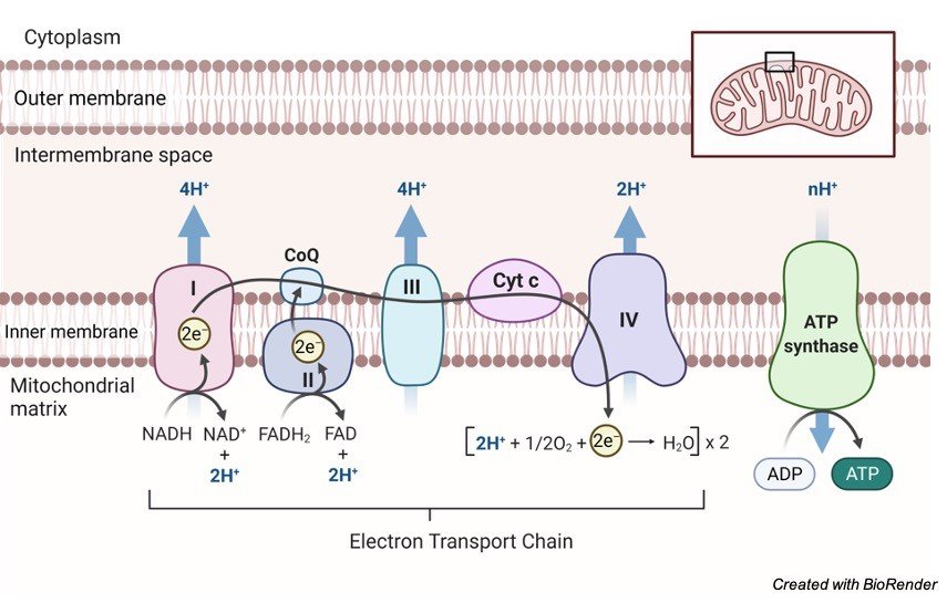
    As a result of pyruvate oxidation, reduced cofactors such a NADH generated which in turn drive the electron transport chain (ETC).

    Complete oxidation generates about 36 moles of ATP from one mole of glucose typically require oxygen–respiring mitochondria. Although, this type of mitochondria exist from unicellular eukaryotes (protists) to mammals.

    However, in many invertebrates such as Fasciola hepatica and mollusks, the mitochondria anaerobically respire and thus generate less amount of ATP as compare to typical aerobic respiration.

    Mitochondria present in a group of unicellular eukaryotes popularly known as “hydrogenosomes” share some enzymatic similarity with typical mitochondrial enzymes involved in ATP homeostasis.

    Human intestinal parasite Entamoeba histolytica possesses small, inconspicuous mitochondria known as Mitosomes, are not involved in ATP synthesis at all.

    Existence of functional landscapes in mitochondria clearly indicates that mitochondrial origin follows phylogenetic evolution.

    However, competing theories about mitochondrial evolution is still paradoxical. In 1970, Lynn Margulis proposed the idea that eukaryotic organelles such as mitochondria and chloroplasts evolved from free-living bacteria via symbiosis within a eukaryotic host cell that was later supported by others in the early 20th century.

    Accumulating number of literature from the recent year supported the endosymbiont hypothesis of organelle origin using various molecular and cell biological experiments.

    The earliest recognized hypothesis suggests that the divergence of alphaproteobacteria leads to mitochondrial evolution.

    According to the endosymbiont hypothesis, once an independent prokaryote but later on mitochondria engulfed into host archaeon and retained as endosymbionts by then.

    As a result of endosymbiosis, mitochondria participated in energy production, particularly ATP synthesis and also involved in reactive oxygen species detoxification for their host archaeon.

    In another hypothesis proposed by Müller and Martin in 1998, popularly known as “hydrogen hypothesis” α-proteobacteria produce carbon source such as hydrogen (H2), carbon dioxide (CO2), and acetate and survival of archaeon was dependent on carbon source generated by α-proteobacteria.

    High survival pressure on archaeon due to the limitation of carbon source leads to fusion of archaeon and α-proteobacteria.

    As a consequence, α-proteobacteria utilize organic compounds provided by archaeon in order to generate H2, CO2, and acetate which support archaeon survival.

    Nutritional-dependency turned-evolution of this metabolic symbiosis develop a mechanism of communication such acetate-mediated generation of acetyl-coA help to regulate enzymatic action via protein acetylation.

    Moreover, acetylation of protein using acetate as a primary carbon source utilized by a variety of organism residing on the phylogenetic tree.

    Moreover, superoxide generation by α-proteobacteria from respiratory chain also utilized as another medium of communication by oxidizing cysteine residue of proteins.

    Over the course of evolution of this metabolic symbiosis, now mitochondria evolved and possess a very efficient machinery to generate energy as well as a variety of TCA metabolites that dictate cell fate.

    Mitochondria as a signaling hub in cancer progression

    Earlier role of mitochondrial function in the cells was only limited to energy production.

    Based on the increased understanding of mitochondrial energetics in the past few decades, it’s now established that mitochondria also function as an important signaling organelle.

    Mitochondrial metabolism not only regulates the bioenergetics, but also dictate cellular fate, and survival.

    Now it has been clear that mitochondria are one of the key factor linking cancer initiation, transformation and progression. Mitochondria could also be a factor linking cancer transformation and progression.

    The importance of mitochondria in cancer also confirms their involvement in the resistance to treatment. During the oncogenesis, the transformed cells not only utilize central bioenergetic functions of mitochondria but they also rely on aberrant mitochondrial metabolism in order to support huge building blocks requirement for tumor anabolism.

    In addition, to a key role in cancer cell anabolism, mitochondrial also control cell-intrinsic and cell-extrinsic mechanisms, redox and calcium homeostasis, participate in transcriptional regulation, and dictate cell survival.

    Thus mitochondrial metabolism serves as a pool of signaling molecule that actively participate and decide the fate of cancer cells.

    Share

    Continue Reading
  • Basic Cell Plating and Cell Culture Maintenance...

    About Cell Plating

    In order to culture cells for standardization of an experiment such as drug dosage, cytotoxicity, transfection or cell migration assays etc., a fairly good number of cells are required.

    In such a case cells are cultured on plates having wells. There are 6/ 24/ 96 well plates used routinely for such experiments.

    Why Cell Plating Required?

    Wide applications of eukaryotic cell culture systems and need of compiling maximum, reliable, reproducible data has given rise to the necessity of miniaturization of this system that led to the development of cell based assays.

    This has become possible by cultivation of cells in small vessels such as 96 well plates or 6 well plates.

    Seeding cells in dishes or multiwell plates is called as cell plating.

    This gives an opportunity to increase number of samples to be assayed with high reproducibility.

    The ability to gather more than one set of data from the same sample (i.e., multiplexing) can contribute to saving time and effort during screening.

    Multiplexing can provide internal normalization controls to confirm the results of other assay methods and eliminate the need to repeat work.

    Cell Plating - Research Tweet 1

    Adopted from BioRender

    Cell Plating Requirements

    Sterile:

    1. Growth medium

    2. Trypsin (0.25%) and1 mM EDTA in PBS

    3. 96, 24, 6 well tissue culture plates

    4. Pipette tips

    5. Reagent reservoir

    6. Multichannel pipette

    7. Tubes, 15 ml, 50 ml

    8. Discard Beaker

    Non Sterile:

    9. 70% IPA

    10. Cotton/tissue paper

    Cell Plating Procedure

    1. View cultures using an inverted microscope to assess the degree of confluency and confirm the absence of bacterial and fungal contaminants.

    2. Remove spent medium. If the cells are in suspension, centrifuge it in 15 or 50 ml sterile tube at 1300 rpm for 7-10 min and proceed to step 6.

    3. Wash the cell monolayer with PBS without Ca+2 Mg+2 using a volume equivalent to half the volume of culture medium. Repeat this wash step if the cells are known to adhere strongly.

    4. Add enough trypsin/EDTA at 37oC to cover the cell layer (~3mL in T75, ~2.0mL in T25).

    5. Incubate for 2 minutes at 37oC in CO2 incubator. Tap occasionally to verify that the cells are releasing. Check in microscope to visualize detachment of cells.

    6. Remove trypsin-EDTA. Add fresh 2 ml of medium and rinse cell layer two or three times to dissociate cells and to dislodge any remaining adherent cells.

    7. Remove 100-200μl aliquote and perform a cell count (Experiment 1.3- cell counting).

    8. Make cell dilution in a reagent reservoir to plate appropriate cell number according to plate used. For instance for 96 well plate 10^4 cells/ well are plated.

    9. By using a multichannel pipette, plate cells in multiwell plates.

    10. Incubate at 37°C in 95% air for 24-hours.

    Cell Plating Citations:

    Share

    Similar Post:

    Continue Reading
  • What Is DOI And How Do I...

    What is DOI?

    A digital object identifier (DOI) is a unique identifier for electronic documents such as journal articles, research articles, book chapters but they can also represent rang of other content such as individual tables, figures, research dataset, and many other items.

    DOI is a permanent and never-changing string of numbers, symbols, and letters assigned to online published articles to identify an article or document.

    DOI system began in 2000 and is managed by the International DOI foundation and its affiliates.

    According to DOI.org, as of 2017, there are 133 million DOI names assigned and over 5 billion DOI resolution per year.

    What is DOI - Research Tweet 3
    DOI can be divided into two parts:
    • Prefix which indicates the registrant
    • Suffix which is chosen by the registrant for that item.
    Where can I find the DOI?
    • In most recently published articles, DOI is clearly visible or printed on the article itself when you open a research article database.
    • DOI usually printed somewhere on the first page, header, or footer.
    • If the DOI is not presented or printed on first page, header, or footer then search DOI on the website CrossRef.org (“Search Metadata” Box to find DOI).

    What is DOI? DOI Example 1

    What is DOI - Research Tweet J1

    What is DOI? DOI Example 2

    What is DOI - Research Tweet J1

    What is DOI? DOI Example 3

    What is DOI - Research Tweet J1
    What is DOI? DOI Example 4
    What is DOI - Research Tweet J1
    What is DOI? DOI Example 5
    What is DOI - Research Tweet J1
    What is DOI? DOI Example 6
    What is DOI - Research Tweet J1
    DOIs format:

    doi:10.1080/02626667.2018.1449

    https://doi.org/10.1111/hex.127

    https://dx.doi.org/10.1080/02626667.2018.1569

    https://doi.org/10.1016/j.jpsychires.2017.11.4

    Why DOI?

    DOI as it indicated by names digital object identifier, is a unique identification number like a Social Security number in United States, Aadhaar Card number is India, or National identification number in China.

    It helps reader to easily retrieve, locate research articles or documents from your citations.

    Using the DOI when referring to an item is more predictable and persistent that using just URL of that article as many times URLs may change.

    What is DOI - Research Tweet
    How can I use a DOI to find the article it refers to?

    Pre-2011: DOIs started with the number 10 and there are article out there still formatted this way.

    You can turn any DOI into a URL by adding

    http://doi.org/ before the DOI.

    For example, http://doi.org/10.3352/jeehp.2013.10.3

    Post-2011: The recommended and most accepted format for DOIs is an active link.

    For example, if your DOI starts with http://

    or

    https://, simply paste it into your web browser.

    How do I cite DOI in a journal article?

    There are three most acceptable format to cite a journal article with a DOI.

    APA format: Prefer DOI whenever possible.

    If DOI is not available, then use source’s URL in the citation.

    Place the DOI or URL at the end of the research article citation.

    A DOI should be represented by a “doi:” label and should always be written in lowercase.

    The APA format od DOI citation allows the use of either older string format doi.org format (“https://doi.org/10.0000/0000”) or

    modern alphanumeric (“doi:0000000/000000000000”).

    If using a URL of research article to cite, include the phrase “Retrieved from…” before the URL.

    Author, A. A., & Author, B. B. (Date of publication). Title of article. Title of Journal, volume number, page range. https://doi.org/10.00/0000

    Author, A. A., & Author, B. B. (Date of publication). Title of article. Title of Journal, volume number, page range. https://doi.org/10.0000/0000

    What is DOI - Research Tweet 1

    MLA format: In MLA style of DOI citation, stable URLs are preferrable to normal URLs.

    Use them if they are available.

    Place the DOI or URL before the access date, which comes at the end of the citation.

    Access dates are optional when using DOIs.

    If using a URL for citation in the research article, do not include the “https://” or “http://” portion of the string.

    Author. “Title.” Title of journal, Other contributors (translators or editors), Number (vol. and/or issue no.), publication year, www.someaddress.com/full/url/ or doi:0000000/00000000000. Accessed dd Mmm. yyyy.

    Author LastName, FirstName, and FirstName LastName. “Article Title.” Journal Name, vol. #, no. #, date, pp. ##-##. Name of Database, doi: 10.00/000.

    AMA/ JAMA format: If you are using a DOI in an AMA citation in your research, do not include an “Accessed” date or a URL.

    Put the DOI at the end of your citation, prefaced with “doi:”

    Author(s). Title. Journal Name. Year;vol(issue no.):page range. https://www.someaddress.com/full/url/ or doi:10.0000000/000000000000

    Author AA, Author BB. Title of article. Name of Jrnl. Year;vol(issue):inclusive pages. doi:10.0000000/000000000000

    Chicago format:

    Lastname, First/middle initials. “Title of Article.” Journal Title volume number, issue no. (Year): page range. https://www.someaddress.com/full/url/ or doi:000/0000000

    Share

    Continue Reading
  • Mitochondrial Electron Transport Chain : Cellular Respiration

    Mitochondrial Electron Transport Chain

    Electron Transport chain is a series of catalysts with rising redox potential.

    The basic mechanism involves collection of the reduced equivalents either hydrogen atom or electrons from the substrates and transferring them in a sequential order to get oxidised with the oxygen molecule resulting in formation of water and energy in the form of ATP.

    The whole process is also called as redox reaction or respiratory chain.

    The electron carriers are assembled in the form of chain within the four membrane-bound enzyme complexes, which are embedded in the inner mitochondrial membrane.

    The main advantage of this system is that the electrons transports in a stepwise manner from substrate to oxygen, thus, generating enthalpy slowly in a systematic way preventing sudden burst of energy making the process efficient and well controlled.

    Component of Electron Transport Chain

    The electron transport chain is composed of:

    A. Hydrogen and electron carriers

    B. Four membrane-bound enzyme complexes

    electron transport chain - Research Tweet

    A. Hydrogen and Electron Carriers of the Electron Transport Chain

    1. Nicotinamide Adenine Dinucleotide (NAD): It is a co-enzyme that carries hydride ion (H-) and thus known as a hydride carrier.

    It gains 2 hydrogen atoms from the substrate of Citric acid cycle like isocitrate, malate, B-hydroxy acyl CoA and B-hydroxy butyrate and form NADH molecule.

    In the process, one electron releases which reduces NADH to NADH+ while passing its hydrogen to flavoprotein containing FMN and iron sulphur protein (FeS).

    2. Flavoproteins (FAD and FMN): In the meanwhile, flavoproteins like FAD and FMN both serves as hydrogen acceptor wherein they tightly bound the hydrogen atom.

    Thus, directly preventing its reduced form to react with oxygen.

    In general, the types of flavoprotein receiving hydrogen passes them to coenzyme Q.

    For example, flavoprotein Fp1 receives 2 hydrogen atoms from reduced NAD+, while flavoprotein Fp2 receiving the same from various substrates like succinate, acyl CoA and choline.

    3. Ubiquinone: It is also known as Coenzyme Q that contains quinone ring. Coenzyme Q is the most common ubiquinone that shows similarity with vitamin K.

    It is lipid soluble with small molecular size thus responsible for free mobility of the molecule in the inner membrane of mitochondria.

    It is the basic carrier of the hydrogen atoms, wherein ubiquinol carries 2 hydrogen atoms while semiquinone carries only one.

    These molecules connect flavoprotein with cytochrome b, where flavoprotein carries 2 hydrogen atoms while cytochrome b carry one.

    Reduction of coenzyme Q leads to passage of electrons to cytochrome b while releasing 2H+ into the mitochondrial matrix.

    The enzymes responsible for the oxidation of ubiquinol involves:

    a): Conduction of electrons to cytochrome c is mainly responsible by ubiquinol (coenzyme Q) dehydrogenase along with coenzymes cyt b, FeS protein and cyt c1.

    b): Another further transfer from cyt c to oxygen is carried out under the presence of cytochrome oxidase along with cyt a and cyt a3 as coenzymes.

    4. Cytochromes are mainly responsible to carry electron from coenzyme Q to oxygen. Thus, exhibits the important role of electron carrier.

    They are categorized with the letter a, b and c.

    The core molecule for all the cytochromes are haemoproteins however they diverge in their redox potential.

    The iron of cytochrome frequently changes their state from oxidation (Fe3+; ferric state) to reduction (Fe2+; Ferrous state) during their normal physiologic action, while that of haemoglobin remains in the reduction state.

    Being mobile components, both coenzyme Q and cytochrome c transport reduced equivalent from the other fixed components.

    5. Iron Sulfur Clusters(FeS): One additional component found is iron sulfur proteins also known as FeS or non-heme iron.

    It is often related with flavoproteins and cytochrome b wherein iron is interchangeably exchange between flavoprotein and cytochrome b resulting in redox reaction.

    B. Enzyme Complexes of the Electron Transport Chain

    In the inner mitochondrial membrane four enzyme complexes are arranged which transport electrons. The following are the enzyme complexes observed:

    Complex I known as NADH dehydrogenase (NADH-ubiquinone oxidoreductase) It is a flavoprotein wherein FMN and FeS protein are the coenzymes.

    It facilitates transport of hydrogen atoms from NADH to ubiquinone in the form of NADH+ H+.

    Complex II is known as Succinate dehydrogenase (succinate-ubiquinone oxidoreductase). 

    It is also a flavoprotein wherein FAD and FeS protein are the coenzymes.

    It transfers hydrogen atoms from succinate to ubiquinone.

    Complex III is known as Ubiquinol dehydrogenase (ubiquinol-cytochrome c oxidoreductase).

    It facilitates transport of electrons from ubiquinol to cytochrome c with the help of coenzymes cyt b and cyt c1.

    Complex IV is known as Cytochrome oxidase (cytochrome-oxygen oxidoreductase).

    It facilitates transport of electrons from cytochrome c to oxygen with the help of coenzymes cyt a and cyt a3.

    Moreover, there is a fifth complex (Complex V) known as ATP synthase and is facilitates the conversion of ADP to ATP using inorganic phosphate.

    Electron Flow in Electron Transport Chain

    The oxidation of substrates with the help of NAD+ or FAD results in production of hydrogen atoms that can pass into the electron chain transfer which further successfully transferred to oxygen to generate energy and water.

    The hydrogen atoms cannot be accepted directly by the cytochrome b.

    Thus, Coenzyme Q ionized the hydrogen atoms to release hydrogen ions and electrons.

    The electrons will reduce the heme group from the cytochrome B, with this starts the transfer chain of electrons from cyt c1, cyt c, cyt a to cyta3.

    Finally, the electrons are transported with the help of cytochrome oxidase towards oxygen to form ionic oxygen.

    ATP Yield in Electron Transport Chain

    The ATP count varies with the catabolism of glucose. Moreover, FAD+ molecules can transport fewer ions resulting in fewer ATP molecules.

    In addition to this, the intermediate components of glucose can be used for different purposes resulting in variation in the ATP count.

    Summary of Electron Transport Chain

    The electron transport chain plays significant role in the aerobic respiration where the free oxygen accepts the last electron produced after glucose catabolism as a result from intermediate compounds.

    The structural configuration of electron transport chain showed four large, multi-protein complexes penetrated in the inner membrane of mitochondria along with this 2 small diffusible electron carrier are present transferring electrons between them.

    During the transfer of the electrons, small amount of energy is lost against the chemiosmosis.

    Thus, high-energy electrons are donated by the NADH or FADH to compensate the energy to finish the chain.

    Water and ATP are the end products of the electron transport chain.

    Moreover, various intermediate products of the tricarboxylic acid cycle can be averted into the anabolism of other biochemical molecules which may serve as the benefactor for glucongenesis.

    Electron Transport Chain Citations:

    Share

    Similar Research Position:

    Continue Reading
  • Monocistronic mRNA: Definition, Importance, and Examples

    What are Monocistronic mRNA?

    Monocistronic mRNA is a mRNA that encodes just a single protein and all eukaryotic mRNAs are monocistronic.

    The improvement of the develop monocistronic eukaryotic record includes a few diverse handling steps.

    Heterogeneous nuclear RNA (hnRNA) refers to the large pre‐mRNAs of various nucleotide sequences transcribed by RNA Polymerase II.

    hnRNA transcription is only a small portion of the transcription which occurs in the nucleus.

    Up to 90% of the transcription is for the production of rRNA. But if we limit our discussion to hnRNA some important observations can be made.

    First, the complexity of the hnRNA is 4 to 10 times greater than that of the mRNA. This suggest two possibilities.

    One, we already know and that is that splicing of genes reduces the size of the final mRNA product in comparison to the original transcript. But this does not account completely for the differences in complexity.

    Monocistronic mRNA Features

    The majority of the difference appears to be accounted for by destruction of hnRNAs before they develop into mature mRNAs.

    hnRNAs are a broad class of products:

    • Average size – 8000 – 10,000 bases

    • Range – 2000 – 14,000 bases

    Since the mean mRNA length is 1800-2000 bases, a sustainable amount of cutting must occur. hnRNA doesn’t really exist in the core as bare RNA however is really connected with protein.

    This complex is assigned hnRNP and is called heteronuclear ribonuclearprotein.

    hnRNP is related with the nuclear matrix. Since chromatin is connected to the nuclear matrix it appears to be intelligent that the essential transcript will likewise get appended to the matrix soon after transcription.

    Processing of Monocistronic mRNA

    The lifetime of hnRNA is very short generally from one minute to one hour and no more. The initial phase in handling is the expansion of a cap.

    The cap is a 5′ methyl guanosine that is added following the beginning of transcript. Capping happens so rapidly that we seldom see the original 5′ base of the message.

    The linkage between the 5′ methyl guanosine isn’t the commonplace 5′- 3′ linkage yet is a 5′- 5′ linkage. The reaction is catalyzed by the enzyme guanylyl transferase.

    The guanosine that is attached is consistently methylated at the 7 situation of the guanine base (7mG). This is called cap 0.

    Likewise, a methyl bunch is added to 2′- OH of the first base in the mRNA. This is catalyzed by 2′- O-methyl-transferase, and this methyl group is alluded to as cap 1.

    Other methylations can takes place, however we will not think about them. About 25% of hnRNA in the end develops in polyadenylated mRNA.

    Not all mRNAs are polyadenylated. The histone mRNAs are a remarkable exemption for the standard. In the event that an inhibitor of polyadenylation is added to a response hnRNA isn’t changed over into mRNA.

    In this way polyadenlyation is a prerequisite for mRNA to show up.

    End of transcription isn’t perceived in any way. One succession that is invariant in eukaryotic mRNA is the sequence 5′- AAUAAA-3′ that is seen around 10-30 bp upstream of the poly A tail.

    The inquiry that this raises is whether this sequence is needed for polyadenylation. Erasures or mutation of this sequence will kill polyadenylation.

    Yet, astounding, point mutation incredibly decreased the quantity of molecules that are divided.

    In any case, those that are separated are polyadenylation. Consequently, this sequence is by all accounts needed for cleavage of the essential transcript.

    The poly-A tail is added by the compound poly(A) polymerase.

    Monocistronic mRNA Characteristic

    Monocistronic mRNAs are the standard mRNAs in eukaryotic cells are perpetually monocistronic, and the size of the mRNA is normally corresponding to the length of the encoded protein.

    This is genuine likewise of most plant and animal viral mRNAs, despite the fact that there are, among viral messenger, a few exemptions for the monocistronic rule.

    These mRNAs are basically polycistronic: they encode at least two overlapping proteins. Anyhow, in every such case, save one, interpretation is restricted to the 5′- proximal cistron.

    (The solitary archived special case is simian virus 40 late 16 SmRNA, in which the 62-aminoacid “agnoprotein” is encoded up-stream from capsid protein VP1 and the two proteins are translated.

    These mRNAs are in this manner adjust to the standard for eukaryotes in that they are practically monocistronic. Few eukaryotic mRNAs comprise a third category.

    These mRNAs encode and express two proteins. Much of the time, these outcomes from commencement at the first and second AUG trios – interestingly with most of eukaryotic mRNAs.

    Monocistronic Rule

    The 5′- proximal AUG works as aninitiator codon. Considering the scarcity, they present just a minor inconsistency to the monocistronic rule.

    The monocistronic rule for gene expression in eucaryotes states not just that a single mRNA coordinates synthesis of a single protein yet in addition that the protein encoded nearest to the 5′ end is the one that gets expressed.

    This remains constant for the entirety of the naturally occurring viral mRNAs recorded, just as for misleadingly constructed messengers that encode two proteins.

    The 5′- terminal restriction is imposed not by the structure of eukaryotic mRNAs yet rather by the properties of eukaryotic ribosomes.

    This was astutely shown by brooding wheat germ ribosomes with a polycistronic mRNA from bacteriophage lambda.

    Though E.coli ribosomes promptly translate the first and second cistrons in that message, wheat germ ribosomes decipher just the first.

    Inability to decipher down-stream cistrons suggests that eukaryotic ribosomes can neither tie straightforwardly to internal initiation sites nor reinitiate at a subsequent cistron in the wake of crossing the first.

    Plainly, eukaryotic ribosomes work under restrictions that don’t happen in procaryotes.

    The failure of eukaryotic ribosomes to start at destinations in the inside of a message implies that eukaryotic cells can’t organize synthesis of a bunch of proteins by grouping the genes behind a single transcriptional promoter.

    Polycistronic transcripts, which are the transcendent regulatory device utilized by procaryotes, won’t work in eucaryotes.

    Taking into account this restriction, it is fascinating to ask what compensatory mechanism eukaryotic cells have formulated to organize gene expression.

    Eukaryotic genes are sometimes fused, hence creating, in lieu of a polycistronic mRNA, a “polyprotein” which is gotten from a single longsome cistron.

    Monocistronic mRNA Example

    Tryptophan synthetase is one such model: the alpha subunit (Mr28,727) and 32 subunit (Mr42,756), which are encoded by isolated genes in E.coli, are fused in S. cerevisiae to form a bifunctional protein of Mr76,000.

    Different instances of fused genes encoding multifunctional proteins are the arom gene cluster in Neurospora, which incorporates five enzymes associated with the polyaromatic biosynthetic pathway, and the his4 gene in S.cerevisiae which encodes a trifunctional protein.

    Maybe the most noteworthy model is mammalian unsaturated fatty acid synthetase (Mr240,000), which relates to a set of 7 individual polypeptides in E.coli (2%).

    Albeit the reasoning behind gene combination is more managable to speculating than to experimentation, it is conceivable that fusion of genes makes up for the failure of eukaryotic ribosomes to utilize polycistronic transcripts for the coordinate synthesis of polypeptides.

    Monocistronic mRNA Citations

    Share

    Similar Post:

    Continue Reading
  • Seed Germination: Definition, Process, Methods, and Major...

    What is Seed Germination?

    Seed Germination is defined as one of the vital process by which all the species of plants got generated from a single seed and how it gets adapted to a particular environment and grow itself as a crop or as an herb or as shrub or a tree.

    The best example that we come across our day-to-day life is sprouting of pulses in our kitchen or young plants developing in our kitchen garden.

    Process of Seed Germination

    When we sow a seed in a mud, first the seed starts absorbing water from its outer environment or directly from the soil, which results in bulging of seed and its seed coat gets moisture and becomes very vulnerable at a particular temperature.

    This stage of the seed which can be seen from one to two days of sowing is commonly referred to as imbibition.

    Seed Dormancy: Germination, Definition, and Mechanism - research tweet 1

    It further resumes its growth process by activating several enzymes and starts its respiration and initiates the production of proteins and stores nutrients in it by metabolising those necessary proteins.

    Hence, it is said that sprouts are rich in proteins.

    This stage of the plant is generally referred to as lag phase. Then the seed coat starts rupturing and gives rise to a small radicle, which later forms the primary root of a plant.

    This helps the plant to absorb the underground water along with other nutrients present in the soil and helps in further growth of the plant.

    The slowly starts growing the plumule of the plant which later develops into the stem portion of the plant.

    As days move on seeds modify itself as seedling, then plumule starts growing upwards giving rise to leaves and other floral parts of the plant.

    Optimum Condition for Seed Germination

    Not all the seeds have the capability to grow at any conditions we sow, certain seeds grow only at certain conditions and certain soil and climatic conditions.

    The things that are necessary for a plant to grow in a good condition are listed below.

    A. Water

    As every living organisms’ water is the most vital component for a seed to germinate.

    Though some seeds do not require much water for their growth they need at least a minimal quality of water at certain intervals.

    It depends on the dry weight of the seed. As discussed above the seed starts its germination process by absorbing water which is necessary for the nourishment of the embryo by providing dissolved oxygen and soluble supplements for their growth.

    The dissolved oxygen helps the seed coat to become softened and converts the seed into a sapling by rupturing the seed coat.

    B. Oxygen

    As water oxygen is also very much important for the seed growth, which is obtained through the water in a dissolved form or through the porous soil.

    But the deep buried seeds are not capable to use the oxygen from the soil pores.

    Oxygen is vital for the metabolic process of the seed in terms of aerobic condition.

    C. Temperature

    If the seed wants to grow in a proper manner without any deficient conditions then the temperature should be maintained at an average of 25 to 30ºC. But in some cases, the seeds need a temperature of 5 to 40ºC.

    Hence the temperature varies according the particular regions where they are native to and other environmental factors.

    D. Light and Dark Cycle

    This depends on the nature of certain seeds, some seeds do not germinate if sunlight falls on them, such kind of seeds are grown in a dark condition and after germination they are brought to the field.

    Whereas most of the plant needs sunlight for the germination and throughout their lifetime.

    Thus, the seed germination needs all these vital factors and generation of plumule and radicle is said to be completion of seed germination.

    Factors Affecting Seed Germination​

    Factors affecting seed growth or germination may be both internal and external and they are listed below as follows.

    Internal Factors

    Seed dormancy is considered as one of the important factors in germination process which means that seeds do not germinate if they met any unfavorable conditions during their process of undergoing their growth.

    During this period the seed coat which allows the passage of water restricts the water to absorb so that the oxygen and nutrients necessary for their growth is limited.

    So that the seeds remain as such without any development. But in some cases, the plant growth regulators help in the growth of the plant.

    However, some seeds germinate within a week where as others take their own time to germinate.

    External Factors

    As mentioned above water, oxygen and temperature are considered as important factors for the germination of a plant.

    a. Water

    Seed growth gets affected when the water is supplied at higher amount than the needed quantity and also when it is supplied in very low quantity or there is no supply of water.

    So, it is important to know the nature of seeds and their adequate flow of water.

    b. Oxygen

    While the seeds start its germination process it intakes large amount of water than other intervals, so it is checking the availability of oxygen for the growth of the plant.

    c. Temperature

    Seeds require only the appropriate temperature necessary for their growth if the temperature is more vigorous than needed then seeds starts shrinking and it will die or degenerate.

    So, the seed must be sown in optimum environmental conditions by considering the environmental factors.

    Seed Germination Citations

    Share

    Similar Post:

    Continue Reading
  • Mendel’s Laws of Inheritance : Types and...

    What is Mendel's Laws of Inheritance or Mendelian genetics?

    Mendelian genetics or Mendelism or Mendelian inheritance is a pack of primary tenets that are affiliated to the relaying of hereditary features from parent organisms to their kids; it is nothing but due to the genetics.

    These tenets were originally extracted from the study of Gregor Mendel published in the year 1865 and 1866, which was founded in 1900.

    It was originally controversial, however later it became core of classic genetics.

    Historical Facts About Mendel's Laws of Inheritance

    Credit of laws of inheritance goes to Gregor Mendel, a monk of nineteen century who performed experiments on hybridization in peas (Pisum sativum).

    Between 1856 and 1863, he grew and tested around 28,000 pea plants and from this he deduced two generalizations that became famous as Mendel’s Laws of Heredity or Mendelian inheritance.

    This is delineated in two sections of the research paper “Experiments on Plant Hybridization”, that was published in the journal in the year 1866.

    What is Inheritance?

    Inheritance can be defined as the procedure of how a child receives genetic information from the parent.

    This process of hereditary is related to the dependence which gives the offspring same trait as of parents.

    Inheritance is which is why, the family members have similar and distinctive features.

    Why Mendel Choose Pea to Study Genetics?

    He chose pea for his experimental procedures because it can be grown easily and are naturally self-pollinating.

    Pea plant not only can be self-pollinated but also can be cross-pollinated.

    It is yearly plant, ergo, many generations can be followed in less duration of time.

    Not only this it also possesses contrasting traits.

    Mendel's Laws of Inheritance

    The two majors’ experiments done by Mendel to establish the laws of inheritance (Mendel’s Laws of Inheritance) were:

    1. Monohybrid Cross Experiment

    2. Dihybrid Cross Experiment 

    During his procedure he found that many features and traits passes to the offspring in a stable way.

    These factors are known as genes. Unit of inheritance is nothing but genes.

    Mendel’s Experiments

    He conducted experiments and contemplated seven major contrasting features in the plants.

    Then, he conducted both the experiments to determine the aforementioned inheritance laws.

    1. Monohybrid Cross

    He took two plants of pea with contradictory traits that is one short and one tall and then crossed them.

    It was noticed by him that the offspring’s of the 1st generation were tall and he termed it as F1 progeny.

    Then he crossed F1 progeny which yield him tall as well as short plants in the ratio 3:1.

    Phenotypic Ratio, Phenotypic, Phenotypic Ratio of dihybrid cross, Phenotypic Ratio definition, Phenotypic Ratio calculation,
    Phenotypic Ratio, Phenotypic, Phenotypic Ratio of dihybrid cross, Phenotypic Ratio definition, Phenotypic Ratio calculation,

    Mendel even did experiment with other contrasting features like green peas vs yellow peas, round vs wrinkled, etc. and he found that the results were the same.

    This led to the discovery and the formulation of the laws of Segregation and Dominance.

    2. Dihybrid Cross

    Mendel in his dihybrid cross trial he took two traits, each having two alleles and then he crossed wrinkled-green seed and round-yellow seeds and noticed that all the first generation progeny (F1 progeny) were round-yellow.

    He therefore concluded that the dominant traits were the round shape and yellow colour.

    Phenotypic Ratio, Phenotypic, Phenotypic Ratio of dihybrid cross, Phenotypic Ratio definition, Phenotypic Ratio calculation,

    He also self-pollinated the F1 progeny and got 4 different features like wrinkled-yellow, round-yellow, wrinkled-green seeds and round-green in the ratio 9:3:3:1.

    Mendel's Laws of Inheritance

    Mendel postulated three laws, which are now called after his name as Mendel’s laws of heredity.

    1. Law of dominance 

    2. Law of segregation

    3. Law of independent assortment

    1. Law of Dominance

    When two homozygous individuals with one or more sets of contrasting characters are crossed, the characters that appear in the F1 hybrids are dominant characters and those do not appear in F1 are recessive characters.

    The dominant and recessive traits of the genes can be dependent on the enzymatic function of the genes of which dominant genes can make functional enzymes which synthesize active polypeptides or proteins.

    On the other hand, the recessive genes (mutant genes) code for incomplete or non-functional polypeptides.

    Therefore, the dominant genes make a distinct phenotype which can be expressed even in the heterozygous condition while the recessive cannot make so.

    Researchers undertook various cross-breeding experiments to authenticate applicability of law of dominance like Correns on peas and maize, Tschermak on peas, and many others.

    They discern plenty of characters in several life form is related as dominant and recessive.

    Importance of Law of Dominance

    In the hybrid trait the normal characteristics shown by dominant gene reveal significant advantages over the dreadful characters exhibited by recessive gene.

    It was noted that diabetes, hemophilia etc. are all traits of recessive characters.

    A hybrid person may possess all these characteristic in addition to this recessive gene can persist for next several generations without expressing themselves.

    In contrast, there is exception to Law of Dominance which is known as Incomplete Dominance.

    Mendel was followed by several scientists in case of blending of characters of two parents.

    This trait shows characteristics which are amalgamated from both the parents with both sets of genes which is commonly known as incomplete dominance or blending inheritance.

    It is the result of expression of genes partially from the dominant trait and partially from the recessive one.

    For example, in four O-clock plant, Mirabilis Jalapa, red (RR)flower plant is crossed with white (rr) colored plant resulting in plant with pink (Rr) color flower.

    On self-pollination with pink color flower the ratio of getting red (RR), pink (Rr) and white (rr) is 1:2:1.

    2. Law of Segregation (Purity of Gametes)

    This law states that when trait of two opposite genes or allelomophs are mixed together the resultant pair stays without being contaminated while gametes separate out from each other and only one enters each gametes.

    As a result, two types of gametes are produced from the heterozygote in equal numerosity. 50% of the gametes possess gene T and other 50% possess gene t.

    Importance of Law of Segregation

    According to cytological studies, the law of segregation holds good to all cases whether it is dominance or no dominance.

    Rare biological generalization has made its applicability far out reaching.

    3. Law of Independent Assortment

    In this law, the study of inheritance of more than one pair of characters is carried our simultaneously, wherein the factors or genes can be sorted independently from its correspondent allele.

    Based on the result of dihybrid cross, Mandel formulated Mendel’s Laws: Independent Assortment .

    Importance of Law of Independent Assortment

    Various cytological studies revealed that those allelomorphs which are assorted independently during cell division, are actually located on different homologous pairs of chromosomes.

    However, if the traits for various characteristics are seen on the same homologous pair of chromosomes, these are passed on to the same gamete.

    This law fails on its universal applicability.

    Biological Significance of Mendel's Laws

    Mendel’s work unfolded after 3 decades of his actual work and the laws he mentioned was widely used by various branched for the purpose of scientific evaluation.

    Many modern genetic modification was seen like getting rust-resistant and disease-resistant varieties of grains and for breeding high grade variety of horses and dogs.

    The science of segregation is the outcome of Mendelism, which deals with the betterment of human race.

    Conclusions from Mendel’s Experiments

    Genotype is defined by the genetic makeup of the organism while phenotype is defined by its physical appearance.

    The genes are transmitted from parents to offspring and is called as allele.

    There is a 50% chance of one of the two alleles to fuse with the other parent during gametogenesis when the chromosomes are halved.

    When the alleles are the same, they are known as homozygous alleles and when they are different they are known as heterozygous alleles.

    Mendel’s law benefited the humankind by generating high grade breed of various plants and animals.

    Moreover, they have higher implication in diagnosing and preventing various genetic disorders.

    Mendel's Laws of Inheritance Citations:

    Share

    Similar Post:

    Continue Reading