Category: Study Materials
-
Microscope, Microscope Parts, Labeled Diagram, and Functions
What is Microscope?
A microscope is a laboratory instrument used to examine objects that are too small to be seen by the naked eye. It is derived from Ancient Greek words and composed of mikrós, “small” and skopeîn,”to look” or “see”.
It is one of the most revolutionized scientific instruments used to observe or examine minute structures not clearly visible from naked eyes.
In 1665, for the first time Robert Hooke made an impressive Micrographic illustration using microscope. Antonie van Leeuwenhoek, another scientist made significant contribution in microscope research by magnifying the simple single lens microscope 300 times.
Structure of Microscope
What Microscope does?
Microscopes magnify or enlarge small objects such as cells, microbes, bacteria, viruses, microorganisms etc. at a viewable scale for examination and analysis.
Microscopes consist of one or more magnification lenses to enlarge the image of the microscopic objects placed in the focal plane. The magnification power of simple laboratory microscope is 1250x.
Types of Microscope
There is various type of microscope such as transmission electron microscopes (TEMs), scanning electron microscopes (SEMs), atomic force microscopes (AFM), near-field scanning optical microscopes (MSOM or SNOM, scanning near-field optical microscopy, and scanning tunneling microscopes (STM).
However, the most common type of microscope is optical microscope.
Microscope Parts and Functions
Microscope Parts Microscope Parts Functions Base Supports the microscope Arm Used to carry the microscope Stage Platform where the slide with the specimen is placed Stage Clips Holds the slide in place on the stage Eyepiece (containing ocular lens) Magnifies the image for the viewer Revolving nose piece Contains the objective lenses; rotates to allow the user to switch between different objective lenses Objective lenses Low-, medium-, and high-power lenses that further magnify the specimen at different intensities Coarse adjustment knob Large knob used for focusing the image under low power (general focusing) Fine adjustment knob Smaller knob used for focusing the image with the medium- and high-power objectives (fine-tuning) Diaphragm Controls the amount of light that passes through the specimen Light source Provides light for viewing the specimen Microscope Parts and their Functions
In general, microscopes are made up of supporting parts to hold the structure of Microscopes and optical parts, consist of lenses used to magnify the specimens.
The description given below summarize the brief description of microscope parts used to visualize the microscopic specimens such as animal cells, plant cells, microbes, bacteria, viruses, microorganisms etc.
The Microscopes parts divided into three different structural parts Head, Base, and Arms.
Head/Body: It contain the optical parts in the upper part of the microscope.
Arm: It supports the tube and connects it to the base.
Base: The bottom of the microscope, used for support.
Optical Components of Microscope
Eyepiece Lens: the lens at the top that you look through, usually 10x or 15x power.
Tube: Connects the eyepiece to the objective lenses.
Illuminator: Illuminator is the most important microscope parts and it serve as light source for a microscope during slide specimen visualization. It is a continuous source of light (110 volts) used in place of a mirror. The mirror of microscope is used to reflect light from the external light source up through the bottom of the stage. Usually, the Illuminator located in the base of the microscope. Most light microscopes use low voltage, halogen bulbs with continuous variable lighting control located within the base.
Stage with Stage Clips: The stage of a microscope is a flat platform where you place your subject slides. Stage clips hold the slides in place. The mechanical stage of your microscope will help you to move the slide around by turning two knobs. One knobs moves it left and right, the other knobs moves it up and down.
Revolving Nosepiece or Turret: Turret is the part of the microscope that holds two or multiple objective lenses and helps to rotate objective lenses and also helps to easily change power.
Objective Lenses: Three are 3 or 4 objective lenses on a microscope. The objective lenses almost always consist of 4x, 10x, 40x and 100x powers. The most common eyepiece lens is 10x and when it coupled with others, total magnification is 40x (4x times 10x), 100x , 400x and 1000x. Objectives can be forward or rear-facing.
Microscope Rack Stop Rack Stop: Rack Stop is an important microscope parts that determines how close the objective lens can get to the slide. It keeps the students from damaging the high power objective lens down into the slide. If you cann’t able to focus on the specimen at high power while using very thin slides then slight adjustment helps you to adjust the focus.
Diaphragm or Iris: Most of the laboratory microscopes have a rotating disk under the stage known as diaphragm or iris. Iris Diaphragm controls the amount of light reaching the specimen. The Iris Diaphragm is located above the condenser lens and below the microscope stage.
The different sized holes in the diaphragm helps to vary the size of the cone and intensity of light that is projected upward into the slide. However, there is no set rule regarding which setting to use for a particular power.
The specimen transparency, degree of contrast and particular objective lens in use decide the Diaphragm or Iris setting. Majority of high quality microscopes used in laboratory include an Abbe condenser with an iris diaphragm. When iris diaphragm is combined with Abbe condenser, it control both the quantity of light applied as well as focus on the specimen.
Aperture: It is the hole in the stage through which the base (transmitted) light reaches the stage.
Condenser: Condenser lenses are used to collect and focus the light from the illuminator on to the specimen. Usually the Condenser lenses are located under the stage in conjunction with an iris diaphragm. Condenser lenses helps in ensuring clear and sharp images are produced with a high magnification of 400X and above.
Magnification power of the condenser is directly related to the image clarity. Most of the sophisticated microscopes in the laboratory come with an Abbe condenser that has a high magnification of about 1000X. Condenser Focus Knob moves the condenser up or down to control the lighting focus on the specimen.
FAQ About Microscope and Microscope Parts
What is Microscope?
Microscopes are instruments that are used in science laboratories, to visualize very minute objects such as cells, microorganisms, giving a contrasting image, that is magnified.
What is the Function of Microscope?
A microscope is usually used for the study of microscopic algae, fungi, and biological specimens.
What is Magnification?
Magnification is the process of maximizing object’s relative size rather than its physical size. This expansion is measured by a calculated number known as “magnification.” Whenever this number is less than one, it corresponds to a reduction in size, also known as minification or de-magnification.
What is Resolution?
The term ‘resolution’ in microscopy refers to a microscope’s ability to differentiate object’s details. In other words, this is the smallest distance at which two distinct points of a specimen can still be seen as separate entities – either by the observer or the microscope camera. A microscope’s resolution is inextricably linked to the numerical aperture (NA) of the optical components as well as the wavelength of light used to examine a specimen.
What is Depth of Field?
The depth of field is defined as the distance between the nearest and farthest object planes that are both in focus at any given moment. In microscopy, the depth of field is how far above and below the sample plane the objective lens and the specimen can be while remaining in perfect focus.
What is Eyepiece?
The eyepiece, also known as the ocular is the part used to look through the microscope. Its found at the top of the microscope. Its standard magnification is 10x with an optional eyepiece having magnifications from 5X – 30X.
What are Objective Lens?
Objective Lens are the major lenses used for specimen visualization. They have a magnification power of 40x-100x. There are about 1- 4 objective lenses placed on one microscope, in that some are rare facing and others face forward.
What is Coarse Adjustment?
The coarse adjustment knob moves the stage up and down to bring the specimen into focus.
What is Fine Adjustment?
The fine adjustment knob brings the specimen into sharp focus under low power and is used for all focusing when using high-power lenses.
What are Condensers Lenses?
Condensers are lenses that are used to collect and focus light from the illuminator into the specimen. They are found under the stage next to the diaphragm of the microscope. They play a major role in ensuring clear sharp images are produced with a high magnification of 400X and above.
What are Abbe Condenser Lenses?
Abbe condenser is a condenser specially designed on high-quality microscopes, which makes the condenser to be movable and allows very high magnification of above 400X. High-quality microscopes normally have a high numerical aperture than objective lenses.
Can I See Germs Under a Microscope?
Due to very small size of germs, an amateur microscope will not allow you to see germs. Germs can only be observed under a microscope with a magnification of at least 1200x. In order to visualize the germs, and the specimen must be stained.
As a result, you’ll need to use a powerful microscope model and special sample preparation techniques. Of course, there are giants among bacteria that can be seen at 900x magnification, but they live at the deepest depths of the ocean and are nearly impossible to obtain.
Can a Light (Optical) Microscope Detect Viruses?
No, they are 1000 times smaller than bacteria, which is the smallest thing an optical microscope can see.
What is Microscopy Staining?
Cell staining is a technique used to enhance the visibility of cells and cell components under a microscope. Different stains can be used to stain specific cell components, such as the nucleus or the cell wall, or the entire cell with different colours.
What are Basic Components of Optical Microscope?
Almost all optical microscopes have a tube, an eyepiece lens, a turret with one or more objective lenses, a light source, an aperture, a condenser, a stage, fine and coarse focus controls, and a stable base.
What is Microscopy in Biology?
In biology, the most important way to gain insight into biological structures and processes is through microscopy, or studying biological forms with an optical microscope.
Why is it Necessary to Begin with 4x Magnification on a Microscope?
The 4x objective lens has the least amount of power and thus the greatest field of view. As a result, locating the specimen on the slide is easier than if you start with a higher power objective.
How to Calculate Magnification of a Microscope?
Simply multiply the magnification of the ocular lens by the magnification of the objective lens to calculate the power of magnification of a microscope. For a typical compound microscope with a 10X ocular lens and objective lenses with magnifications of 4X, 10X, 40X, and 100X, your microscope will have magnifications of 40X, 100X, 400X, and 1000X depending on which objective lens you use. The same principle applies to stereo microscopes; a 10X eyepiece combined with a 4X objective lens produces a magnification of 40X.
Some stereo microscopes have a continuous zoom objective lens with magnification ranging from 0.75X to 7.5X. When combined with a 10X ocular lens, the total magnification will be 7.5X to 75X. When combined with a 25X ocular lens, the total magnification will be 18.75X to 187.5X.
What is Field of View (FOV)?
When looking through a microscope, the FOV is the diameter of the circle of light that you see. The field of view shrinks as magnification power increases, and vice versa.
How Does the Diopter Adjustment Work?"
To compensate for the difference in vision between your two eyes, the diopter adjustment allows you to focus one eyepiece independently of the other. When the diopter adjustment is correct, both eyes are at ease during observations.
How do Achromatic, Plan Achromatic, Semi-Apochromatic, and Apochromatic Objectives Differ?
The main distinction between microscope objectives is in their design, image quality, as well as research methods.
Spherical and chromatic aberrations are reduced in achromatic objectives. This objective type has a flat field of focus in the centre of the 65 percent field of view. The delivered image may have a hint of blue-red.
Color aberrations can be corrected by planning achromatic objectives. They flatten the field of view to provide a very sharp image across the entire image. These macro objective lenses are fantastic for close-up photography.
The image quality of semi-apochromatic objectives is average (in comparison to achromatic and plan achromatic objectives). These objectives’ optical elements contain fluorite, allowing them to be used in fluorescence microscopy.
Apochromatic objectives (as opposed to achromatic objectives) produce a sharper image with more vibrant colours. This is accomplished by enlarging the spectral region.
How to Calculate Total Magnification?
Magnification refers to how much larger an object appears when viewed through a microscope versus the naked eye. The multiplication symbol (X) on the eye piece and objective lenses indicates how many times the lens of each microscope part magnifies an object.
Multiply the magnification of the eyepiece (ocular lens) by the magnification of the objective lens in use to calculate the total magnification of any object viewed under the microscope. This can be demonstrated using the formula.
Total magnification = ocular lens x objective lens
For example, if the ocular lens magnifies the image 4X and the low-power objective lens magnifies the image 10X, the total magnification of the object at low magnification is 40X (4×10=40X). The total magnification would be 160X (4 x 40= 160X) with the same ocular lens and a high – power objective that magnifies 40X.
How to Focus Your Microscope?
The proper way to focus a microscope is to start with the lowest power objective lens and crank the lens down as close to the specimen as possible without touching it while looking from the eye piece.
Examine the image through the eyepiece lens and focus only upward until it is sharp.
If you can’t get it to focus, go through the process again. Once the image with the low power lens is sharp, you should be able to simply click in the next power lens and make minor adjustments with the fine adjustment knob.
If your microscope has a fine focus adjustment, a slight turn should suffice. Continue with the next objective lens, fine-tuning the focus each time.
When looking through the microscope, keep both eyes open. This reduces eye fatigue caused by keeping the nonviewing eye closed.
It takes some practice to keep both eyes open, but it is highly recommended. Also, never let your eye come into contact with the ocular lens. You are too close if your eyelashes touch the lens.
When using a microscope, always remove your glasses. If your eyeglass lens comes into contact with the microscope, it may be scratched.
What is the Best Way to Use a Microscope?
• Step 1: Connect the light microscope to a power source in step one. You can skip this step if your microscope has a mirror instead of an illuminator. Instead, look for a location with plenty of natural light.
• Step 2: Rotate the revolving nosepiece so that the lowest objective lens is in place.
• Step 3: Install your specimen on the stage. But first, make sure your specimen is adequately protected by placing a coverslip on top of it.
• Step 4: Use the metal clips to secure your slide. Make sure the specimen is in the centre, directly beneath the lowest objective lens.
• Step 5: Looking through the eyepiece, slowly turn the coarse adjustment knob to bring your specimen into focus. Make sure the slide does not come into contact with the lens.
• Step 6: Set the condenser to produce the most light possible. Because you’re using a low power objective, you may need to reduce the illumination. To make adjustments, use the diaphragm beneath the stage.
• Step 7: Slowly turn the fine adjustment knob until you have a clearer image of your specimen.
• Step 8: Inspect your specimen.
• Step 9: Once you’ve finished viewing with the low-power objective, switch to the medium-power objective and re-adjust the focus with the fine adjustment knob.
• Step 10: Once you’ve focused the low-power objective, move on to the high-power objective.
Safety Precautions for Using Microscopes
Carrying: Carry your microscope with two hands, one grasping the arm or back slot and the other supporting the base.
Table Placement: Position the microscope on flat, solid support that will not be easily knocked off. To avoid tripping over the cord, coil it.
Cleaning: Lenses must be clean in order for resolution to be achieved. Only lens paper or gauze and cleaning solution should be used. Never clean your lenses with your finger, handkerchief, paper towels, or spit. Remove no parts for cleaning; doing so allows dust to enter the microscope.
Electron Microscopes vs Light Microscopes
Feature Light Microscope Electron Microscope Size Small Very large Training required Little; suitable for middle-school students as well as older Extensive training necessary: only laboratory personnel in research institutions will have access Viewing live specimens Live specimens can be viewed Live specimens cannot be viewed Can see in color? Yes, since light is used to illuminate the specimen No, because electrons are used to generate an image. Any colors seen are computer-generated for clarity Total magnification 1000X maximum up to around 10,000,000X (some can view atoms) Clarity/resolution Vague; generally poor resolution Very clear; very high resolution and clarity MCQ on Microscopy
1. What is the overall magnification of a light microscope if the eyepiece magnification is x10 and the objective magnification is x40?
a) x10
b) x40
c) x400
d) x4
2. What is the purpose of the condenser on a light microscope?
a) To focus the light source
b) To diffuse the light source
c) To provide the light source
d) To control the light source
3. What is the proper name for the microscope lens in the eyepiece?
a) Ocular
b) Binocular
c) Objective
d) Condenser
4. What is the proper name for the primary microscope lens that focuses the image?
a) Ocular
b) Binocular
c) Objective
d) Condenser
Microscope Slide Preparation
Microscope slides are pieces of clear glass or plastic that hold a sample and allow it to be viewed through a light microscope. Because there are various types of microscopes and samples, there is more than one way of preparing a microscope slide.
i. Wet Mount Slide
Wet mounts are used for living samples, clear liquids, and aquatic samples. A wet mount is similar to a sandwich. The slide is the bottom layer. The liquid sample comes next. To minimise evaporation and protect the microscope lens from sample exposure, a small square of clear glass or plastic (a coverslip) is placed on top of the liquid.
1. Collect a clean microscope slide and a coverslip (a thin piece of plastic covering). Fill the centre of the microscope slide with a drop or two of water.
2. Submerge the specimen in a drop of water.
3. Grab the coverslip by the edges. Do not touch the coverslip’s surface. Place the coverslip on its edge alongside the drop of water. Allow the water drop to run along the edge of the coverslip. This will assist in preventing air bubbles from forming beneath the coverslip.
4. Gently lower the coverslip over the water drop and specimen.
5. Ensure that the object is completely submerged in water. If not, remove the coverslip, add a little more water, and replace it.
6. If excess water seeps out from underneath the coverslip, gently blot it away with a small piece of paper towel.
7. The slide is now ready to be viewed. When you’re finished viewing, properly dispose of the specimen and clean the slide and coverslip as directed by your teacher.
ii. Dry Mounting
Dry mount slides can be made up of either a sample placed on a slide or a sample covered with a coverslip. The size of the object isn’t important for a low-power microscope, such as a dissection scope, because its surface will be examined.
1. Lay the slide down on a flat surface.
2. Place the sample on the slide with tweezers or forceps.
3. Place the sample on top of the coverslip. In some cases, viewing the sample without a coverslip is acceptable, as long as care is taken not to bump the sample into the microscope lens. If the sample is soft, create a “squash slide” by gently pressing down on the coverslip.
iii. Blood Smear Slide
1. On a flat surface, place a clean glass slide. One small drop of blood should be added to one end.
2. Take another clean slide and, holding at a 45-degree angle, touch the blood with one end of the slide, allowing the blood to run along the edge of the slide via capillary action. To make a thin smear of blood, carefully push along the length of the first slide.
3. Make two smears, let them air dry, and label them clearly. Place the dried slides in the slide transport containers provided.
v. Staining Slides
1. Microscope cell staining is a technique used to improve the visibility of cells and cell parts under a microscope. A nucleus or a cell wall can be seen more clearly by using different stains.
2. Iodine, crystal violet, and methylene blue are examples of simple stains.
3. Make a wet or dry mount with a coverslip.
4. Apply a small amount of stain to the coverslip’s edge.
5. Place the opposite edge of the coverslip with a tissue or paper towel. To stain the specimen, capillary action will pull the dye across the slide.
Typical Microscope Examinable Objects
Many everyday foods and objects make fascinating slide subjects. Food slides should be wet mount. Dry mount slides are ideal for use with dry chemicals. The following are some examples of appropriate subjects:
• Table salt
• Epsom Salt
• Alum
• Dishwashing detergent powder
• Sugar
• Mold from bread or fruit
• Thin slices of fruits or vegetables
• Human or pet hair • Pond water
• Garden soil (as a wet mount)
• Yogurt
• Dust
Microscope Citations:
Share
Similar Post:
-
Drosophila melanogaster (Fruit Flies): Overview and Life...
Drosophila melanogaster
Drosophila melanogaster, which is commonly known as fruit fly and comes under the family Diptera.
It is mostly seen in all regions, where it flies above the fruits and undergoes its life cycle mostly in ripened fruits such as banana.
This is mostly used as a specimen in biology to undergo a study or research in its lifecycle.
It is also used in the field of genetics, pathology and microbiology for research purposes.
Drosophila melanogaster is most commonly used in the field of research as it has a shorter life span and also consisting of only four pairs of chromosomes, and it produces a greater number of off springs in a quitter period of time.
It is known as the African species having all the non-African lineages.
More than a research specimen it is generally a common pest or insect in restaurant, home and in road side food stalls.
Scientific Classification of Drosophila melanogaster
Kingdom : Animalia
Phylum : Arthropoda
Class : Insecta
Order : Diptera
Family : Drosophilidae
Genus : Drosophila
Species : D. melanogaster
Physical Characteristics of Drosophila melanogaster
Drosophila melanogaster is considered as a wild type of flies, and they are usually colored with yellow and brown along with black transverse rings in the region of their abdomen.
The eyes are brick red in color. And this color is due to the presence of pigments xanthommatin.
These flies have their sexual dimorphism such that females have their backs longer than males.
Males have dark rounded short backs. However, they are easily distinguished; as males have their abdomen darker than females and they also contain spiky hair around their reproductive regions.
These flies are also capable of sensing air currents which makes their hair move along the air currents and as well their eyes are also sensitive to intense light, so that they are away from predators when a shadow falls on them.
Life Cycle of Drosophila melanogaster
Generally, these flies have a growth range of about 50 days from the stage of egg to death.
However, the development depends upon the temperature, humidity and other notable factors.
Considering the shortest growth rate i.e., from egg to adult it is of 7 days, and the temperature noted here is of 28ºC.
Under increased temperature and at crowded areas the development of these flies increases but it results in smaller flies.
Life cycle of Drosophila melanogaster with stages is shown below
Where as Drosophila melanogaster is an holometabolous insect, which means that it has the capability to undergo full metamorphosis.
The life cycle of Drosophila melanogaster is divided into four stages as embryo/ egg, larva, pupa and adult.
The eggs hatch after half a day that is from 12 to 15 hours of laying which are about 0.5 mm in its size.
The larva which comes out of the embryo has its duration of about 4 days, during this larval stage mounting undergoes twice which results in instars as second and third instar.
During these stages they feed on decomposed matter such as decayed mushrooms, microorganisms and in fruits and enters through the pupal stage where it undergoes metamorphosis for 4 days at 25º C and after this stage come the adult fly.
Reproduction in Drosophila melanogaster
Males Drosophila melanogaster perform a behavioral pattern to attract female Drosophila melanogaster, where males place themselves extending their wings horizontally and starts singing a courtship song by vibrating their wings, then male make their position beyond the female’s abdomen and undergoes copulation.
If females are not interested, they kick the males out.
During copulation males transfer their hundreds of long sperm cells into females, then females store their sperm cells in the seminal vesicle which is present between spermathecae.
Sperm cells compete and undergoes fertilization, then it is being followed by fertilization after several copulation processes.
Female lays about five eggs at a time and almost 400 eggs in its lifetime.
The female mates with first mater throughout its life as it has its sensory neurons in their uterus; which responds to it, by sending signals to the hypothalamus.
Why Drosophila melanogaster is a Model Organism in Genetics?
Drosophila melanogaster is the most commonly used specimen in the life study of genetics and has its own importance in the field of developmental biology too.
It is used to study the environmental mutagenesis.
These were considered as the first organism to use in research field as fruit flies help in the better understanding of transcription and replication in all living eukaryotes inclusive of humans.
Thomas Hunt Morgan, who was the first researcher to use fruit fly in his hereditary experiments, at Columbia University in a Fly room, where the scholars used milk bottle to capture the flies and rear them and as well, they started observing their traits in a microscope which results in better observations.
Only through this study Morgan and his colleagues found out the basic theories and principles of heredity such as gene mapping, epistasis, multiple alleles and sex-linked inheritance.
They are being used since the historical period to study the patterns of inheritance and to know about mutagenesis particularly about environmental mutagenesis.
There are several valuable reasons why we use these flies in laboratory and few of them are:
There is no need for any large culture mediums or equipment’s to carry out experiments with them.
It can also be anesthetized just by giving ether or carbon dioxide as it does not require any medications or vaccines.
Its morphology can be read easily in anaesthetized flies.
Since it has a short life span it helps us to read the mutations and hereditary variations that it undergoes genetically for generations.
It has also had the capability to produce more individuals at a short period of time.
It can also be cultured easily without spending any funds against it.
Females can also be identified easily as, virgin or not and thus helps in experimenting cross genetic techniques.
Where as the mature larva contains a giant chromosome in their salivary glands which is also known as polytene chromosomes.
It also has a puff like structure in the center which helps us to identify the region where transcription takes place.
These flies show its developmental period in a shorter sequence of time. So, it is very helpful in reading developmental biology.
Citations
- Drosophila melanogaster as an experimental organism. Science . 1988 Jun 10;240(4858):1453-9.
- Drosophila melanogaster: a model organism for controlling Dipteran vectors and pests. J Enzyme Inhib Med Chem . 2015 Jun;30(3):505-13.
- Drosophila melanogaster, a new model for exercise research. Acta Physiol (Oxf) . 2019 Nov;227(3):e13352.
- Drosophila melanogaster as a model organism of brain diseases. Int J Mol Sci . 2009 Feb;10(2):407-40.
Share
Similar Post:
-
Mitochondrial Electron Transport Chain : Cellular Respiration
Mitochondrial Electron Transport Chain
Electron Transport chain is a series of catalysts with rising redox potential.
The basic mechanism involves collection of the reduced equivalents either hydrogen atom or electrons from the substrates and transferring them in a sequential order to get oxidised with the oxygen molecule resulting in formation of water and energy in the form of ATP.
The whole process is also called as redox reaction or respiratory chain.
The electron carriers are assembled in the form of chain within the four membrane-bound enzyme complexes, which are embedded in the inner mitochondrial membrane.
The main advantage of this system is that the electrons transports in a stepwise manner from substrate to oxygen, thus, generating enthalpy slowly in a systematic way preventing sudden burst of energy making the process efficient and well controlled.
Component of Electron Transport Chain
The electron transport chain is composed of:
A. Hydrogen and electron carriers
B. Four membrane-bound enzyme complexes
A. Hydrogen and Electron Carriers of the Electron Transport Chain
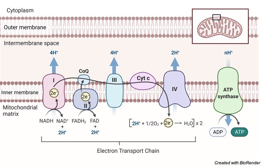
1. Nicotinamide Adenine Dinucleotide (NAD): It is a co-enzyme that carries hydride ion (H-) and thus known as a hydride carrier.
It gains 2 hydrogen atoms from the substrate of Citric acid cycle like isocitrate, malate, B-hydroxy acyl CoA and B-hydroxy butyrate and form NADH molecule.
In the process, one electron releases which reduces NADH to NADH+ while passing its hydrogen to flavoprotein containing FMN and iron sulphur protein (FeS).
2. Flavoproteins (FAD and FMN): In the meanwhile, flavoproteins like FAD and FMN both serves as hydrogen acceptor wherein they tightly bound the hydrogen atom.
Thus, directly preventing its reduced form to react with oxygen.
In general, the types of flavoprotein receiving hydrogen passes them to coenzyme Q.
For example, flavoprotein Fp1 receives 2 hydrogen atoms from reduced NAD+, while flavoprotein Fp2 receiving the same from various substrates like succinate, acyl CoA and choline.
3. Ubiquinone: It is also known as Coenzyme Q that contains quinone ring. Coenzyme Q is the most common ubiquinone that shows similarity with vitamin K.
It is lipid soluble with small molecular size thus responsible for free mobility of the molecule in the inner membrane of mitochondria.
It is the basic carrier of the hydrogen atoms, wherein ubiquinol carries 2 hydrogen atoms while semiquinone carries only one.
These molecules connect flavoprotein with cytochrome b, where flavoprotein carries 2 hydrogen atoms while cytochrome b carry one.
Reduction of coenzyme Q leads to passage of electrons to cytochrome b while releasing 2H+ into the mitochondrial matrix.
The enzymes responsible for the oxidation of ubiquinol involves:
a): Conduction of electrons to cytochrome c is mainly responsible by ubiquinol (coenzyme Q) dehydrogenase along with coenzymes cyt b, FeS protein and cyt c1.
b): Another further transfer from cyt c to oxygen is carried out under the presence of cytochrome oxidase along with cyt a and cyt a3 as coenzymes.
4. Cytochromes are mainly responsible to carry electron from coenzyme Q to oxygen. Thus, exhibits the important role of electron carrier.
They are categorized with the letter a, b and c.
The core molecule for all the cytochromes are haemoproteins however they diverge in their redox potential.
The iron of cytochrome frequently changes their state from oxidation (Fe3+; ferric state) to reduction (Fe2+; Ferrous state) during their normal physiologic action, while that of haemoglobin remains in the reduction state.
Being mobile components, both coenzyme Q and cytochrome c transport reduced equivalent from the other fixed components.
5. Iron Sulfur Clusters(FeS): One additional component found is iron sulfur proteins also known as FeS or non-heme iron.
It is often related with flavoproteins and cytochrome b wherein iron is interchangeably exchange between flavoprotein and cytochrome b resulting in redox reaction.
B. Enzyme Complexes of the Electron Transport Chain
In the inner mitochondrial membrane four enzyme complexes are arranged which transport electrons. The following are the enzyme complexes observed:
Complex I known as NADH dehydrogenase (NADH-ubiquinone oxidoreductase) It is a flavoprotein wherein FMN and FeS protein are the coenzymes.
It facilitates transport of hydrogen atoms from NADH to ubiquinone in the form of NADH+ H+.
Complex II is known as Succinate dehydrogenase (succinate-ubiquinone oxidoreductase).
It is also a flavoprotein wherein FAD and FeS protein are the coenzymes.
It transfers hydrogen atoms from succinate to ubiquinone.
Complex III is known as Ubiquinol dehydrogenase (ubiquinol-cytochrome c oxidoreductase).
It facilitates transport of electrons from ubiquinol to cytochrome c with the help of coenzymes cyt b and cyt c1.
Complex IV is known as Cytochrome oxidase (cytochrome-oxygen oxidoreductase).
It facilitates transport of electrons from cytochrome c to oxygen with the help of coenzymes cyt a and cyt a3.
Moreover, there is a fifth complex (Complex V) known as ATP synthase and is facilitates the conversion of ADP to ATP using inorganic phosphate.
Electron Flow in Electron Transport Chain
The oxidation of substrates with the help of NAD+ or FAD results in production of hydrogen atoms that can pass into the electron chain transfer which further successfully transferred to oxygen to generate energy and water.
The hydrogen atoms cannot be accepted directly by the cytochrome b.
Thus, Coenzyme Q ionized the hydrogen atoms to release hydrogen ions and electrons.
The electrons will reduce the heme group from the cytochrome B, with this starts the transfer chain of electrons from cyt c1, cyt c, cyt a to cyta3.
Finally, the electrons are transported with the help of cytochrome oxidase towards oxygen to form ionic oxygen.
ATP Yield in Electron Transport Chain
The ATP count varies with the catabolism of glucose. Moreover, FAD+ molecules can transport fewer ions resulting in fewer ATP molecules.
In addition to this, the intermediate components of glucose can be used for different purposes resulting in variation in the ATP count.
Summary of Electron Transport Chain
The electron transport chain plays significant role in the aerobic respiration where the free oxygen accepts the last electron produced after glucose catabolism as a result from intermediate compounds.
The structural configuration of electron transport chain showed four large, multi-protein complexes penetrated in the inner membrane of mitochondria along with this 2 small diffusible electron carrier are present transferring electrons between them.
During the transfer of the electrons, small amount of energy is lost against the chemiosmosis.
Thus, high-energy electrons are donated by the NADH or FADH to compensate the energy to finish the chain.
Water and ATP are the end products of the electron transport chain.
Moreover, various intermediate products of the tricarboxylic acid cycle can be averted into the anabolism of other biochemical molecules which may serve as the benefactor for glucongenesis.
Electron Transport Chain Citations:
- Mitochondrial electron transport chain: Oxidative phosphorylation, oxidant production, and methods of measurement. Redox Biol . 2020 Oct;37:101674.
- Respiratory metabolism: glycolysis, the TCA cycle and mitochondrial electron transport. Curr Opin Plant Biol . 2004 Jun;7(3):254-61.
- The mitochondrial respiratory chain. Essays Biochem. 2010;47:1-23.
- Bacterial electron transport chains. Annu Rev Biochem . 1988;57:101-32.
- Kinetic modeling of the photosynthetic electron transport chain. Bioelectrochemistry . 2001 Jan;53(1):35-53.
- Electron transport chain defects in Alzheimer’s disease. Neurology . 1995 Mar;45(3 Pt 1):599-600.
- Bioflavonoid effects on the mitochondrial respiratory electron transport chain and cytochrome c redox state. Redox Rep . 1999;4(1-2):35-41.
- Electron transport in xanthine oxidase. A model for other biological electron transport chains. Biophys J . 1976 Aug;16(8):939-51.
Share
Similar Research Position:
-
Monocistronic mRNA: Definition, Importance, and Examples
What are Monocistronic mRNA?
Monocistronic mRNA is a mRNA that encodes just a single protein and all eukaryotic mRNAs are monocistronic.
The improvement of the develop monocistronic eukaryotic record includes a few diverse handling steps.
Heterogeneous nuclear RNA (hnRNA) refers to the large pre‐mRNAs of various nucleotide sequences transcribed by RNA Polymerase II.
hnRNA transcription is only a small portion of the transcription which occurs in the nucleus.
Up to 90% of the transcription is for the production of rRNA. But if we limit our discussion to hnRNA some important observations can be made.
First, the complexity of the hnRNA is 4 to 10 times greater than that of the mRNA. This suggest two possibilities.
One, we already know and that is that splicing of genes reduces the size of the final mRNA product in comparison to the original transcript. But this does not account completely for the differences in complexity.
Monocistronic mRNA Features
The majority of the difference appears to be accounted for by destruction of hnRNAs before they develop into mature mRNAs.
hnRNAs are a broad class of products:
• Average size – 8000 – 10,000 bases
• Range – 2000 – 14,000 bases
Since the mean mRNA length is 1800-2000 bases, a sustainable amount of cutting must occur. hnRNA doesn’t really exist in the core as bare RNA however is really connected with protein.
This complex is assigned hnRNP and is called heteronuclear ribonuclearprotein.
hnRNP is related with the nuclear matrix. Since chromatin is connected to the nuclear matrix it appears to be intelligent that the essential transcript will likewise get appended to the matrix soon after transcription.
Processing of Monocistronic mRNA
The lifetime of hnRNA is very short generally from one minute to one hour and no more. The initial phase in handling is the expansion of a cap.
The cap is a 5′ methyl guanosine that is added following the beginning of transcript. Capping happens so rapidly that we seldom see the original 5′ base of the message.
The linkage between the 5′ methyl guanosine isn’t the commonplace 5′- 3′ linkage yet is a 5′- 5′ linkage. The reaction is catalyzed by the enzyme guanylyl transferase.
The guanosine that is attached is consistently methylated at the 7 situation of the guanine base (7mG). This is called cap 0.
Likewise, a methyl bunch is added to 2′- OH of the first base in the mRNA. This is catalyzed by 2′- O-methyl-transferase, and this methyl group is alluded to as cap 1.
Other methylations can takes place, however we will not think about them. About 25% of hnRNA in the end develops in polyadenylated mRNA.
Not all mRNAs are polyadenylated. The histone mRNAs are a remarkable exemption for the standard. In the event that an inhibitor of polyadenylation is added to a response hnRNA isn’t changed over into mRNA.
In this way polyadenlyation is a prerequisite for mRNA to show up.
End of transcription isn’t perceived in any way. One succession that is invariant in eukaryotic mRNA is the sequence 5′- AAUAAA-3′ that is seen around 10-30 bp upstream of the poly A tail.
The inquiry that this raises is whether this sequence is needed for polyadenylation. Erasures or mutation of this sequence will kill polyadenylation.
Yet, astounding, point mutation incredibly decreased the quantity of molecules that are divided.
In any case, those that are separated are polyadenylation. Consequently, this sequence is by all accounts needed for cleavage of the essential transcript.
The poly-A tail is added by the compound poly(A) polymerase.
Monocistronic mRNA Characteristic
Monocistronic mRNAs are the standard mRNAs in eukaryotic cells are perpetually monocistronic, and the size of the mRNA is normally corresponding to the length of the encoded protein.
This is genuine likewise of most plant and animal viral mRNAs, despite the fact that there are, among viral messenger, a few exemptions for the monocistronic rule.
These mRNAs are basically polycistronic: they encode at least two overlapping proteins. Anyhow, in every such case, save one, interpretation is restricted to the 5′- proximal cistron.
(The solitary archived special case is simian virus 40 late 16 SmRNA, in which the 62-aminoacid “agnoprotein” is encoded up-stream from capsid protein VP1 and the two proteins are translated.
These mRNAs are in this manner adjust to the standard for eukaryotes in that they are practically monocistronic. Few eukaryotic mRNAs comprise a third category.
These mRNAs encode and express two proteins. Much of the time, these outcomes from commencement at the first and second AUG trios – interestingly with most of eukaryotic mRNAs.
Monocistronic Rule
The 5′- proximal AUG works as aninitiator codon. Considering the scarcity, they present just a minor inconsistency to the monocistronic rule.
The monocistronic rule for gene expression in eucaryotes states not just that a single mRNA coordinates synthesis of a single protein yet in addition that the protein encoded nearest to the 5′ end is the one that gets expressed.
This remains constant for the entirety of the naturally occurring viral mRNAs recorded, just as for misleadingly constructed messengers that encode two proteins.
The 5′- terminal restriction is imposed not by the structure of eukaryotic mRNAs yet rather by the properties of eukaryotic ribosomes.
This was astutely shown by brooding wheat germ ribosomes with a polycistronic mRNA from bacteriophage lambda.
Though E.coli ribosomes promptly translate the first and second cistrons in that message, wheat germ ribosomes decipher just the first.
Inability to decipher down-stream cistrons suggests that eukaryotic ribosomes can neither tie straightforwardly to internal initiation sites nor reinitiate at a subsequent cistron in the wake of crossing the first.
Plainly, eukaryotic ribosomes work under restrictions that don’t happen in procaryotes.
The failure of eukaryotic ribosomes to start at destinations in the inside of a message implies that eukaryotic cells can’t organize synthesis of a bunch of proteins by grouping the genes behind a single transcriptional promoter.
Polycistronic transcripts, which are the transcendent regulatory device utilized by procaryotes, won’t work in eucaryotes.
Taking into account this restriction, it is fascinating to ask what compensatory mechanism eukaryotic cells have formulated to organize gene expression.
Eukaryotic genes are sometimes fused, hence creating, in lieu of a polycistronic mRNA, a “polyprotein” which is gotten from a single longsome cistron.
Monocistronic mRNA Example
Tryptophan synthetase is one such model: the alpha subunit (Mr28,727) and 32 subunit (Mr42,756), which are encoded by isolated genes in E.coli, are fused in S. cerevisiae to form a bifunctional protein of Mr76,000.
Different instances of fused genes encoding multifunctional proteins are the arom gene cluster in Neurospora, which incorporates five enzymes associated with the polyaromatic biosynthetic pathway, and the his4 gene in S.cerevisiae which encodes a trifunctional protein.
Maybe the most noteworthy model is mammalian unsaturated fatty acid synthetase (Mr240,000), which relates to a set of 7 individual polypeptides in E.coli (2%).
Albeit the reasoning behind gene combination is more managable to speculating than to experimentation, it is conceivable that fusion of genes makes up for the failure of eukaryotic ribosomes to utilize polycistronic transcripts for the coordinate synthesis of polypeptides.
Monocistronic mRNA Citations
- Translation of the RNAs of brome mosaic virus: the monocistronic nature of RNA1 and RNA2. J Mol Biol . 1976 May 5;103(1):77-88.
- Gene Gangs of the Chloroviruses: Conserved Clusters of Collinear Monocistronic Genes. Viruses . 2018 Oct 20;10(10):576.
- Identification of novel monocistronic HTLV-1 mRNAs encoding functional Rex isoforms. Retrovirology . 2015 Jul 2;12:58.
- A promoter within the E6 ORF of human papillomavirus type 16 contributes to the expression of the E7 oncoprotein from a monocistronic mRNA. J Gen Virol . 2003 Dec;84(Pt 12):3429-3441.
Share
Similar Post:
-
Seed Germination: Definition, Process, Methods, and Major...
What is Seed Germination?
Seed Germination is defined as one of the vital process by which all the species of plants got generated from a single seed and how it gets adapted to a particular environment and grow itself as a crop or as an herb or as shrub or a tree.
The best example that we come across our day-to-day life is sprouting of pulses in our kitchen or young plants developing in our kitchen garden.
Process of Seed Germination
When we sow a seed in a mud, first the seed starts absorbing water from its outer environment or directly from the soil, which results in bulging of seed and its seed coat gets moisture and becomes very vulnerable at a particular temperature.
This stage of the seed which can be seen from one to two days of sowing is commonly referred to as imbibition.
It further resumes its growth process by activating several enzymes and starts its respiration and initiates the production of proteins and stores nutrients in it by metabolising those necessary proteins.
Hence, it is said that sprouts are rich in proteins.
This stage of the plant is generally referred to as lag phase. Then the seed coat starts rupturing and gives rise to a small radicle, which later forms the primary root of a plant.
This helps the plant to absorb the underground water along with other nutrients present in the soil and helps in further growth of the plant.
The slowly starts growing the plumule of the plant which later develops into the stem portion of the plant.
As days move on seeds modify itself as seedling, then plumule starts growing upwards giving rise to leaves and other floral parts of the plant.
Optimum Condition for Seed Germination
Not all the seeds have the capability to grow at any conditions we sow, certain seeds grow only at certain conditions and certain soil and climatic conditions.
The things that are necessary for a plant to grow in a good condition are listed below.
A. Water
As every living organisms’ water is the most vital component for a seed to germinate.
Though some seeds do not require much water for their growth they need at least a minimal quality of water at certain intervals.
It depends on the dry weight of the seed. As discussed above the seed starts its germination process by absorbing water which is necessary for the nourishment of the embryo by providing dissolved oxygen and soluble supplements for their growth.
The dissolved oxygen helps the seed coat to become softened and converts the seed into a sapling by rupturing the seed coat.
B. Oxygen
As water oxygen is also very much important for the seed growth, which is obtained through the water in a dissolved form or through the porous soil.
But the deep buried seeds are not capable to use the oxygen from the soil pores.
Oxygen is vital for the metabolic process of the seed in terms of aerobic condition.
C. Temperature
If the seed wants to grow in a proper manner without any deficient conditions then the temperature should be maintained at an average of 25 to 30ºC. But in some cases, the seeds need a temperature of 5 to 40ºC.
Hence the temperature varies according the particular regions where they are native to and other environmental factors.
D. Light and Dark Cycle
This depends on the nature of certain seeds, some seeds do not germinate if sunlight falls on them, such kind of seeds are grown in a dark condition and after germination they are brought to the field.
Whereas most of the plant needs sunlight for the germination and throughout their lifetime.
Thus, the seed germination needs all these vital factors and generation of plumule and radicle is said to be completion of seed germination.
Factors Affecting Seed Germination
Factors affecting seed growth or germination may be both internal and external and they are listed below as follows.
Internal Factors
Seed dormancy is considered as one of the important factors in germination process which means that seeds do not germinate if they met any unfavorable conditions during their process of undergoing their growth.
During this period the seed coat which allows the passage of water restricts the water to absorb so that the oxygen and nutrients necessary for their growth is limited.
So that the seeds remain as such without any development. But in some cases, the plant growth regulators help in the growth of the plant.
However, some seeds germinate within a week where as others take their own time to germinate.
External Factors
As mentioned above water, oxygen and temperature are considered as important factors for the germination of a plant.
a. Water
Seed growth gets affected when the water is supplied at higher amount than the needed quantity and also when it is supplied in very low quantity or there is no supply of water.
So, it is important to know the nature of seeds and their adequate flow of water.
b. Oxygen
While the seeds start its germination process it intakes large amount of water than other intervals, so it is checking the availability of oxygen for the growth of the plant.
c. Temperature
Seeds require only the appropriate temperature necessary for their growth if the temperature is more vigorous than needed then seeds starts shrinking and it will die or degenerate.
So, the seed must be sown in optimum environmental conditions by considering the environmental factors.
Seed Germination Citations
- Seed dormancy and germination. Curr Biol . 2017 Sep 11;27(17):R874-R878.
- Seed germination traits shape community assembly along a hydroperiod gradient. Ann Bot . 2020 Jan 8;125(1):67-78.
- Phytochrome Regulation of Seed Germination. Methods Mol Biol . 2019;2026:149-156.
- Seed germination and dormancy: The classic story, new puzzles, and evolution. J Integr Plant Biol . 2019 May;61(5):541-563.
- The biomechanics of seed germination. J Exp Bot . 2017 Feb 1;68(4):765-783.
- Studies on the molecular mechanisms of seed germination. Proteomics . 2015 May;15(10):1671-9.
Share
Similar Post:
-
Mendel’s Laws of Inheritance : Types and...
What is Mendel's Laws of Inheritance or Mendelian genetics?
Mendelian genetics or Mendelism or Mendelian inheritance is a pack of primary tenets that are affiliated to the relaying of hereditary features from parent organisms to their kids; it is nothing but due to the genetics.
These tenets were originally extracted from the study of Gregor Mendel published in the year 1865 and 1866, which was founded in 1900.
It was originally controversial, however later it became core of classic genetics.
Historical Facts About Mendel's Laws of Inheritance
Credit of laws of inheritance goes to Gregor Mendel, a monk of nineteen century who performed experiments on hybridization in peas (Pisum sativum).
Between 1856 and 1863, he grew and tested around 28,000 pea plants and from this he deduced two generalizations that became famous as Mendel’s Laws of Heredity or Mendelian inheritance.
This is delineated in two sections of the research paper “Experiments on Plant Hybridization”, that was published in the journal in the year 1866.
What is Inheritance?
Inheritance can be defined as the procedure of how a child receives genetic information from the parent.
This process of hereditary is related to the dependence which gives the offspring same trait as of parents.
Inheritance is which is why, the family members have similar and distinctive features.
Why Mendel Choose Pea to Study Genetics?
He chose pea for his experimental procedures because it can be grown easily and are naturally self-pollinating.
Pea plant not only can be self-pollinated but also can be cross-pollinated.
It is yearly plant, ergo, many generations can be followed in less duration of time.
Not only this it also possesses contrasting traits.
Mendel's Laws of Inheritance
The two majors’ experiments done by Mendel to establish the laws of inheritance (Mendel’s Laws of Inheritance) were:
1. Monohybrid Cross Experiment
2. Dihybrid Cross Experiment
During his procedure he found that many features and traits passes to the offspring in a stable way.
These factors are known as genes. Unit of inheritance is nothing but genes.
Mendel’s Experiments
He conducted experiments and contemplated seven major contrasting features in the plants.
Then, he conducted both the experiments to determine the aforementioned inheritance laws.
1. Monohybrid Cross
He took two plants of pea with contradictory traits that is one short and one tall and then crossed them.
It was noticed by him that the offspring’s of the 1st generation were tall and he termed it as F1 progeny.
Then he crossed F1 progeny which yield him tall as well as short plants in the ratio 3:1.
Mendel even did experiment with other contrasting features like green peas vs yellow peas, round vs wrinkled, etc. and he found that the results were the same.
This led to the discovery and the formulation of the laws of Segregation and Dominance.
2. Dihybrid Cross
Mendel in his dihybrid cross trial he took two traits, each having two alleles and then he crossed wrinkled-green seed and round-yellow seeds and noticed that all the first generation progeny (F1 progeny) were round-yellow.
He therefore concluded that the dominant traits were the round shape and yellow colour.
He also self-pollinated the F1 progeny and got 4 different features like wrinkled-yellow, round-yellow, wrinkled-green seeds and round-green in the ratio 9:3:3:1.
Mendel's Laws of Inheritance
Mendel postulated three laws, which are now called after his name as Mendel’s laws of heredity.
1. Law of dominance
2. Law of segregation
3. Law of independent assortment
1. Law of Dominance
When two homozygous individuals with one or more sets of contrasting characters are crossed, the characters that appear in the F1 hybrids are dominant characters and those do not appear in F1 are recessive characters.
The dominant and recessive traits of the genes can be dependent on the enzymatic function of the genes of which dominant genes can make functional enzymes which synthesize active polypeptides or proteins.
On the other hand, the recessive genes (mutant genes) code for incomplete or non-functional polypeptides.
Therefore, the dominant genes make a distinct phenotype which can be expressed even in the heterozygous condition while the recessive cannot make so.
Researchers undertook various cross-breeding experiments to authenticate applicability of law of dominance like Correns on peas and maize, Tschermak on peas, and many others.
They discern plenty of characters in several life form is related as dominant and recessive.
Importance of Law of Dominance
In the hybrid trait the normal characteristics shown by dominant gene reveal significant advantages over the dreadful characters exhibited by recessive gene.
It was noted that diabetes, hemophilia etc. are all traits of recessive characters.
A hybrid person may possess all these characteristic in addition to this recessive gene can persist for next several generations without expressing themselves.
In contrast, there is exception to Law of Dominance which is known as Incomplete Dominance.
Mendel was followed by several scientists in case of blending of characters of two parents.
This trait shows characteristics which are amalgamated from both the parents with both sets of genes which is commonly known as incomplete dominance or blending inheritance.
It is the result of expression of genes partially from the dominant trait and partially from the recessive one.
For example, in four O-clock plant, Mirabilis Jalapa, red (RR)flower plant is crossed with white (rr) colored plant resulting in plant with pink (Rr) color flower.
On self-pollination with pink color flower the ratio of getting red (RR), pink (Rr) and white (rr) is 1:2:1.
2. Law of Segregation (Purity of Gametes)
This law states that when trait of two opposite genes or allelomophs are mixed together the resultant pair stays without being contaminated while gametes separate out from each other and only one enters each gametes.
As a result, two types of gametes are produced from the heterozygote in equal numerosity. 50% of the gametes possess gene T and other 50% possess gene t.
Importance of Law of Segregation
According to cytological studies, the law of segregation holds good to all cases whether it is dominance or no dominance.
Rare biological generalization has made its applicability far out reaching.
3. Law of Independent Assortment
In this law, the study of inheritance of more than one pair of characters is carried our simultaneously, wherein the factors or genes can be sorted independently from its correspondent allele.
Based on the result of dihybrid cross, Mandel formulated Mendel’s Laws: Independent Assortment .
Importance of Law of Independent Assortment
Various cytological studies revealed that those allelomorphs which are assorted independently during cell division, are actually located on different homologous pairs of chromosomes.
However, if the traits for various characteristics are seen on the same homologous pair of chromosomes, these are passed on to the same gamete.
This law fails on its universal applicability.
Biological Significance of Mendel's Laws
Mendel’s work unfolded after 3 decades of his actual work and the laws he mentioned was widely used by various branched for the purpose of scientific evaluation.
Many modern genetic modification was seen like getting rust-resistant and disease-resistant varieties of grains and for breeding high grade variety of horses and dogs.
The science of segregation is the outcome of Mendelism, which deals with the betterment of human race.
Conclusions from Mendel’s Experiments
Genotype is defined by the genetic makeup of the organism while phenotype is defined by its physical appearance.
The genes are transmitted from parents to offspring and is called as allele.
There is a 50% chance of one of the two alleles to fuse with the other parent during gametogenesis when the chromosomes are halved.
When the alleles are the same, they are known as homozygous alleles and when they are different they are known as heterozygous alleles.
Mendel’s law benefited the humankind by generating high grade breed of various plants and animals.
Moreover, they have higher implication in diagnosing and preventing various genetic disorders.
Mendel's Laws of Inheritance Citations:
- Mendel’s laws, Mendelian randomization and causal inference in observational data: substantive and nomenclatural issues. Eur J Epidemiol . 2020 Feb;35(2):99-111.
- Limits of imagination: the 150th Anniversary of Mendel’s Laws, and why Mendel failed to see the importance of his discovery for Darwin’s theory of evolution. Fly (Austin) . Jan-Mar 2013;7(1):28-38.
- Mendel’s law reveals fatal flaws in Bateman’s 1948 study of mating and fitness. Fly (Austin) . Jan-Mar 2013;7(1):28-38.
- ‘Rediscovery’ revised – the cooperation of Erich and Armin von Tschermak-Seysenegg in the context of the ‘rediscovery’ of Mendel’s laws in 1899-1901. Plant Biol (Stuttg) . 2011 Nov;13(6):835-41.
- Historical study: Johann Gregor Mendel 1822-1884. Am J Med Genet . 1991 Jul 1;40(1):1-25; discussion 26.
- A female contribution to early genetics: Tine Tammes and Mendel’s laws for continuous characters. J Hist Biol . Fall 1995;28(3):495-531.
Share
Similar Post:
-
Amphibolic Pathway: Definition and Example I Research...
What is Amphibolic Pathway?
Survival of the organism depends on the nutrient uptake from the environment and complete utilization of nutrients.
This process is vital and forms a dynamic nature of the cell. The conversion of external nutrient to an internal biomolecule is a main function of any biosystem present and is achieved through the process of metabolism.
Metabolism is the process by which nutrient are converted to desirable products for synthesis and breakdown of biochemical components
"A pathway which participates in Catabolism and anabolism is termed as amphibolic pathway"
Metabolism is otherwise termed as Catabolism + Anabolism. Catabolism is a process of breaking down of Complex food molecules to simpler compounds.
Anabolism is mainly concerned with the biosynthesis of Chemical compounds which are essential for storage and other functions from smaller molecules.
Synthesis (Anabolism) and degradation (Catabolism) of any biomolecule takes place in a series of process which are distinct in its own mechanism and have specific pathway.
But there are certain pathways which can act both catabolic an anabolic under appropriate circumstances.
A pathway which participates in Catabolism and anabolism is termed as amphibolic pathway.
Amphibolic Pathway involves many intermediates which forms new compounds (biosynthesis) such as amino acid, fats, glucose etc., or they get oxidized to produce energy.
An ideal example for Amphibolic Pathway is the CITRIC ACID CYCLE. The citric acid cycle is Amphibolic because it oxidizes Carbohydrates, Fats, Proteins to yield energy and it is the common oxidative pathway for any biochemical compound.
Any biochemical degradation either directly yields acetyl Co – A or through pyruvate and other components of citric acid cycle.
Similarly, the intermediates from the citric acid cycle becomes precursor for the synthesis of Glucose, fats, Amino Acid etc.,
The catabolism and anabolism of the intermediates present in the citric cycle depends upon the internal cues of the body.
Internal cues can be the requirement of synthesis or breakdown of a compound and is dependent on many other factors.
Amphibolic Pathway Characteristics
The main characteristics of amphibolic pathway is the involvement of both catabolic and anabolic pathway and Citric Acid Cycle becomes an ideal cycle to understand the Amphibolic Pathway
Catabolic Nature of Citric Acid Cycle
In cellular respiration, citric acid cycle is the common oxidative pathway where the Acetyl Co – A breakdown yielding NADH, FADH and ATP in plants GTP in Animals.
The basic biological compound namely Proteins, Fats and Glucose oxidizes to form Acetyl Co – A directly or through Pyruvate enters the common pathway of oxidation.
From the basic cellular respiration, we know that the substrates of Citric Acid Cycle are not completely derived from glycolysis but the amino acids and lipids also contribute to the substrate.
Catabolic activities of citric acid cycle are,
1. Oxidation of Glucose
2. Transamination of Amino Acids
3. Oxidation of Fatty acids
Glucose is broken down to pyruvate by Glycolysis.
Fatty Acids on Beta – Oxidation provides Acetyl Co – A directly
Amino Acid Catabolism
Essential and non – essential amino acids are present in body for the proper functioning of the living system.
Among all of them each amino acids do not form acetyl Co – A directly.
Few forms other intermediates of the Citric Acid Cycle by the process of Transamination or Aminotransferase. The Aminotransferase reaction involves the transfer of alpha – amino group from the carbon skeleton and transforming the skeleton to Amphibolic intermediate to produce energy.
Few amino acids oxidize to become pyruvate and Acetyl Co -A also.
For example:
I. Glutamine is transformed to Glutamate with enzyme involving Glutaminase. Glutamate then by transamination becomes alpha – ketoglutarate.
II. Similarly, Asparagine becomes Aspartate on transamination’s yields oxaloacetate and Certain Amino acids like alanine, Serine etc., directly oxidizes to produce pyruvate
Anabolic Nature of Citric Acid Cycle
Biosynthesis of glucose, fatty Acids, Amino Acids, Nucleic Acids are produced from the intermediates of the Krebs cycle.
The intermediates form many precursors corresponding to their respective substrates from which the biosynthesis takes place.
Anabolic activities are;
I. Citrate from mitochondria enters cytosol to oxidize to form Acetyl Co – A, this initiates the Biosynthesis of Fatty Acids.
II. α-ketoglutarate is the precursor for Amino Acid Glutamate which on Transamination provides Glutamine, Arginine and Proline and provides Purines.
III. Succinyl Co – A becomes a precursor for Porphyrin and Heme forming oxygen carriers in Blood.
IV. Oxaloacetate is the precursor for Aspartate and they form Asparagine to produce pyrimidines and other Amino acids that acts as an intermediate for Gluconeogenesis
Amphibolic Pathway Citations
Share
Similar Post:
-
What is Lactic Acid? Where Does it...
What is Lactic acid?
Do you know what type of acid is present in all the milk products including cheese, milk, curd and butter.
Yeah, absolutely it’s a Lactic acid.
Lactic acid is generally considered as an organic acid and it is produced by a fermentation process which occurs in milk sugar.
It was first identified by Carl Wilhelm in 1780 in milk. It is a colorless water-soluble substance which has been widely used in food and pharmaceuticals and it has also been synthesized by our body in many metabolic conditions.
The First Demonstration of Lactic Acid
Lactic acid was first identified by Scheele in sour milk in the year 1780. Further when Pasteur was undergoing research on pasteurization, he found the microorganism which is necessary for lactic acid production.
Lactic acid is also being commercially sold for many purposes.
How Lactic Acid is Utilized by Human Body?
Lactic acid is used for the process of conversion of glucose into energy when our body has very low oxygen content.
We often release a painful sensation when we exercise or do heavy activities suddenly or after a long interval, it is due to the accumulation of lactic acid in the muscles which results in muscle cramps at a certain area in our body.
It does not cause any serious issues as liver has the capability to breakdown the extra lactate.
How Lactic Acid Build up in Our Body?
In some cases, our muscles do not have enough oxygen to breakdown glycogen into glucose for the synthesis of energy molecules which is known as ATP, at this time lactic acid gets accumulated in a certain area where enough oxygen is not available which is often referred to as anaerobic metabolic process.
When lactate gets accumulated it leaves to formation of lactic acidosis.
It may also upgrade at the times of heart attack, sudden shock, and at the cases of lung diseases.
Lactic acid forms in many varieties at each area according to their nature.
When they are being accumulated at muscles and blood tissues they are known as dextrorotary form.
The lactate which is obtained as a result of sucrose formation is known as levorotary form, when the same lactate if produced by bacteria during fermentation they are known as racemic form.
Thus, each of its form has its origin from the process of their accumulation or synthesis.
To explain in detail about how our body produces lactic acid, as said earlier lactic acid gets accumulate in our body when we perform heavy works or exercises during a sudden period of time.
This because when we perform powerful activities, we take a long and faster breathes which leads to the usage of more oxygen by the muscles, which tend to perform that action, which results in synthesizing energy anaerobically.
We generally get energy by the process of glycolysis, where glucose is reduced to pyruvic acid and release of ATP molecules occurs through further process which is important for the energy source.
This is a general process, but when our muscles are in a state with absence or lack of oxygen the pyruvate which is produced through a process of glycolysis convert itself into a substance namely lactate and supplies energy in the anaerobic form which leads to the accumulation of lactate at higher levels.
But this is performed by our body, for a longer time it leads to side effects by the accumulation of lactate at higher levels in the tissues which also disrupts the other metabolic activities of the muscle and creates an acidic environment around the muscle cells.
So, it results in degrading a working capability of the muscle and cannot perform any heavy or powerful activities further.
But on considering an immune response of our body, natural immune barriers prevent the muscle tissues from permanent damage and initiates the cells to work normally by upcoming the situation and by performing regular muscle contraction improves the lactic acid accumulation.
This is because our body gets slow during these conditions and allows the reverse mechanism of conversion of lactate into pyruvate and normal aerobic respiration is followed by the tissues.
Fermentation of Lactic Acid
Lactic acids are also synthesized in industries for various purposes through fermentation.
Several microorganisms are involved in the production of lactic acid, each of it has the mechanism of producing either D (-) lactic acid or L (+) lactic acid depending on their nature.
The racemic form is formed due to the presence or production of an enzyme known as racemase, the lactic acid usually be in an active form unless it is deactivated by any other enzymes.
The two most important steps involved in the production of lactic acid are as follows,
1. Homo-fermentative Process
This type of process involves the utilization of certain bacteria such as Lactobacillus delbruckii, L.casei, L.leichmanii, streptococcus lactis etc. which utilizes its EMP pathway to synthesis pyruvic acid which helps in the production of lactic acid by reducing lactate dehydrogenase.
Though these microbes can serve under anaerobic conditions they are known an anaerobic microbe.
2. Heterofermentative Process
This process involves the synthesis of lactic acid, ethanol, acetic acid, carbon dioxide and water with the help of Leuconostoc mesenteroids.
Manufacturing of Lactic Acid
The medium should be prepared such that it contains masses of molasses, maltose, lactose, sucrose, calcium carbonate, hydrogen and phosphate which are kept at a pH of 5.5 to 6.5.
As lactic acid is soft and delicate in nature the mixture of metals is avoided in the culture medium and fermenters are nature based and mostly wood fermenters are used.
The use of Thermophilic clostridia in the culture results in the formation of major and important constituents of lactic acid namely butanol and butyric acid.
Uses of Lactic Acid
Considering lactic acid as a commercial one it can also be used in polymer production since it has a property of weak acid.
It is also being used in food and other industries such as production of beverages in preservation as it has an acidic property which is due to the anaerobic property of that which prevents the food from spoilage.
It is also being used in leather industries for de-lining and also in laundry and textiles for the treatment of fabric.
Calcium lactate is being used as a source of calcium in pharmaceuticals and also in baking units.
Lactic Acid Citations
- Lactic acid and exercise performance : culprit or friend? Sports Med . 2006;36(4):279-91.
- Lactic acid fermentation of human excreta for agricultural application. J Environ Manage . 2018 Jan 15;206:890-900.
- Biosynthesis of D-lactic acid from lignocellulosic biomass. Biotechnol Lett . 2018 Aug;40(8):1167-1179.
Share
Similar Research Position:
-
What is Sex Determination System? Definition and...
Sex Determination Overview
By nature, we all look every living organism in this environment as male or female, considering from a plant, pets and we the humans; because these characters are externally shown and they can be easily differentiated into separate individuals.
We will be discussing here about how each sexes are determined, either through gene or through other external features.
Usually, all organisms in this living environment are classified as two sexes namely male and females depending upon their gametes and sexual characters and differentiation among them.
Accordingly, the Latin term sex means separation or section. Some organisms of lowest forms contain more than two sexes, where the species of protozoa which is known as Paramecium bursaria has eight sexes.
The organisms which usually posses two sex organs in a same individual are referred as hermaphrodite individuals.
Earthworms are hermaphrodites: each carries male and female sex organs.
Where as in flowers the staminate or anther is seen in males and pistillate is being seen in females but they are present in the same plant hence the term monoecious is used here.
But most of the flowers has both male and female organs in a same plant and it is called as the original or a perfect flower.
As such for animals the organisms which produce male and female gametes individually are referred by the term dioecious.
However, these sex cells and reproductive organs and their respective hormones play an important role in determining their primary and secondary sexual characters.
Mechanism of Sex Determination
Mostly our sexes are determined in a genetical manner and they are classified into four categories as follows,
1. Heterogamesis or sex chromosome mechanism
2. Genic balance mechanism
3. Male haploidy or haplodiploidy mechanism
4. Single gene effects.
1. Heterogamesis
Generally, in dioecious organisms two types of chromosomes are present such as autosomes and sex chromosomes.
The sex chromosomes form two types of sex determination depending on two systems namely heterogametic females and heterogametic males.
In heterogametic sex, females have two x chromosomes where as the males have one x and one y chromosomes with 50 percent of the gametes.
The heterogametic male contains XX-XO type in plants and XX-XY type in mammals.
Where as the heterogametic female contains ZZ-ZO types in moths and ZW-ZZ type in fishes and certain insects
2. Genic Balance
This mechanism explains that some genes are being carried by sex chromosome which is responsible for the determination of sex in individuals.
There will be mostly an inherited mechanism of sexes in both individuals considering from masculine features in male to feminine characters in female.
This was discovered by Wilson, Bridges and Goldschmidt in different organisms.
If an individual is found intermediate between both sexes they are referred to as intersexes.
Sex Determination in Drosophila
The Y chromosomes present in a drosophila are very much important for maintaining the fertility in male flies but they do not have much importance in determining the sex.
The sex of the flies are determined polygenically, where the genes are situated in a distributive manner such that autosomes determine the male character and where as in female it is determined by X chromosome.
Bridges crossed the polyploid flies of experimentally produced female drosophila which has triploid set of chromosomes (3AA:3XX) to that of the diploid male chromosomes.
Which resulted in the formation of intersexes, superfemales and super males and also a normal diploid male along with the triploid females.
From this experiment he concluded that autosomes also carry the genes which are responsible for sex determination.
The intersexes obtained were sterile with both male and female phenotypic characters. This proves the genic balance mechanism.
Sex Determination in Human
Like Drosophila, sex determination can also be seen in humans, where male has XX-XY pair of chromosomes, here Y trait has a potential to bring all male determining characters, where as X chromosome is considered as a feminine character determiner.
Whereas the male having one X and one Y chromosomes can be a normal male by overcoming the feminine characters of X chromosome until there is no chances of chromosomal abnormalities or abbretions, which results in syndromes such as Turner and Klinefelter’s which causes feminine characters in male; like inhibiting sperm production, lack of masculine features; such as bread growth and body hairs, etc. which leads to many physical and mental abnormalities.
Where in Turners syndrome the lack of female characters tends to occur, such as shield chest, short stature, congenital malformations and webbing of neck etc,.
The individual who has addition of both type of chromosomes that is extra X and Y chromosomes, shows true hermaphroditism which have both ovarian and testicular tissues and genitalia appears to be intersexual.
3. Male Haploidy or Haplodiploidy
This is considered as one of the types of parthenogenesis mechanism which is commonly seen in hymenopterous insects.
These types of insects include ants, bees, wasps and sawflies.
Where fertilise eggs develop into diploid females and also as unfertilised eggs are developed into haploid males.
So, this is also considered as a form of sex determination. Considering an example of honey bee, where they can lay two varieties of eggs.
Such that it produces a fertilised egg by controlling a splinter of her sperm receptacle and an unfertilized one. Here the fertilised egg has diploid zygote and develops into a female and an unfertilised egg has only 16 zygotes and develops into a male.
Later the females are grown up as a honey bee or worker bee depending upon the food they consume or diet they undertake and the one which is developed from unfertilised eggs are males.
Single Gene Control of Sex
In certain organisms like Neurospora, Chlamydomonas, asparagus, yeast, maize etc, the single gene plays a very important role in determination of sexes and also in expressing the genes hence it is known as single gene control of sex.
Sex Determination System Citations
- Putting the heat on sex determination. Genetica . 1992;87(1):1-6.
- Prenatal sex determination and selection. Hum Reprod . 1993 Oct;8(10):1545-9.
- Genetic sex determination of mice by simplex PCR. Biol Sex Differ . 2017 Oct 17;8(1):31.
- Sonographic fetal sex determination. Obstet Gynecol Surv . 2009 Jan;64(1):50-7.
- Polygenic sex determination. Experientia . 1964 Apr 15;20(4):190-9.
- Sex Determination and Sex Chromosomes in Amphibia. Sex Dev . 2017;11(5-6):298-306.
Share
Similar Post:
-
Cytoplasm: Definition, Function, and Example
What is Cytoplasm?
We all know that cells are the basic, structural and functional units of all living organisms.
Cells are made up of many organelles and they are bound embedded in the cytoplasm.
It plays a very important role in functioning of the cell and also contains the genetic information of the cell, as it contains chromosomes in it.
The term cytoplasm was first coined by Rudolf Von Kolliker, a Swiss biologist.
"Cytoplasm is contained within cells in the space between the cell membrane and the nuclear membrane"
Cytoplasm is considered as the fluid compartment of the cell, which contains cytosolic filaments, ion substances, proteins and other macromolecular substances. Along which the other cell organelles are found suspended.
The Eukaryotic cytoplasm contains all the cell organelles found embedded in it except the nucleus.
The prokaryotic cytoplasm is found throughout the cell carrying its genetic material as the prokaryotic organism does not have its well- defined nucleus.
Does Cytoplasm Have Any Structure?
Cytoplasm doesn’t have a specific structure its just a jelly or glassy liquid like substance which fills up the space in each cells volume and make the other cell organelles being embedded in it and provides a better environment for the organelles and genetic materials to carry out their function.
Cytoplasm Vs Cytosol
In biology, Cytoplasm is often confused with cytosol due to their similarity in understanding of these words.
Cytoplasm is the liquid part of the cell which is found outside or around the nucleus.
Where as Cytosol is denoted as the substances or proteins which are found in the cytoplasm apart from other cell organelles.
Protoplasm Vs Cytoplasm
The protoplasm is jelly like substance, which is composed of macromolecules and water.
It is actually a colorless living part of the cell. It can be generally known as the organic or inorganic substance which is made up of cytoplasm, nucleus, mitochondria and plastids of a cell, which play a major role in functioning of the cell.
Thus, cytoplasm is a part of a protoplasm which is placed in between the nucleus and the cell membrane in all eukaryotic cells. Where the organelles are embedded in it.
Function of Cytoplasm
Cytoplasm provides a suitable environment for all other cell organelles to maintain their turgidity which helps them to maintain their shape.
The fluid form of the cytoplasm is made up of salts and water which form a jelly like structure and helps the other cell organelles to embed themselves in that space.
The Cytoplasm also acts as a center for other cell organelles to perform their function, as it contains many enzymes and molecules which helps them to perform their metabolic reactions.
As cytoplasm helps the cell organelles to fit it into, so that they cannot move here and there and fix themselves in their origins, if not there is a high chance of mixing up of molecules of one organelle into the other which results in change in body functioning.
The fluid matrix of the cytoplasm which is known as cytosol has no organelle, so it fills up the space which is left out by the organelles.
Cytoplasm also paves the role for many cellular functions to occur in it where the ribosomes synthesize their proteins and the process of cellular respiration takes by initiating glycolysis and reproduction of cell phases like mitosis and meiosis occurs.
Cytoplasmic Streaming
Cytoplasmic streaming is often referred to as cyclosis by which the molecules or substances present in the cell are circulating within the cell.
Cyclosis mostly occur in all types of cells such as Plant cells, in bacteria and fungi.
But these movements change accordingly with the type of chemicals and hormones present in it and also according to the temperature and humidity of the cell.
Plants perform cyclosis to settle the chloroplast in the area, where we can get suitable sunlight.
Whereas chloroplast is a very important and essential component in performing the photosynthesis which help the plant to stay alive.
Whereas in protists such as slime molds and amoeba the cyclosis movement is used for the locomotion.
These temporary movement of cells is termed as pseudopodia which helps the cell in capturing the food.
Cyclosis also helps the cell in cell division processes where the daughter cells are divided by the process of mitosis and meiosis.
Other Features of Cytoplasm
As discussed above cytoplasm is a glassy liquid substance which contains all the cell organelles and other substances except the nucleus.
The eukaryotic cytoplasm is present between the cell membrane and the nucleus of the cell, where as in prokaryotes cytoplasm just acts as a covering to all the substances in a cell membrane.
However, in both the cases the role of cytoplasm is to carry all the metabolism and growth and functions of all other organelles present in a cell.
The organelles present in the cytoplasm perform various functions, where as the nucleus contains the genetic information and helps in caring our hereditary characters throughout the generations.
Mitochondria which is found embedded in the cytoplasm acts as a powerhouse of the cell producing Adenosine tri phosphate.
The chloroplast present in plants cell helps in performing photosynthesis.
Likewise, each cell organelles perform various functions, but without the help of cytoplasm which provides them a good outer condition by providing proper humidity and other conditions, these cells cannot undergo their functions.
The cytosol which acts as a fluid part of the cell, consisting of 70% of water and other molecules including ion particles such as chloride, potassium, bicarbonate magnesium and calcium serves as a site for many chemical reactions.
Cytosol also plays an important role in cell signaling and osmoregulation by inducing action potentials such that of nerve and muscle cells.
Cytoplasmic Biochemical Reactions
Basically, cytoplasm acts as a center for many biochemical reactions, the biochemical reactions are thereby classified as anabolic and catabolic reactions, where catabolism refer to a breakdown of complex molecules and anabolism refers to a synthesis of biomolecular substances with the help of ATP (Adenosine Triphosphate) to form a complex substance.
Biological Functions of Cytoplasm
Cytoplasm acts as a center for both growth and metabolism, as said earlier it helps in both generation and degradation processes.
Whereas glycolysis, which is the first process in the cellular respiration occurs at a cytosol, the fluid component of the cell and this process for further followed by oxidative decarboxylation reaction and Krebs cycle or citric acid cycle and followed by electron transport chain.
Citations
Share
Similar Post: