Category: Study Materials

Category: Study Materials

  • Haemophilia: Cause, Diagnosis, Symptoms, and Treatment

    What is Haemophilia?

    Haemophilia is one kind of inherited genetic disorder. This is the condition where body does not have the capability to clot the blood cells during a case of an injury or any other accidents.

    This leads to the higher risk in people due to its blood accumulation in the joints or at the central nervous system.

    The individuals who are not suffering so much and has only mild symptoms after an injury or an accident does not cause any serious issues.

    Where as bleeding in joints results in permanent damage to the bone cells or cartilage, and bleeding in brain can cause severe headache, seizures, unconsciousness and some times it may also lead to mortality of an individual.

    Types of Haemophilia

    Haemophilia is generally classified into two types based on the clotting factors such as follows;

    Haemophilia A occurs due to a condition where there is a production or synthesis of low amount of clotting factor VIII.

    Haemophilia B is caused due to the low production of clotting factor IX.

    This is considered as an inherited disorder because they are passed down from their parents where the parent carrying X chromosome with defects passes the defected X chromosome to an offspring.

    It is rare that the person is affected by his self without carrying inherited gene due to the result of mutation or any other defects in the chromosome or this may also happen when the self-antibodies are reacting against the body’s own clotting factors.

    The other type which is said to be haemophilia C is due to the lower levels of clotting factor X and the other type known as Para haemophilia, which is due to the minimal amounts of clotting factor V.

    The haemophilia which are due to acquired conditions causes cancers, autoimmune disorders etc.

    To find the ability of blood to clot is the only source of diagnosis for this disorder.

    Characteristics of Haemophilia

    Most of the sex-linked genes are present on the X chromosome because for a simple reason that X chromosomes are larger than Y chromosomes.

    Haemophilia is also an X-linked disorder, it is a type of bleeding disease which is due to the absence of clotting factors in the blood.

    People who are affected with this condition have a long day bleeding or oozing of blood from the place of injury or surgery or even when the tooth is picked off.

    When haemophilia undergoes a serious condition, it leads to continuous bleeding even when we met with small minor injuries, these may also result in the bleeding of joints and muscles, brain or other internal organs.

    This above condition happens often in haemophilia A which is also known as classic haemophilia or factor VIII deficiency and haemophilia B condition is referred to as factor IX deficiency or Christmas disease.

    These two types are almost similar in showing their symptoms and both are inherited through X- linked inherited recessive pattern.

    The genes responsible for this condition are located on the X chromosome which is one of the types of allosome.

    As males have only one X chromosome passing of one defective allele or mutation in one of the alleles in X chromosome can cause this condition easily, where as the females have two X chromosomes in their allosomes; So it is necessary for a female individual that both the chromosomes to get mutated or inherited to get this condition.

    If only one of the chromosomes has got mutated then the respective female is considered as carrier female.

    This is the reason why males are affected more than the females. It is also because the fathers do not pass the X-linked traits to their son is also to be considered.

    The below graph shows how the haemophilia is being inherited.

    Haemophilia - research tweet 1
    How to Prevent Haemophilia?

    We know that since it is an inherited disorder there is no chances of preventing this condition, but it can be diagnosed before the child has been given birth by the process of amniocentesis.

    Where the parents are led to under a counselling to understand the risks of having a baby with this disorder and it is left to their decision whether to brought up a child carefully or terminate it.

    It is the better option to consult a physician if the parents are grandparents have the condition of haemophilia. So they can come to know the results earlier.

    According to research it is said that if 50 percentage if chance where the son will have haemophilia and the other 50 percentage chance is that his daughter will be carrier.

    Symptoms of Haemophilia

    Usually, the symptoms are based on levels of clotting factors that is present in our blood, which are responsible for clotting the blood after bleeding due to an injury or an accident.

    These factors are identified during any surgical operations or while overcoming an injury with bleeding.

    Symptoms of spontaneous bleeding include large and deep bruises joint pain along with swelling, unexplained bleeding and bruises, sometimes there may also a blood in urine or stools.

    Nose bleeds often without having an appropriate reason, excessive bleeding and pain in the gums of the teeth and bleeding may occurs often after vaccinations.

    Haemophilia Citations

    Share

    Similar Post:

    Continue Reading
  • Genetic Disorders: Definition, Development, and Examples

    What are Genetic Disorders

    Genetic disorder is a condition where there is a mutation in a genetic material of an organism which leads to certain abnormalities.

    We know that each cell in our body contains its one molecule of genetic material, which is known as DNA.

    It encodes the cell to perform its particular function by giving instructions to the cell.

    If there is any defect in the genetic material it leads to mutation or any other infections which leads to genetic disorders.

    Cause of Genetic Disorders

    Genetic disorders occurs when there is change in particular segment of a DNA or change or loss of a particular part of DNA or a whole chromosome, which is present within the body cells.

    Almost all cells in our body contains an elongated strand of DNA. Each DNA is made of nucleosides and phosphate groups which encodes the cells to work in an appropriate manner so that the functioning of body remains stable.

    DNA’s are placed in a chromosome, where each chromosome contains the small segment of DNA which is called as Genes.

    It provides instructions to the body. Human body consists of about 23 pair of chromosomes where each parent sends their copy of one pair to their offspring’s, when such genes are passed to a next generation, a defect or mutation in a particular gene causes genetic disorder.

    If the defect is passed from the past generations, it is said to be inherited. Only few people have this condition of getting inherited disorders.

    Genetic disorders are classified into various types depending upon the defect in a chromosome or in a genome.

    Before knowing about the disease-causing factors of gene it is important to about human genome.

    Human Genome

    All the gene and DNA which is required to build a human is referred to as human genome. Human Genome Project (HGP) is one of the global research projects which is used to map the human genome.

    This project helps in sequencing the genes and to know about their different function.

    HGP found that there are about 20,000 to 25,000 genes in the human genome. It also found the genomes and their appropriate functions which helped us to find the disease-causing factor of the genes depending upon the mutation in the bases of the DNA, such as adenine, thymine, guanine and cytosine.

    Each DNA molecule is made up of two strands of DNA Which are coiled around each other in a form of a spiral ladder.

    The bases are present in between the two strands, in a combination of adenine with thymine or cytosine with guanine.

    A change in these combinations also leads to mutation. This change in order of these base pairs affect the instructions that are provided to the body to function.

    DNA sequencing is referred too as reading of these base pairs. Thus, sequencing of genomes provides a better understanding in causative organism of diseases.

    A change or fault in this condition of a DNA causes a genetic condition.

    Since this genetic information is passed to their offspring’s through parents they are considered as inherited.

    But not everyone from this generation will be affected by this because it also depends upon the passing of traits.

    Genetic disorder has the capability to affect any of the genomes which involves many symptoms and causative factors.

    Development of Genetic Disorders

    Genetic characteristics pass from past generation through future generation between the families.

    When parents pass their traits to the children, in some cases they tend to pass their disorders too. Each parent; both father and mother pass half a copy of their genes to their children which is commonly called as allele.

    When two form of alleles are passed from parent to the children, the cells in the body of a offspring take the information from only one pair of that allele, which is known as dominant allele, and the other pair of alleles which is least concerned is referred to as recessive allele.

    In such cases the person develops a genetic disorder if he gets either one of the dominant alleles or both the recessive allele from the infected parent.

    Factors of Genetic Disorders

    Genetic disorders are caused by many factors such as single gene inheritance, multifactorial inheritance, chromosomal inheritance and mitochondrial inheritance.

    Single Gene Genetic Disorders

    Single gene disorder is also known as monogenic disorder.

    It is caused due to the mutations in a single gene caused in an individual and they are passed to the upcoming generations due to various factors such as Genomic imprinting and uniparental disomy.

    Sometimes these conditions may also cause due to invitro fertilization.

    In many cases, the congenital metabolic disorders, which are also known as inborn errors of metabolism are also due to single gene defects.

    Single genes disorders also result in autosomal dominant disorders such as Huntington’s disease or Autosomal recessive disorders such as sickle cell disease, cystic fibrosis, phenylketonuria and thalassemia. 

    It also results in X-linked and Y- linked inherited disorders such as turners’ disorder.

    Genetic Disorder - research tweet 1
    Multifactorial Inheritance

    Multifactorial inheritance is caused as a result of combination of both genetic factors and environment influences.

    The non-genetic factors that influence the individual is smoking, alcohol, cancer, diabetes, multiple sclerosis and Alzheimer’s disease, etc.

    Chromosomal Inheritance

    Chromosomal abnormalities are due to the mutation or change in a chromosome of an individual such as having a lesser number of chromosomes than usual, having extra chromosomes or change in structure of any of the chromosomes.

    Downs syndrome is one of the examples of chromosomal abnormality.

    Genetic Disorder - research tweet 2
    Mitochondrial Inheritance

    This is also due to the single gene inherited disorder, it is also known as maternal inheritance and it occurs in rare cases than usual disorders.

    This condition occurs as a result of defect in any of the 13 genes that are encoded by mitochondrial DNA, As the developing embryo gets its mitochondria from the egg cells.

    Genetic Disorder - research tweet 3

    In case the carrying mother is affected this disorder can pass to the offspring.

    One of the examples of this type of disorder is Leber’s Hereditary optic neuropathy.

    It is also important to know that many mitochondrial disorders are due to defect in the nuclear gene which resembles as such of autosomal recessive inheritance.

    Genetic Disorders Diagnosis and Treatment

    It is also to be clear that not every genetic disorder leads to death of a progeny. Even though there are no remedies for genetic disorders some can be diagnosed at an early stage.

    The genetic disorders such as downs syndrome, and Muscular dystrophy shows no signs until turning into adult.

    Even though there is no treatment there is chances of improving the quality of life than degradation such as physiological therapies and management of pain and choosing alternative medications.

    Though the treatment of genetic disorders is not yet discovered or it is state of ongoing battle.

    However, gene therapy plays an important role in bringing the normal healthy gene into a patient and removing the defective one.

    Genetic Disorders Citations

    Share

    Similar Post:

    Continue Reading
  • Codominance: an Overview, Definition, and Examples

    Law of Dominance: Codominance

    In hybridization techniques, two alleles are considered one as dominant character and other as recessive character when these alleles are let to undergo fertilization by crossing techniques the expression of dominant allele will be high compared to that of recessive allele phenotypically.

    Hence the dominant character will be expressed Hence it is known as law of dominance. Law of dominance is also said to be Mendel’s first law of inheritance.

    Mechanism of Dominance

    On undergoing various experiments Mendel evidenced himself that there will be a difference in their genetic characters, even though the phenotypic character resembles as such as their parents.

    Because the character does not remain the same as it is being for their parents.

    Therefore, there will be something which controls all these characters which are later found as genes which are the units of DNA (Deoxyribo nucleic acid) or RNA (Ribo nucleic acids) accordingly which are present in the chromosomes of an individual and are passed on to the next generation through parents via gametes of male and female.

    Hence the individual has 23 pair of chromosomes each pair from one of the parents, each chromosome consists of genes as their functional unit which consists of contrasting characters made up of alleles.

    If there are two or more contrasting pair of alleles then it is said to be as allelomorph.

    These alleles are produced as the effect of mutation in a wild gene. For example, let us consider a pea plant, where homozygous tall plant has two alleles such as TT on their gene loci in their homologous chromosomes and homologous dwarf plants is represented by the allele tt.

    During the process of gametogenesis, the two homologous alleles TT and tt are separated and each chromosome contains a single allele as T and t and it is passed via gametes.

    These alleles which are passed through gametes of both the parents (father and mother) combine together during fertilisation.

    Thus, the new individual in the F1 generation has two different alleles and it is referred to as heterozygous in condition.

    The dominant character is being expressed and the recessive character of the individual gets suppressed this the mechanism why only dominant characters are expressed though the individual has both the alleles.

    Variation in Dominance

    Mendel studied the dominant and recessive characters in pea plants which helped him to identify seven pairs of genes showing different phenotypes in homozygous and heterozygous condition with lot of variation.

    Variation in dominant character is further classified into two types as Incomplete and complete dominance.

    Codominance

    In some cases, both the alleles in a heterozygote lacks the character of being both dominant and recessive.

    Which means that each trait is capable of obtaining some degree of phenotypic expression from their parents, hence it can also be considered that there will no dominance between two alleles, and they be in equilibrium condition to express their traits.

    1. Example of Codominance

    Here the coat colour of cattle breeds is taken into account, Where the coat colour of the cattle is chosen as Black and white.

    The cattle with Black colour coat have its allele as BB and the white cattle’s coat is denoted as WW.

    When these two alleles are crossed the resultant allele obtained is considered as BW, where the coat colour obtained is Spotted (mixture of both).

    Where the white colour hair is spread throughout the coat and black patches is scattered on the coat.

    In the filial 2 generation the coat colour of the cattle appears to be spotted and also the parental characters of black and white also appears.

    CoDominance-Incomplete Dominance - Definition and Examples - research tweet 1

    2. Example of Codominance

    The best example of co-dominance in humans is ABO blood group, it was first discovered by Landsteiner and Levine.

    The alleles here are represented as A and B accordingly.

    Here three groups are possible which are denoted as A, B and AB which have their alleles as AA, BB and AB accordingly.

    The genotype and its characteristic antigen and antibody are listed below.

    Allele, Allele Definition, What is Allele, Allele Examples,
    Codominance Citations

    Share

    Similar Post:

    Continue Reading
  • Polycistronic mRNA: Definition, Examples, Types, Advantage

    What is Polycistronic mRNA?

    Polycistronic mRNA is a mRNA that deciphers various proteins and is an attribute of many prokaryotic bacterial and chloroplast mRNAs.

    For instance, if a bacterial cell desires to consume lactose as a source of energy, it will duplicate an mRNA molecule that encrypts several protein products needed for lactose metabolism.

    In contrast, eukaryotes possess monocistronic mRNA that only encrypts for a single protein product per mRNA molecule.

    Polycistronic mRNA comprises a leader sequence which pave the way for the first gene.

    The gene is trailed by an intercistronic zone and then towards another gene. A tail end of the amino acids follows the terminal gene in the mRNA.

    Example of Polycistronic mRNA

    Instances of a polycistronic records are found in the chloroplast.

    One area that displays various polycistronic messages from a similar area is the psbb/ psbH/ petB/ petD locale.

    The accompanying points records the qualities, their items and the complex of which the item is a section.

    • Gene psbB leads to production of 51 kilo Dalton chl a binding protein of complex PSII.

    • Gene psbH leads to production of 10 kilo Dalton phosphoprotein of complex PSII.

    • Gene petB leads to production of cytochrome b6 of complex Cytochrome.

    • Gene petD leads to production of subunit 4 of cytochrome b6/f of Cytochrome.

    Albeit the transcripts are co-deciphered, the proportion of the two complex differs in the lit and unlit just as between the mesophyll and the bundle sheath cells.

    In this way some kind of guideline should exist. Something like 15 distinct mRNAs are created from this gene group.

    Polycistronic mRNA: One mRNA, Multiple Polypeptides

    A mRNA atom is supposed to be monocistronic when it contains the hereditary data to decipher just a solitary protein chain (polypeptide).

    This is the situation for a large portion of the eukaryotic mRNAs.

    On the other hand, polycistronic mRNA conveys a few open reading frame (ORFs), every one of which is converted into a polypeptide.

    These polypeptides normally have a connected capacity (they frequently are the subunits creating a last unpredictable protein) and their coding succession is gathered and controlled together in an administrative locale, containing a promoter and an operator.

    The majority of the mRNA found in microorganisms and archaea is polycistronic, just like the human mitochondrial genome.

    Dicistronic or bicistronic mRNA encodes just two proteins.

    Virtually all positive-sense RNA viruses have genomic RNAs that encode different protein items as forerunner polyproteins that are then prepared to the utilitarian polypeptides utilized by the virus during contamination.

    A segment of these virus like human rhinovirus, hepatitis C infection, cricket paralysis virus possesses no less than one open reading frame (ORF) anteceded by IRES structures.

    In any case, there is developing proof that some cell and vertebrate mRNAs likewise have IRES-like designs further downstream of the 5′- UTR, inside or after the 5′ proximal ORF, empowering the expression of proteins from pair or overlapping ORFs.

    As with prokaryotic polycistronic qualities, standard interpretation normally starts close the 5’ends of mRNAs of vertebrate bicistronic genes.

    In any case, in a couple of cases it has been recorded that the production of a subsequent protein is started through an IRES component found downstream of or inside the first open reading frame.

    Polycistronic mRNA: a Cellular Genes

    Because of different systems of elective gene expression and translation in eukaryotic cells, the recognition of mRNAs holding onto true blue IRES sequences requires various tough models that should be fulfilled.

    Cryptic promoters in columnist plasmids and elective splicing occasions that can prompt unmistakable records and optional protein items should be precluded.

    Also, various systems of secondary protein translation involving ribosomal scanning, re-initiation, stop codon read-through, or translational frameshifting might be found in a similar gene.

    Indisputable proof for the presence of an IRES requires prohibition of these systems and a various utilitarian examination.

    Thirteen polycistronic genes recognized through the writing.

    Genes were excluded that simply communicated shortened types of a similar protein with basically a similar capacity.

    Genes for which a cap-independent expression mechanism had not been upheld tentatively were likewise eliminated.

    In the following segments, we give instances of the four useful classes of polycistronic genes and the biology/ gene expression designs associated with them.

    1. 2 subunits of a multi-subunit complex where the expression is directed in one transcript.

    2. Functionally same gene outcomes that are distinctively co-expressed.

    3. Functionally different gene outcomes that have programmatically-connected expression

    4. Signaling proteins produced by stimulus-coupled protease severe or by cap-independent translation.

    Benefits of Polycistronic mRNA

    Polycistronic mRNA or genes have various benefits for co—ordinated gene expression, 4 particular classes have been arranged showing expression mechanism of each polycistronic gene.

    Besides, while polycistronic gene association permits an exceptional and specific component for control of protein expression, within the sight of hereditary transformations or dysregulation of the IRES this hereditary methodology has a various possible antagonistic clinical results.

    Definite mutations of polycistronic genes lead to intricate and various phenotypes, potentially on account of their consequences for either ORF or the IRES sequence itself.

    Thus, polycistronic gene could likewise make the way for novel treatments.

    Polycistronic mRNA Summary

    The organization and expression of particular protein from vertebrate polycistronic mRNAs appears to give a same layer of coordinated expression control to that used fundamentally by invertebrates and protozoans.

    Therefore, an additional comprehension of the control of polycistronic gene expression in mammalian tissues should dispense new understanding into several human genotype-phenotype correlations along with therapies of human disorder and disease.

    Polycistronic mRNA Citations

    Share

    Similar Post:

    Continue Reading
  • Respiratory Quotient: Definition and Examples I Research...

    What is Respiratory Quotient?

    Energy is the driving force for all living organism. Energy in biosystem is present in the form of ATP.

    Energy is obtained when the biomolecules are broken down. The process of oxidizing the biomolecules for the energy is termed as respiration.

    Internal and External respiration are two types of respiration involving the interaction of external environment with the internal environment through responsible organs.

    The air filled with oxygen is transported to the internal tissues which is absorbed by the cell to drive the metabolic pathway that yield carbon dioxide and energy as the end products.

    This carbon dioxide is released from the tissues to the external environment.

    The amount of O2 consumed and CO2 released will provide the metabolic rate of any organisms along with the nature of component can also be detected.

    To measure the metabolic rate of any organism the oxygen consumption and carbon dioxide release is experimentally determined.

    The ratio between the O2 consumption and CO2 release is the respiratory quotient of the particular substrate uptake. Expressed as:

    RQ = volume of CO2 released / volume of O2 consumed.

    The quotient lacks dimension and unit. The respiratory quotient is also termed as Respiratory Ratio.

    Features of Respiratory Quotient

    1. RQ is substrate specific and species specific

    2. RQ is dimensionless hence lacks unit.

    3. RQ changes according to the external environmental factors such as pH, Temperature etc.,

    4. RQ determines type of respiration – aerobic or anaerobic

    5. Basal Metabolic Rate of the body can be determined.

    Plants and animal utilize oxygen at different rate according to their needs and external environment to produce energy. But the basic concept of respiratory quotient and substrate specificity remains the same.

    Condition and Interpretation of Respiratory Quotient

    There are certain condition and interpretation for RQ.

    1. When, RQ = 1

    Considering Glucose molecule,

    C6H12O6 + 6 O2 → 6 CO2 + 6 H2O

    RQ = 6/6 = 1

    Interpretation: Respiration is aerobic.

    2. When, RQ < 1

    Considering Triolein,

    C57H104O6 + 80 O2 → 57 CO2 + 52 H2O

    RQ = 57/80 = 0.7

    Interpretation: Respiration is still aerobic but the substrate is either fat or protein

    3. When, RQ = 0

    Carbohydrates are transformed to organic acids consuming Oxygen with no release of CO2.

    2C6H1206 + 302 → 3C4H605 + 3H20

    RQ = 0/3 = 0

    Interpretation: carbon dioxide is not released This takes place in succulents during night.

    4. When, RQ > 1

    C4H60 + 3 02 → 4C02 + 3H20

    RQ = 4/3 = 1.3

    Interpretation: Organic acids breakdown under aerobic

    C6H12O6 → C4H5OH + 2 CO2

    RQ= 2/0 = ∞

    Interpretation: Anaerobic respiration taking place

    The respiratory quotient of any animal is of the average 0.8. this is because any organism will not consume either one of the biomolecules at a particular time. The substrate hence remains mixed this yields the reduced RQ.

    Factors Affecting Respiratory Quotient

    1. Role of diet: From the above conditions the RQ for carbohydrate remains is one indicating the O2 consumption and CO2 release are the same. In fats they consume a lot oxygen and reduce the CO2 release rate. Hence diet influences the RQ.

    2. Effect of Interconversion: In interconversion of glucose to fat and fat to glucose. The interconversion increases High CO2 and Low O2

    3. Alkalosis and Acidosis: In alkalosis reduced less CO2 is released. Acidosis will increase the O2 consumption

    4. Rise of Body Temperature: Results in excessive loss of CO2

    Importance of Respiratory Quotient

    1. Low value of RQ means

    a. Aerobic respiration takes place

    b. CO2 is absorbed in them.

    2. High value of RQ means

    a. Anaerobic respiration

    b. Carbohydrates converts to fats

    c. Food storage process takes place.

    3. Respiratory Quotient is used to determine the BMR – Basal Metabolic Rate

    4. Quality of respiratory organ

    Respiratory Quotient Citations

    Share

    Similar Post:

    Continue Reading
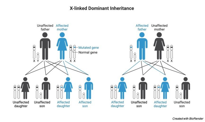
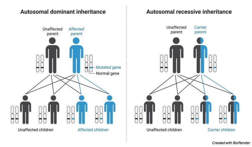
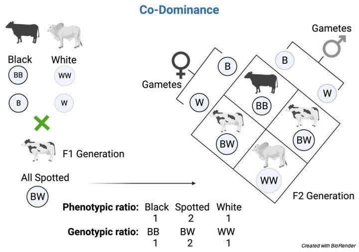
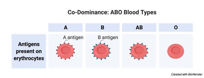
  • Incomplete Dominance: Definition and Examples

    Incomplete Dominance: Introduction We all know that Mendel’s contribution is very important…

    Continue Reading
  • Krebs Cycle: Cellular Respiration, Full Steps With...

    Krebs Cycle Have you ever imagined why living system (plants) synthesize food?…

    Continue Reading
  • Restriction Enzymes: Definition, Function, & Types

    What are Restriction Enzymes? Restriction enzymes also known as restriction endonuclease is…

    Continue Reading
  • ribosomal RNA (rRNA): Definition, Classification, and Function

    What is Ribosomal RiboNucleic Acid (rRNA)? In ribosomes, the Ribosomal ribonucleic acid…

    Continue Reading
  • Mendel’s Law of Segregation Mechanism, Examples, and...

    Law of Segregation The law of segregation had been put forth by…

    Continue Reading