Category: Study Materials
-

Avogadro’s Law: Formula, Calculation, Definition, and Examples
Continue ReadingAvogadro's Law
In 1811, Avogadro’s law was described by a famous Italian chemist and physicist named Amedeo Avogadro. Avogadro’s law is a relationship between volume of gas and the number of moles.
This law states that at a constant temperature and pressure the total number of atoms or molecules present in a gas is directly proportional to the volume occupied by that gas.
Avogadro's Law Formula
This equation is given by;
V ∝ n
V=kn or k = V/n
V = volume of gas
n = Number of moles present in given gas
k = proportionality constant
The graphical representation of Avogadro’s law is given below;
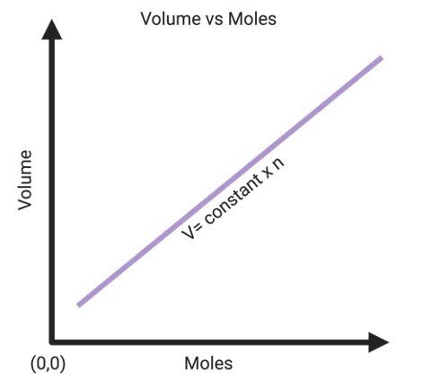
The straight line indicates that the two quantities that are volume and number of moles present in gas are directly proportional to each other.
Thus, the straight line that passes through the origin signifies that 0 moles for gas will occupy zero space.
Avogadro's Law Calculation
Avogadro’s law can be derived from the ideal gas equation, which can be written as follows:
PV = nRT
• ‘P’ is defined as the pressure applied by the gas on the walls of container
• ‘V’ is defined as the volume occupied by the gas
• ‘n’ is defined as number of moles of gas present in given molecule
• ‘R’ is defined as the universal gas constant
• ‘T’ is defined as the absolute temperature of the gas
By reorganizing the ideal gas equation, the following equation can be obtained.
V/n = (RT)/P
Here, the value of (RT)/P is constant because pressure and temperature are constant according to Avogadro’s hypothesis and R is the universal constant.
Therefore,
V/n = k
Thus, the proportionality between the volume occupied by a gas and the number of molecules present in given gas is confirmed.
Avogadro’s Law Examples
Given below are few examples of Avogadro’s law;
I. Breathing
One of the finest example of Avogadro’s law is breathing. When we inhale, our lungs enlarge because they are filled with air. Similarly, when we exhale, the lungs let the air out and thus shrink in size. This change in volume is observed, which is proportional to the amount or the number of molecules of air confined by the lungs.
II. Inflating Tyres
The shape of a flat tyres gets distorted in the absence of air inside it. As soon as the flat tyres is filled with the required amount of air, it gets back to its original shape. Hence, the inflation of flat tyres is a clear example of Avogadro’s law in our everyday life.
III. Bicycle Pump Action
The pump abstracts the air from the environment and pushes it inside a flattened object. This increase in the amount of gas molecules in the object congruently changes its shape and helps it to enlarge. This example is clearly explained by Avogadro’s law.
IV. Pool Tube
A flattened pool tube becomes transportable as the number of air particles inside the tube is decreased which in turn reduces its volume and makes it compact.
During inflation, when the tube is filled with air, increasing the number of air molecules in it which in turn increases the volume and size of the pool tube. Henceforth, Avogadro’s law can be applied to inflate or deflate the given pool tube as per our necessity.
Avogadro's Number
Avogadro’s number is defined as the number of molecules of gas present in one mole that is huge (6.02×10²³).
The unit for Avogadro number is mole-1.
Avogadro’s number is generally symbolized by N.
Avogadro’s number is a unit used by chemists for easy calculations all over the world.
Significance of Avogadro’s Law
After realizing that the volume of a gas is directly proportional the number of particles present in the gaseous molecules, this formula established a vital relationship for simple molecules at that time when the distinction between atoms and molecules was not visibly understood.
Specifically, the existence of diatomic molecules such as that H2, O2 and Cl2 was not identified until the results of researches involving the volume of gas molecules was discovered.
Limitations of Avogadro’s Law
This law is also known as Avogadro’s principle or Avogadro’s hypothesis. This law is only relevant to ideal gases and gives an estimated result for the real gases.
The gases with light molecules for instance helium, hydrogen, etc., follow Avogadro’s law more precisely as compared to the gases with heavy molecules.
Avogadro's Law Citations
- AVOGADRO’S LAW AND THE ABSORPTION OF WATER BY ANIMAL TISSUES IN CRYSTALLOID AND COLLOID SOLUTIONS. Science . 1913 Mar 21;37(951):427-39.
- Avogadro’s Hypothesis (or Law). Nature volume 82, page338 (1910)
- AVOGADRO’S LAW AND THE ABSORPTION OF WATER BY ANIMAL TISSUES IN CRYSTALLOID AND COLLOID SOLUTIONS. Science 21 Mar 1913: Vol. 37, Issue 951, pp. 427-439
Share
Similar Post:
-

Transport in Plants: Definition, Mechanism, and Types
Continue ReadingTransport in Plants: Introduction
Plants – Sessile, well developed tissue level grade of organization are the primary food producers for all the living organisms with least exceptions.
The energy of the sun and our waste respiratory product CO2 along with water becomes essential to produce sugars and other nutrient; vitamins and minerals which are obtained from the soil in which the plants are stationed to produce energy to plant dependent organism.
For a sessile organism to survive plants has developed many mechanisms to attain the basic needs for survival.
Tissue level organization is primitive even in higher plants with simpler but efficient structure to transport nutrients and water to the plants.
Uptake of nutrients and water is the key role in plant’s physiological process involving energy and carbon assimilation to produce Oxygen as a product.
Transportation in plants becomes essential and significant role in their physiological functions for the sedentary mode of life time employing simpler and number of techniques in combination to take nutrients from soil and water.
Specialized network of tissues connects the plants from its origin aids in nutrients translocation and distribution.
The specialized tissues are segregated and are present in all parts of body forming a connection between the nutrient receptor, storage, and synthesis regions of plants distinctly.
Major transportation in plants are water, minerals, and nutrients; distinctive pathway is defined for every transportable element; water and minerals are transported through XYLEM; Phloem another tissue transports and distributes organic matter to the sites of synthesis and their end products from synthesis site are distributed to energy utilizing site either as ATP or other sugar compounds.
Transportation in broader sense involves xylem and phloem – the vascular tissues provide a network to transfer the nutrients from source to the receiver site involves physics and chemistry to move evenly to sustain the plant.
Narrowing down; the cells also play a significant role in transportation and in energy utilization and storage.
Transport in cells plays a key role in the dynamics of cell were each cell functioning as a single unit integrates with neighbor’s cell wall and cell in integrating the flow of nutrient throughout the plant.
Transport in plant is extensively studied in earlier times by many scientists who experimented and identified the network of transporters laid out many theories for the transportation in plants and in trees.
History of Transport in Plants
The study of plant transport started back in 18th century by many scientists to understand the internal structure, circulation, and mechanical support for the plants.
It was an intriguing question on how a plant transports soil nutrients and water to the apex of the shoot and how does they synthesis their food?
Observing circulation in animals’ circulation in plants were also researched to understand the mechanism behind.
In earlier, 18th and 19th centaury many scientists observed various transport in plants by dissection and other methods and formulated theories of transportation of water and other elements.
The theory “Cohesion – Adhesion” developed by H. H. Dixon gave an acceptable mechanism in transport of materials in a general term of “Ascent of Sap”.
Transport in Plants Theories
Many theories have been put forward to understand the transport of water and other materials combinedly called as sap from the root to tip of the plant or a tree – Ascent of Sap. The theories were classified into three topics based on the ideology.
They are grouped into: 3 theories namely vital force; root pressure; physical force theories.
I. Vital Force Theory
The theory defines the movement of water or sap from root to other parts by the pulse created by the cortical cells which absorbs water from the outer side and pump them to the openings of xylem vessels.
The theory was put forth by J. C. Bose in 1923 also called a pulsation theory.
The theory stated that the cortical cells to be main cells to pump the water from the soil; which is proven wrong by eliminating roots from plant and the plant; stem in specific; still absorbed water.
II. Root Pressure Theory
The theory defines the movement of water or sap from root to other parts by the pulse created by the cortical cells which absorbs water from the outer side and pump them to the openings of xylem vessels.
The theory was put forth by J. C. Bose in 1923 also called a pulsation theory.
The theory stated that the cortical cells to be main cells to pump the water from the soil; which is proven wrong by eliminating roots from plant and the plant; stem in specific; still absorbed water.
III. Passive or Physical Force Theories
Theories corresponding to passive forces such as capillary force, collision force, atmospheric pressure, imbibition, diffusion pressure are factors upon which these theories are built upon.
Numerous scientists over a period have produced theories for identifying the transport.
They are:
a) Capillary Theory, 1863 by Boehm
Capillary movement is transport of substance over a small surface area over a small distance is acted upon by a small capillary force sufficient to pull water to a small extent.
Concept of capillary theory is based on the small force created by a narrow surface area to move a small amount of water.
The force is passive based on the physical properties of pressure and force and does not cost any energy.
Plant vascular system consists of tracheid and trachea which are longitudinal tubes of vascular bundles which when empty builds a force against gravitational force to pull the water from the roots and transport it to stem.
But this force is not sufficient to move to water column to the entire length of the plant.
b) Imbibition Theory, 1868 by Unger
Imbibition absorbs water on contact with water; swells by the pressure built up in a packed wood increase in surface area.
The pressure and increase in surface area pull the water but the force built up is exceedingly small to water to ascend the apex.
c) Cohesion - Tension Theory, 1894 by Dixon and Jolly
This is also called as Cohesion – Tension Transpiration Pull Theory.
The cohesive force and Transpiration pull combines to attract the water and other elements to move through the column of vascular tissues are now moved to the apex of the plant.
The cohesive force between the walls of xylem and hydrophilic water molecules attracts the molecules to the xylem wall.
Alongside xylem cohesive pull; water transpires when stomata open; which attracts water from nearby cells causing a Diffusion Pressure Deficit gradient between the guard cells and surrounding cells which causes diffusion from water filled cells to water deficit cells.
This theory was widely accepted by the scientific community thereby resolving a question of how a plant conducts water along its length.
Vascular Tissues of Transportation
Vascular tissues comprising xylem and phloem provides mechanical strength and support the plant in its sedentary lifestyle by transporting and distributing the assimilates from the site of synthesis to other parts.
The primary and secondary growth provides development of the vascular tissues to support even larger plants like trees where they reach a greater height.
The basic mechanism in transport is same in all organisms but distributing the synthesis and the network differs from species to species.
Phloem transports organic substance and nutrients from synthesis site and then to other storage sites and assimilation sites with network of vascular bundles which are well differentiated into various components such as fibers and other storage parenchymal cells.
Xylem and Phloem which are present in vascular bundles has many components.
Phloem comprised sieve tubes, companion cells, parenchymal cells, and fibers; transport organic substances for energy synthesis.
Xylem costs of tracheid’s, vessels, parenchyma, and fibers responsible for water and mineral uptake.
Transportion Classification
Transport in plants is classified to short distance transportation and long-distance transportation.
Short Distance Transport
The plants transport nutrients or water over a small distance.
Example: Water from soil is taken up by root hairs present in the soil by the process of diffusion where the root hairs are rich in solutes and when water is high in the soil; the difference in the concentration between root and exerts a pull for water from soil to the root hairs. The water then travels through the cells enter xylem.
Long Distance Transport
The plants and the organizing tissues transports water and nutrients to longer distance.
For Example: Water entering root xylem by diffusion is carried to the shoot apex of the plant by combined efforts of the diffusion, imbibition, transpiration, cohesion, and adhesion. Transpiration is the determining factor directly proportional to uptake of water.
Mechanism of Transportion
Mechanism of transport in plants as classified are of 2 main vascular bundles which transports water, minerals and other organic compounds by xylem and phloem, respectively.
Phloem transports organic compounds such as Sucrose and other carbohydrates from the region of synthesis; distributed to other plant parts of requirement to store or to be utilized in the process of energy production for growth and development.
For Example: Carbohydrates; sucrose synthesized in Leaves of plants; rich in chloroplasts and stomata assimilates CO2 and converts into carbohydrates (Sucrose) carried again through phloem to other parts of the plants such as stem, flower and to developing shoots through network of vascular bundles connecting plant parts.
The phloem vessels and cells present from the root hair to the apex facilitates the assimilate transport from the synthesis point to other regions of plant.
Phloem transports organic materials by a mechanism of mass flow of substance through circulation.
Similarly; Xylem transports water and minerals from roots to the apex of the plants through various mechanisms in combination with Cohesive and Transpiration to other parts of plant.
Xylem transports water and minerals unidirectionally; varies from phloem circulating the metabolic products bidirectionally (i.e.) Root to Apex, Leaves; Leaves to Stem, flower, root.
Water is taken from the soil through root hairs through the process of osmosis; from roots the water is further taken up by the combined action of Transpiration, Capillary Action, and root pressure.
Changes in any one factor has a respective contribution and determining role in uptake of nutrients.
Minerals are also taken up by xylem by a different action of Diffusion; uptake of nutrients passively by the difference between the concentration gradient; and Active Transport utilize ATP, an energy molecule to move a molecule using transmembrane protein from extracellular matrix to the cell’s internal environment to mediate many metabolic processes.
Each mechanism and processes have its significant role in uptake of nutrients and act as a limiting factor in controlling the transport.
Transport in Plants Citations
- Molecular mechanisms of boron transport in plants: involvement of Arabidopsis NIP5;1 and NIP6;1. Adv Exp Med Biol . 2010;679:83-96.
- Molecular mechanisms of urea transport in plants. J Membr Biol . 2006;212(2):83-91.
- Strategies for optimization of mineral nutrient transport in plants: multilevel regulation of nutrient-dependent dynamics of root architecture and transporter activity. Plant Cell Physiol . 2014 Dec;55(12):2027-36.
Share
Similar Post:
-

Mitosis vs Meiosis: Chart, Definition, and Diagram
Continue ReadingMitosis vs Meiosis: Introduction
Both are type of cell division taking place in every eukaryote. But the main difference is Mitosis produces Diploid Somatic cells identical to parent and Meiosis give rise to cells which forms a new progeny which are genetically different from the parent by producing haploid gametes.
The differences vary mitosis from meiosis producing regular somatic cell and differentiated gametes of future progeny respectively.
Out of 2 cellular divisions in meiosis; Meiosis II is more similar to mitosis with the difference in the end products of haploid gametes and regular diploid somatic cells respectively.
Mitosis vs Meiosis
Mitosis Meiosis Two daughter cells are produced from a single cell Four daughter cells are produced from a single cell Chromosomal segregation separates equal number of chromosomes to the progeny Chromosomal segregation separates only half chromosomes of the single cell Infinite cell cycle rotation varies in different cells Single rotational cell cycle A cell cycle involves 2 Gap phases and one synthesis and Mitotic phase Cell cycle involves 2 continuous Meiosis preceded by single gap phase and synthesis phase No homologous chromosomes are found Homologous chromosomes are attached during Prophase Recombination is not required as the division involves a replication of unmodified genetic material Recombination is the main specialty of meiosis where the genetic material is non – sister chromatids are exchanged with each other Daughter cells are identical to parent Daughter cells are varied from the parent During development the division produces number of differentiated cell lineage based on the location, hormone regulation etc., During division it produces single lineage of cells Chromosome divides during Anaphase Chromosomal division is during Anaphase II The cells can be Haploid or Diploid The cells undergoing such division contains only diploid number of chromosomes and above Cells which undergo such division are called as Somatic cells Such division gives rise to sex cells called Gametes Mitosis vs Meiosis Diagram
I. Mitosis Diagram

II. Meiosis in Oocyte

III. Meiosis in Sperm

Why We Need Meiosis?
o Eukaryotes are evolved and well distinguished for the various adaptive features to survive the dynamic changes and evolved along the changes.
o Changes in the environment was followed by the adaptation in organism for the respective environment was motivated by an act of survival and to ensure a progeny to develop the lineage thereby sustaining life on earth.
o Environmental modification led changes in animals and plants; regulated the diversification and population growth of all organism by frequent modifications in niche where the organism functions to its fullest; challenging the organisms to promote change in functionalities for the survival factor.
o Charles Darwin pointed out the Theory of “Natural Selection” where the organisms are naturally and randomly selected by nature to develop lineage.
o He called it the ” Survival of Fittest” where the organisms suitable for the environment survived and others which couldn’t survive became extinct.
o The Survivors became the key organism for the variability among the genus, species level which on further adaptions evolved to give rise to new classes and phyla.
o Changes governing evolution are inherited from parent to offspring; is sexually selected – another phenomenon of evolution and diversification; mediated by special type of cell division Meiosis.
o Gametes of meiosis inherits the changes from parent during fertilization to produce competitive offspring for better survival and variability from parents by giving it a unique identification and distinction from the parent.
o Differentiation, compatibility to the environment is genetically marked and inscribed in the genes from parents separately before the fertilization.
o The inheritance of the qualities is determined post fertilization in zygote involves principles of genetics while exhibiting features of an organism.
o Such unique, distinctive characteristics of an organism predetermined in gametes of parents; formed by a reductive division.
o Meiosis revolves around the components of cell cycle and molecular activation by an organism.
o The cell of meiosis initially are a result of mitosis and meiosis is the smallest but significant part in life history of any organism.
o The specialized division becomes more significant for its role in determining the characteristics of future progeny and aids in variability which on long run evolves the organism.
o Meiotic steps are crucial in determining half feature of an organism by each parent to make up the whole organism.
Significance of Meiosis
1. Evolution of sexual reproduction in eukaryotes are marked by Meiosis. Sexual reproduction becomes and important characteristic feature of meiotic division; involves maternal gametes and paternal gametes to determine the ability of an organism.
Sexual reproduction involves fertilization between same species with distinct genetic characters originated from their unrelated immediate ancestors to produce viable gametes to compete and accustomate to the environmental challenges for survival.
2. Meiosis is conserved in broader terms among all sexually reproducing eukaryotes. Broadly, the mechanism of meiosis is similar in all eukaryotes involving same steps and function.
Changes from the preset is that each organism takes different time to proceed the cycle of meiosis and end products can be different from each species in structure and function.
3. Meiosis maintains a constant chromosome number. Advantage of using meiosis over mitosis for gamete production is to maintain the chromosome number.
On mitotic division; the chromosomes produce daughter cells with diploid chromosome number; if the same prevails; for meiosis the chromosomes double on each fertilization resulting in doubling at each generation.
For example: 2n –> 4n –> 8n –> 16n…….. infinity.
4. Recombination of paternal and maternal chromosomes produces genetic variability, diversity and in turn evolution.
Recombination is a process of exchange of genetic material between maternal and paternal chromosomes with the progeny to get a combination of both traits and not either of the traits causing reduced viability in offspring.
Crossing over is a step-in prophase where the homologous chromosomes pairs by a synaptonemal complex and crosses each other in further steps by a chiasma; providing a bridge for the exchange of the genetic material.
5. Recombination takes place with rules and by the process of law of independent assortment. The law stated by Mendel says that the genetic material of maternal or paternal chromosomes are independently crossed over to recombine with each other to produce variabilities in the haploid gametes to express traits of both maternal and paternal in origin.
6. Positive adaptation caused by mutation suitable for the environment is naturally selected and carried over to next generation. Meiosis is the process of random selection and fusion of male and female gametes to future generation.
Any changes in the germ cell chromosomes are inherited to the next generation and are exposed to the environment; surviving with the mutation in ease might take the changes to generations further and undesirable change caused by mutation might be eliminated rapidly because of the in functionality of the process.
7. The segregation, crossing over and recombination of the chromosomal components ensures the variable inheritance of parental chromosomes. The recombination produces diversity among the population on long run produces variability and then evolve to new organism or to any environment.
8. After the meiosis the post – miotic process or the end product might vary from species to species. Plants after meiosis produces 4 microspores in anthers and 4 megaspores in Female. The microspores 3 degenerates one fuse with one megaspore.
Later, the remaining haploid megaspores combine to form endosperm in one stage to provide a developmental part of other fusion.
Mitosis vs Meiosis Citations
- Recombination, Pairing, and Synapsis of Homologs during Meiosis. Cold Spring Harb Perspect Biol . 2015 May 18;7(6):a016626.
- Meiosis: the chromosomal foundation of reproduction. Biol Reprod . 2018 Jul 1;99(1):112-126.
- Abnormal mitosis in reactive astrocytes. Acta Neuropathol Commun . 2020 Apr 15;8(1):47.
- Mitosis in vertebrates: the G2/M and M/A transitions and their associated checkpoints. Chromosome Res . 2011 Apr;19(3):291-306.
Share
Similar Post:
-
Meiosis II: Definition, Stages, Phases, and Diagram
Continue ReadingMeiosis II: Introduction
o The living world has seen many numbers of generations and successions of organism evolved adaptively to the pertaining environmental changes; sustaining the diverse lifeforms on The Earth.
o Sustenance of a species and its lineage is the cumulative adaptations of the ancestors inherited over a million years to reach the present morpho – functional form of beingness.
o The inherited changes from parent to offspring, its proliferation supported by external growth factors and the environment; developed the organism to become fit and ensure viability for further generations to sustain.
o The fundamental cellular process supporting the growth and development is the inheritance of expressive genes from the parents to offspring mediated by the process of Meiosis.
o Cell division that leads to the contribution of one half of the chromosomes from each of the sexually reproducing parent to offspring is meiosis.
o Sexual reproduction is possible among the same species with same number of chromosomes and are equally contributed to the offspring so that half of maternal and paternal genes are contributed to the progeny.
o Meiosis is specialized because of the variability it provides to the offspring by the recombination of the homologous genetic material and the production of reduced chromosome where a diploid cell gives haploid daughter; on fusion of the male gamete (Sperm in animals and pollen in plants) and female gametes Eggs (plants and animals) produces diploid zygote proceeds with mitosis to develop into new organism.
Mechanism of Meiosis II
o Significance of meiosis is that it takes place twice to segregate the homologous pair and give 4 haploid cells in continuous division without any gap between the two division.
o At the end of Meiosis I, the single gamete on induction becomes divided into 2 diploid cells with varied genetic expression due to the recombination enter the meiosis II.
Meiosis Diagram in Sperm
o Meiosis II is a replica of Mitosis with a difference in chromosomal composition of the somatic cells and the meiotic cell.
o The Meiosis I and Meiosis II has no gap phase between them to duplicate the 46 chromosomes present in the cell at the end of the Telophase I.
o Other components are similar to that of mitosis; wherein prophase II has the same rule where the chromatin condenses faster to become a chromosome.
o Metaphase II includes the appearance of the spindle fibers from the organizing center attaches to the sister kinetochores of the same chromosomes; a difference from the METAPHASE I where one of the sister chromatids of the homologous are attached.
Meiosis Diagram in Oocytes
o The chromosomes are attached to opposite end fibers of the other sister kinetochores and aligns in the center of the cell forming equatorial plate.
o This phase has a regulator where all the chromosomes must be attached at the sister kinetochores of the opposite poles to initiate Anaphase where the real separation of haploid chromosomes takes place.
o At Anaphase II the sister chromosomes held by cohesins are removed by the cascade activity of separase; chromosomes when bound induces APC/C complex which eliminates the repressor of separase and separase on removal of repressor cleaves cohesins along the length of the sister chromatids including centromere.
o Cohesin degradation releases the tension maintained when held at the equator which pulls the chromatids to opposite ends separating the haploids to the opposite ends.
o Reaching poles, the last Meiosis II stage starts by forming a deep furrow between cells and formation of cell organelles and nuclear membrane and separates.
o Post Meiotic II involves maturation of the germ cells and re – organization of the structural and functional capability of the cell.
o In female, the cell matures to eggs and in male the cells undergo structural modification to attain a sperm with head, neck and tail and the transformation varies for different groups of eukaryotes.
Meiosis II Citations
- Polo is not solo in meiosis. Cell Cycle . 2018;17(3):273-274.
- The histone codes for meiosis. Reproduction . 2017 Sep;154(3):R65-R79.
- Cell Cycle-specific Measurement of γH2AX and Apoptosis After Genotoxic Stress by Flow Cytometry. J Vis Exp . 2019 Sep 1;(151).
- Flow Cytometry Analysis of Cell Cycle and Specific Cell Synchronization with Butyrate. Methods Mol Biol . 2017;1524:149-159.
- Novel insights into cell cycle regulation of cell fate determination. J Zhejiang Univ Sci B . 2019 Jun;20(6):467-475.
- Cell Cycle-Dependent Control and Roles of DNA Topoisomerase II. Genes (Basel) . 2019 Oct 30;10(11):859.
Share
Similar Post:
-

Meiosis I: Definition, Stages, Phases, and Diagram
Continue ReadingMeiosis I: Introduction
Sexual reproduction is a stepwise evolution of eukaryotes; varying from species to species. The basic mechanism of zygote fusion and meiosis regulators are conserved in all eukaryotes; the difference is seen in the method the gametes meet and evolved along the groups.
Pollination in flowering plants is the means of gametic fusion and produces seeds; which germinates on suitable condition to produce plants.
Plants are exposed to varied errors in cell division and few species under adverse conditions; produces spores which on exposure to suitable conditions proliferates.
Eggs and pollens of plants naturally depends on external mechanical factors for the initial fusion of gametes.
The development and maturation of the plants takes place in a sequential manner on appropriate time and nutrient availabilities.
The basic principles of meiosis include the law of independent assortment with which the plant acquires genetic diversity and variability by crossing over forming a recombinant.
Crossing Over and recombination followed by continuous double cellular division give rise to haploid daughter cells; changes to gametes of male and female in respective parts of a flower of same plant or a different plant.
The reductive division from which the name meiosis was given; in Greek meiosis is to diminish, recombination by crossing over where homologous chromosomes come together to interchange genetic material, and independent assortment of genes to the progeny are the main features a meiotic cell provides to make the process more unique and provide genetic diversity.
Meiosis History
Meiosis was first described by Oscar Hertwig, German Biologist in 1876 further research took place in the field where Theodor Boveri, 1888 reported it in Roundworm.
The reason behind such division was later pointed by August Weismann as to maintain equal amount of genetic material transfer from parent to progeny because if the reproductive division follows mitosis a diploid cell becomes tetraploid and increase exponentially over generation and have resulted in the formation of species with infinite genetic material.
Thomas Hunt Morgan in Drosophila found the recombination of the chromosomes (genetic material) to provide evolutionary variability to organism.
Mitosis vs Meiosis
Both are type of cell division taking place in every eukaryote. But the main difference is, Mitosis produces Diploid Somatic cells identical to parent and Meiosis give rise to cells which forms a new progeny which are genetically different from the parent by producing haploid gametes.
In general; all cells are mitotic and undergoes mitotic cell division; even the gametes before meiosis are divided mitotically from their progenitor and enter meiosis.
“The Molecular Switch” present in every cell when induced on appropriate nutrients makes the cell competent to enter meiotic cycle; is the mechanism to “turn on” meiosis at the beginning of Phase G2.
From G2 phase the meiosis proceeds by 2 continuous cell division.
Further; the phases of meiotic cell division differ from the mitotic phase to support complex changes during meiosis.
Meiosis is divided into MEIOSIS I and MEIOSIS II; each meiotic phase has sequence of Prophase, Metaphase, Anaphase and Telophase.
The Prophase of Meiosis I is more significant where the primary feature of meiosis takes place: Pre – leptonema, Leptonema, Zygonema, Pachynema, Diplonema, Diakinesis and are absent in Prophase of Meiosis II.
Meiosis I
Somatic cells and germ line cells are differentiated in this method; specialized and prominent switching of process from mitotic to meiotic phase in testes and ovaries; takes place in Prophase I.
Meiosis generally skips the G2 Phase as soon as the Switch is “ON” to Prophase I.
Significant modification in the genetic material is well accounted in different phases of PROPHASE I; other cellular changes are similar in that of mitosis wherein the nuclear membrane starts disintegrating in the prophase and disintegration of other membrane bound organelles.
The specialized phases of the prophases and the events at each stage are given below:
Prophase I: Pre-Leptonema
The chromosomes are extremely thin to be identified except for the differentiated sex chromosomes which has Heter pyknotic bodies.
Heter pyknotic bodies are regions of either tightly or loosely bound chromatin fibers which are stained more or very less from the rest of the chromosomes.
Prophase I: Leptonema
o Leptonema in Greek means thin thread like structures.
o The chromosomes in the phase are characterized by thin appearance even after the replication.
o Out of all phases of meiosis the PROPHASE I is longer in all eukaryotes with time variation in different species. The phase also constitutes other changes:
o Nucleus enlarges in size occupying most of the cytoplasm signifying the increased genetic content in cells.
o Chromatin starts to form a loop of 5 – 22 µm DNA.
o DNA appear single rather than double as in mitosis because of this the phase has its name LEPTONEMA with thin chromosomal appearance.
o The chromosome starts condensing and has bead like thickened structures called chromomeres present irregularly in a chromosome and the number of chromomeres are not constant.
o Prophase chromosomes forms a telomere bouquet which orients the chromosomes theoretically to form homologous pairs.
o These telomere bouquets attach the chromosome to the inner nuclear membrane making chromosomes easier to pair.
o Synaptonemal Complexes are initiated to form in this phase as a preparatory part of next phase.
Meiosis I Prophase Diagram

Prophase I: Zygonema
o Zygonema in Greek means Adjoining. The threaded chromosomes pairs with its homologous chromosome.
o Maternal chromosome and paternal chromosome of same functions are segregated and paired to each other for recombination in next phases.
o Chromatin loops concentrate further in ZYGONEMA.
o To make sure the homologous pairing corresponds with the similar DNA sequences in both homologs.
o Recombination Complex breaks the double strand at specific sites and join the similar part of the chromosome.
o This process takes place before the synapsis of the 2 chromosomes called presynaptic complex.
o Chromosomal pairing by the synaptonemal complex.
o The homologous chromosomes pairing causes the formation of synapsis.
Synaptonemal Complex
o Synaptonemal Complex is a highly complexed structure involving proteins similar to histones to form rail road like or zipper like filaments across both chromosomes along their peripheral axis.
o Synaptonemal Complex has two kinds of filaments two Lateral and 2 transverse or Medial filaments.
o Synaptonemal Complex prevents the complete fusion of homologous chromosome with 100nm gap.
o The initiation of the complex is random and starts at any point of the pair; guided by the telomere bouquet attachment to the inner nuclear membrane.
o At the point of attachment of telomere to the nuclear membrane the Synaptonemal Complex deposits to form a thick fixation plate.
o These fixation plate attracts the re – formation of nuclear pore annuli at the region of attachment.
o For a homologous chromosome to form the process must be made possible by multitudinal involvement of various parts and process of the nuclear complex and Synaptonemal Complex forms a skeleton to support the complex formation and recombination of the chromosome providing stability.
Prophase I: Pachynema
o Pachynema refers to thick chromosome in Greek. The stage is significant because of the crossing over and recombination of the maternal and paternal chromosome results in genetically different species.
o SC is complete
o Chromatin loop is well concentrated making the genetic material to have brush like appearance.
o The number of chromosomes reduces to half forming bivalents or tetrads.
o The region of connection between the bivalents are termed as chiasma where the homologs forms X – shaped connection to hold each paternal and maternal chromosome.
o Chromatids of homologue becomes 8 with 8 kinetochores on each chromatid.
o SC ensures the homologous pairing of all chromosomes in the nucleus before proceeding to next process.
o The SC remains intact throughout the pachytene.
o Crossing over between homologous pairs takes place.
o Crossing over is regulated by components and are determined to provide structural support and genetic variability and diversity among the species.
o Crossing over regulations takes before crossing over ensures the chromosome to attain more than one recombination and restricts the closely related genes from crossing over.
o A separate rule prevails to conserve the integrity of the chromosome.
o The chromosome is divided into “Hotspots” and “Cold spots” based on the recombination sites.
o Telomeric and heterochromatin centromere regions are prevented from crossing over.
o Other regions are exposed to the crossing over for the recombination of the genetic materials among the maternal and paternal genes.
o Recombination also takes place in Pachytene stages indicated by the formation of Recombination Nodules which has intact SC to ensure the cuts which are produced to recombine does not eliminate the region from the chromosome which leads to errors in cell division.
o The recombination nodule forms a bar like structure across the chromatids to reach its corresponding pair and exchange its DNA material.
Prophase I: Diplonema
o The crossed over chromosomes are separated in the phase but are held by chiasmata.
o SC is removed at the stage after the crossing over.
o Chiasmata is intact and separation of paternal maternal chromosomes at most of the sites takes place.
o The removal of cross overs and recombination sites leaving a single site makes the four tetrads are visible.
o Diplonema is more significant because of long duration it takes for all chromosomes to separate.
o In certain species; the chromosomes have a specialized appearance of lamp brush.
Prophase I: Diakinesis
o Diakinesis is the terminal process of prophase I where the chromosome’s chiasmata are completely lost except at the end region, this process is the TERMINALIZATION.
o Diakinesis in Greek means breaking across; where the chromosomes are cut across each other and marks the end of Prophase I
Pro-Metaphase
o Disintegration of Nuclear membrane
o Complete condensation of chromosomes
o The kinetochores of homologous chromosomes attach to microtubules
o Sister kinetochores maintains the integrity as a functional unit.
Meiosis I Diagram

Metaphase
o Similar to mitosis Metaphase I has similar functions
o The metaphase arranges the bivalents at the equator by the microtubules
o The main difference is the chiasma between the homologous chromosome remains intact
Prophase I: Anaphase I
o The Cohesins in the chromosomal arms are removed to break free the homologous pairing and tension created because of the separation makes the microtubule to contract towards the organizing center.
o The chromosomes are segregated to the poles.
Prophase I: Telophase I
Telophase is simple and marked by formation of the nuclear membrane by furrow in animals and phragmoplasts in plants and the cell separation – Cytokinesis.
Meiosis I Citations
- SnapShot: Meiosis – Prophase I. Cell . 2020 Jun 11;181(6):1442-1442.e1.
- LINC complexes as regulators of meiosis. Curr Opin Cell Biol . 2018 Jun;52:22-29.
- Conservation and Variability of Meiosis Across the Eukaryotes. Annu Rev Genet . 2016 Nov 23;50:293-316.
- Mixing and Matching Chromosomes during Female Meiosis. Cells . 2020 Mar 12;9(3):696.
- Elevated Mutagenicity in Meiosis and Its Mechanism. Bioessays . 2019 Apr;41(4):e1800235.
- The molecular biology of meiosis in plants. Annu Rev Plant Biol . 2015;66:297-327.
- Recombination, Pairing, and Synapsis of Homologs during Meiosis. Cold Spring Harb Perspect Biol . 2015 May 18;7(6):a016626.
- Meiosis: the chromosomal foundation of reproduction. Biol Reprod . 2018 Jul 1;99(1):112-126.
- The cohesin complex in mammalian meiosis. Genes Cells . 2019 Jan;24(1):6-30.
Share
Similar Post:
-

Significance of Mitosis: Definition, Mitotic Phase, and...
Continue ReadingSignificance of Mitosis: Introduction
Growth and development are essential in to an organism to survive in dynamic environment.
Development and growth are inevitable changes which requires and needs a specific mechanism to grow in an adverse condition and ensure a healthy progeny to do the same.
Cell division in general is the process where an organism grows and develops in the ever-changing environment.
To ensure the line of a species they reproduce in which half of parent’s genetic material is transferred to the young one by the process of reduced division of cell; plays a primary and minimal role in developing a progeny.
In an organism’s life the cell division plays a major role in development; determining the structure, function and capacity according to the genetic expression.
The patterning and molding are mediated by exponential cell division known as the mitosis.
Changes in cell division which is not inherited to the progeny but affects the regular functions are observed in Somatic cells – divides through mitosis.
Mechanism of Mitosis
Mitosis is one of the phases in a cell cycle; is a process which produces two identical daughter cells from a single parent cell; induced when proper nutrient requirement and hormonal internal cues are synchronized.
Mitosis is preceded by 2 Gap phases and single Synthesis phase where diploid cells replicate and becomes tetraploid chromosomes with 4 arms and a centromere – highly condensed region during initial mitosis stage – Late prophase and Metaphase.
The commitment to enter the cell cycle is given by the expression of protein dependent kinases – dimers in cell cycle control; Cyclin and Cyclin Dependent Kinases.
Preparatory interphase makes a cell capable of self-division and replication of cellular function from the preexisting cells.
Mitosis itself proceeds through different phases of prophase, metaphase, anaphase and telophase; arranging and segregation the chromosomes and other cell materials and organelles (precursors) from the existing cells.
Pre-Mitotic Phase
The regulatory proteins make essential measures and fulfills cell’s need to enter mitosis in a steady state.
At each stage the cells gain an important feature to undergo cell division and divide genetic and other cell component evenly to each daughter cell.
During Interphase; it is the non – dividing phase of a cell where the cells undergo synthesis of RNA, Proteins and genetic material to proceed the cycle; are frequently regulated by protein kinases phosphorylating inhibitors to repress few genes and enhance the gene expression of few others to synthesis DNA.
Checkpoints at each phase makes the cell cycle error free and maintains the integrity of the cycle.
The G1 phase has a checkpoint which when crossed; the cells get committed to mitosis irreversibly is governed by the CDK – CYC complexes in eukaryotes.
The G1 checkpoint makes sure the cell undergoing mitosis has essential nutrients to proceed throughout the cell cycle.
The synthesis phase is initiated when the CDKA – CYC D of G1 produces transcriptional factors (ORC – Origin Replication Complex) which sits on the replication origin region of the chromatin fibers and initiate DNA Replication in S Phase.
When the cell completes the S Phase, they are auto regulated by same Transcriptional factors induced G1 phase protein kinases which limits replication when the cells have an ideal DNA for mitosis.
The cells enter G2 Phase. CDKA – CYCA expression increases in G2 Phase produces transcriptional factors which completes gene expression for other cellular mitotic requirements and when the expression reaches a particular threshold activates other G2 transcriptional factors which repress the gene expression and promotes.
Condensation of the chromatin fibers and prepare the cell to enter mitosis.
Mitotic Phase
Mitosis is regulated by CYCB – CDKB from late G2 phase and are then dominated by CYCD – CDKA where each phase has their significant feature starting from prophase; the chromatin condenses to chromosome but not completely; includes the disintegration of many cell organelles and nuclear membrane and in cytoplasm many microtubules are formed.
In Plants a specialized pre – prophase band starts to form.
Metaphase marks the complete elimination of nuclear membrane and condensed chromosomes are formed.
Later the preprophase band in plants attach to the kinetochores and make the chromosome to arrange at metaphase equatorial plate after all chromosomes are arranged by the spindle fibers of opposite poles the cell enters Anaphase.
The sister chromatids re separated by breaking Cohesins binding the chromosomes by proteolysis and drags the sister chromatids to opposite poles.
In Telophase; the daughter chromatids on opposite ends starts decondensation; reappearance of Golgi complex mitochondria chloroplast and other cell organelles are formed.
The cell cycle when complete develops the cell wall and remains adjacent to each other; when induced enters the cell cycle again or remains same in the G1 phase or enter G0 phase for maturation till they are eliminated.
Significance of Mitosis
o Mitosis is the primary mechanism for an organism to grow and helps in development.
o After birth the child develops both quantitatively and qualitatively.
o Mitosis produces new cells from existing cells and increases exponentially to increase the whole growth of the organism.
o Division is from preexisting cells. Every cell in our body is a result of a division of a single cell.
o Cells which enter the cell cycle take up essential nutrients and when an organism undergoes a development or to replace older ones; an induction signal and certain modifying factors makes the cell enter the cell cycle from G0 Phase to produce new cells; is the first type.
o The other type includes ever dividing meristematic cells in plants for growth and cells of intestine and skin of animals where the dead cells must frequently be removed to protect the internal organs from direct exposure to acid and sun.
o Mitosis takes place at appropriate time and appropriate internal cues.
o All cells are not capable of cell division and enter into a G0 Phase where they remain the same all their period of life.
o A nerve cell cannot regenerate or repair any damage caused to it even with stimulus and nutrients; unlike enterocytes, skin cells in animals and meristematic cells in plants which on appropriate nutrition provide continuous growth for the plant.
o In the absence of the appropriate conditions the plant parts under the condition become dormant or temporarily stopped for a particular time and develop when appropriate conditions rise.
o Protects the body from errors of DNA Replication.
o The checkpoints of cell division at each stage of cell cycle from G1 Phase checks primary availability of all essential components such as nutrients, DNA defects, cell size and hormonal influence before the entry of cell into cell cycle.
o G2 phase ensures the health of DNA and DNA replication in Synthesis phase when such defects are noticed, cell cycle halts and repair mechanisms for damaged DNA or extend time for complete DNA Replication.
o Replenish the tissue cell pool and organization.
o In animals’ specific tissues exposed for heavy work are subjected to wear and tear; makes the cell lose its ability to continue the role and efficiency is reduced.
o For Example: Red Blood Cells carrying Oxygen to the tissues, Enterocyte’s exposure to acidic bolus, Stem cells, Skin cell’s exposure to external mechanical stressors such as sun light, pressure etc., requires set of cells to be exposed to and these regions are given the genetic activation of continuous cell division to maintain the tissue pool and sufficient function.
o Provides structural integrity and stability to an organism.
o Cells make up the whole organismal existence which must develop over a period of time to maintain its structure and shape for proper function and survival of the organism.
o A proper cell function is essential to maintain a Homeostasis.
o A small malfunction in a cell caused by external source factors might disrupt the homeostasis causing deviation from the ideal cell.
o These deviations are identified and rectified by the checkpoints of the cell cycle.
o Mitosis is conserved over evolution in eukaryotes.
o The mechanism of how a cell divide is the same in all eukaryotes.
o The main difference will be the signals, nutrients and the proteins involved in regulation of cell cycle.
o Ratio between the cytoplasm and nucleoplasm are maintained.
o The cytoplasm and nucleoplasm are derived from similar components and have the same function of storing components for cell functioning.
o The quantity and few other features such as viscosity differs in cells are maintained during the mitosis at the end of cycle.
o The ratios must be maintained constantly where the deviations will make the cell dysfunctional.
Defects of Mitosis
o Cell division in somatic cells is not inherited by progeny but can be fatal when the regulatory functions does not work properly.
o Most of the time when a cell encounters a somatic DNA errors the body has developed many mechanisms to eliminate the divergent to maintain the homeostasis of a system.
o But the errors when not detected might lead to tumor and cancer which is fatal.
o Reasons for errors in cell division is not clearly known; it might be a result of mutation caused by exposure to carcinogenic gasses, X – rays or other chemical substances.
o Defects of mitosis in plants leads to aneuploidy and does not have fatal effects as in animals and humans.
Few defects in Mitosis are:
1. SAC (Spindle Assembly Checkpoint) defects
2. Cohesins defect
3. Merotelic Attachments
4. K – MT Stability
5. Centrosome amplification
6. Tetra ploidy
Most of the defects are common and are predominantly found in animals.
Impacts in plants are usually less and mostly results in aneuploidy; which is not inherited to the off springs and not fatal.
Significance of Mitosis Citations
- Mitosis in filamentous fungi: how we got where we are. Fungal Genet Biol . 1999 Jun;27(1):1-25.
- Abnormal mitosis in reactive astrocytes. Acta Neuropathol Commun . 2020 Apr 15;8(1):47.
- Mitosis in vertebrates: the G2/M and M/A transitions and their associated checkpoints. Chromosome Res . 2011 Apr;19(3):291-306.
Share
Similar Post:
-

Cytokinesis: Definition, Checkpoints, Diagram, and Examples
Continue ReadingCell Cycle, Cell Cycle Phases, and Cytokinesis
Cell cycle is the sequential process taking place to regulate the growth of organism; cell divides to produce a genetic replica and enters the stage of cell growth.
Cell growth involves the synthesis of organic material and integrates information across its counter parts for synchronous development of the whole body.
The cell synthesis phase lasts till a cell reaches its maturity; on initiation the cell again divides to produce new cell and the process continues. Cell cycle is a sequential development of cell between two cell divisions.
The cycle is genetically controlled and are programmed in every cell and are specific for each region.
Varied species has variable time length of cell cycle decided by physiological and influences pertaining to their niche.
Two Phases of cell cycle are: INTERPHASE and MITOTIC PHASE. INTERPHASE involves G1, synthesis and G2 phase; chromosomal replication and development is regulated by the phase; determines the quality and quantity of chromosome entering the daughter cells and a balance is maintained by the phase.
Karyokinesis and Cytokinesis division, segregation of chromosome and cell takes place during MITOTIC PHASE.
The notable feature of cell cycle is eukaryotic organisms though diverse and distinct have a common type of cell division over the Kingdom of Eukaryotes is a scientific wonder and research have emphasized that timing of a cell entering the cell cycle is essential in cell cycle regulation.
History of Cell Cycle
The history of mitosis dates back to 18th and 19th century where the aid from microscope and visibility of cell division under the microscope supported the discovery of the cell division.
The discovery was earlier than the DNA discovery was a breakthrough in the scientific community as it answered the most intriguing question of Humans “How do we grow? Develop? Reproduce? What is the driving factor for the growth? How do we resemble our parents?” etc.,
Before the discovery of cell division there were many theories on how the cells are related to the overall development of an organism’s lifecycle.
One of which was Rudalph Virchow’s theory of cell: “Omnis cellula e cellula” which states that a cell originates from a pre-existing cell.
Walther Flemming discovered and published a detailed book on cell division in 1882 after discovering cell division in 1879.
He named it “Mitosis” after the Greek word Mito – “Wrapping thread” owing to the thread like appearances of the chromosomes.
He conducted a detailed study and deduced staining techniques to understand cell cycle and named the stages of each division as Prophase, metaphase, Anaphase and Telophase.
The cell division was identified in Salamander’s embryo. Flemming supported Virchow’s cell theory with precision and stated that “Omnis nucleus e nucleo” which states that a nucleus origin from a pre – existing nucleus and highlighting the chromosomal segregation and laid foundation for the theory of inheritance where the chromosomes play an important role carrying the genetic information from the parent to the offspring.
Cell Cycle Phases
The cell cycle is common for all eukaryotic organisms; travelling through 2 major phases based on the cell division:
INTERPHASE and MITOTIC PHASE. Interphase consists of 3 phases Gap 1 phase, Synthesis Phase and Gap 2 Phase.
Similarly, mitosis has four phases Prophase, metaphase, anaphase, and telophase.
The development of cells through these phases are influenced and facilitated by heterodimeric protein kinases – Cyclin and Cyclin Dependent Kinases.
Mitotic Phase
The changes in above phases are minimal or not clearly visible in microscopes whereas the changes in M Phase are easily detectable.
The phase has 4 parts in which the division takes place systematically and continuously.
The cell stages are easily visible in plant parts as the specialized dividing region – MERISTEM is prevalent in roots and shoots are continuously dividing providing a mechanical support and functional integrity to the plants.
The 4 phases are: Prophase, Metaphase, Anaphase and Telophase.
Each phase has a distinctive change to be identified and Eukaryotic cells replicates in the same order in most of the organisms.
Prophase: The prophase is marked by chromosomal condensation and disintegration of cellular components and assembly of cytoskeletons for cell division. RNA synthesis is inhibited.
Metaphase: Nuclear membrane is eliminated completely chromosomes are completely condensed. The cytoskeleton – spindle fibers attach to the kinetochores. The chromosomes are aligned in the equatorial plate.
Anaphase: Chromosomal split forms daughter chromatids; travels to the opposite poles. The chromosomes are V – Shaped as they are dragged to the opposite sites.
Telophase: Microtubules disappear and chromosomes decondense to chromatin mass. Nuclear envelope starts to form. The disintegrated organelles form again.
Cytokinesis
Next part of cell cycle is Cytokinesis where the duplicated sister chromosome is designated a separate functional unit by the formation of microtubules to form the rigid cell membrane.
Cytokinesis in Higher Plants
Cell division is complete when the daughter chromatids are segregated and are given the status of independent functioning.
This is achieved by cell cleavage and cell wall formation of the dividing cells; marked by cytokinesis.
Cell formation from the previous cells and the division of nucleus and chromosome segregation is known as karyokinesis.
Cytokinesis completion is required for a cell to attain maturity by entering the Interphase of the next cell cycle.
Predominant transfer of membrane bound organelles. But the initiation of the phase takes place in late anaphase and in Telophase with Preprophase Band (PPB). Contrast to animals; plants lack Microtubule Organizing centers (MTOC) – centrosomes which is supported by PPB formed at the median plate perpendicular to the equatorial plate.
Tubulins and Dynein’s segregates chromosomes; whereas Actin filaments guide the cell wall formation between the cells as cell division predominantly produces daughter cells in adjacent sides.
Cell Wall formation: Phragmoblast
Cell wall formation separates the daughter cells is the main event in CYTOKINESIS.
Cellulosic cell wall formation is semi – rigid and are guided by PHRAGMOPLASTS.
Phragmoplasts arise from the PPB after Telophase formed by the interzonal or interpolar microtubules; at the middle region of the cell will use microtubules and Golgi vesicles to form a cell plate.
The cell plate formation takes place centrifugally referred as Nascent Cell Plate.
The nascent cell plate forms the initial semi rigid cell wall extends and reach the adjacent cell wall from the center.
Cell communication becomes essential in a tissue where the availability of the nutrients and other information’s are passed through certain pores namely Plasmodesmata are formed by disintegrated Golgi vesicles carrying pectin.
Pectin of one vesicle when fuses with pectin of other vesicle they components mix and forms pores.
These early cells plate / nascent plate formation is supported by Golgi vesicles which contains glycoproteins and polysaccharides for the cell wall formation.
The microtubules forms semi – crystalline lattice on both side of the daughter cell.
Later they mature to form a rigid cell wall by G1 PHASE.
Segregation of Membrane Bound Organelles
By the time of cell division (i.e.) Cytokinesis the cell organelles are equally divided among the daughter cells.
The cell organelles which are bound by a membrane are transported to the cells by motor proteins to the poles.
Generally, mitochondria and chloroplasts are many in number which is sufficient for each cell is multiplied during the mitosis and transported before cytokinesis.
Endoplasmic reticulum which is a part of nucleus cuts off during cell plate formation to the daughter cell.
The chromatids completely condense and are bound by nuclear membrane.
Cytokinesis Citations
- Cytokinesis and cancer. FEBS Lett . 2010 Jun 18;584(12):2652-61.
- Update on plant cytokinesis: rule and divide. Curr Opin Plant Biol . 2019 Dec;52:97-105.
- Positioning cytokinesis. Genes Dev . 2009 Mar 15;23(6):660-74.
- Cytokinesis in trypanosomes. Cytoskeleton (Hoboken) . 2012 Nov;69(11):931-41.
- Mechanics of cell division and cytokinesis. Mol Biol Cell . 2018 Mar 15;29(6):685-686.
- Cytokinesis defects and cancer. Nat Rev Cancer . 2019 Jan;19(1):32-45.
Share
Similar Post:
-

Telophase: Definition, Checkpoints, Diagram, and Examples
Continue ReadingCell Cycle, Cell Cycle Phases, and Telophase
Cell cycle is the sequential process taking place to regulate the growth of organism; cell divides to produce a genetic replica and enters the stage of cell growth.
Cell growth involves the synthesis of organic material and integrates information across its counter parts for synchronous development of the whole body.
The cell synthesis phase lasts till a cell reaches its maturity; on initiation the cell again divides to produce new cell and the process continues. Cell cycle is a sequential development of cell between two cell divisions.
The cycle is genetically controlled and are programmed in every cell and are specific for each region.
Varied species has variable time length of cell cycle decided by physiological and influences pertaining to their niche.
Two Phases of cell cycle are: INTERPHASE and MITOTIC PHASE. INTERPHASE involves G1, synthesis and G2 phase; chromosomal replication and development is regulated by the phase; determines the quality and quantity of chromosome entering the daughter cells and a balance is maintained by the phase.
Karyokinesis and Cytokinesis division, segregation of chromosome and cell takes place during MITOTIC PHASE.
The notable feature of cell cycle is eukaryotic organisms though diverse and distinct have a common type of cell division over the Kingdom of Eukaryotes is a scientific wonder and research have emphasized that timing of a cell entering the cell cycle is essential in cell cycle regulation.
History of Cell Cycle
The history of mitosis dates back to 18th and 19th century where the aid from microscope and visibility of cell division under the microscope supported the discovery of the cell division.
The discovery was earlier than the DNA discovery was a breakthrough in the scientific community as it answered the most intriguing question of Humans “How do we grow? Develop? Reproduce? What is the driving factor for the growth? How do we resemble our parents?” etc.,
Before the discovery of cell division there were many theories on how the cells are related to the overall development of an organism’s lifecycle.
One of which was Rudalph Virchow’s theory of cell: “Omnis cellula e cellula” which states that a cell originates from a pre-existing cell.
Walther Flemming discovered and published a detailed book on cell division in 1882 after discovering cell division in 1879.
He named it “Mitosis” after the Greek word Mito – “Wrapping thread” owing to the thread like appearances of the chromosomes.
He conducted a detailed study and deduced staining techniques to understand cell cycle and named the stages of each division as Prophase, metaphase, Anaphase and Telophase.
The cell division was identified in Salamander’s embryo. Flemming supported Virchow’s cell theory with precision and stated that “Omnis nucleus e nucleo” which states that a nucleus origin from a pre – existing nucleus and highlighting the chromosomal segregation and laid foundation for the theory of inheritance where the chromosomes play an important role carrying the genetic information from the parent to the offspring.
Cell Cycle Phases
The cell cycle is common for all eukaryotic organisms; travelling through 2 major phases based on the cell division:
INTERPHASE and MITOTIC PHASE. Interphase consists of 3 phases Gap 1 phase, Synthesis Phase and Gap 2 Phase.
Similarly, mitosis has four phases Prophase, metaphase, anaphase, and telophase.
The development of cells through these phases are influenced and facilitated by heterodimeric protein kinases – Cyclin and Cyclin Dependent Kinases.
Mitotic Phase
The changes in above phases are minimal or not clearly visible in microscopes whereas the changes in M Phase are easily detectable.
The phase has 4 parts in which the division takes place systematically and continuously.
The cell stages are easily visible in plant parts as the specialized dividing region – MERISTEM is prevalent in roots and shoots are continuously dividing providing a mechanical support and functional integrity to the plants.
The 4 phases are: Prophase, Metaphase, Anaphase and Telophase.
Each phase has a distinctive change to be identified and Eukaryotic cells replicates in the same order in most of the organisms.
Prophase: The prophase is marked by chromosomal condensation and disintegration of cellular components and assembly of cytoskeletons for cell division. RNA synthesis is inhibited.
Metaphase: Nuclear membrane is eliminated completely chromosomes are completely condensed. The cytoskeleton – spindle fibers attach to the kinetochores. The chromosomes are aligned in the equatorial plate.
Anaphase: Chromosomal split forms daughter chromatids; travels to the opposite poles. The chromosomes are V – Shaped as they are dragged to the opposite sites.
Telophase: Microtubules disappear and chromosomes decondense to chromatin mass. Nuclear envelope starts to form. The disintegrated organelles form again.
Characteristic of Telophase
The phase starts when the equal segregation of chromatids takes place in the daughter cells and the lost organelles from the nuclear envelope to other organelles are re – formed as the phosphorylated proteins are still intact and evenly distributed among the cells.
The mechanism involves dephosphorylation of the CDK – CYC complexes to do following activity:
1. De-condensation of chromosomes to chromatin allowing gene transcription
2. Nuclear envelope formation along with lamina – Dephosphorylation. Initially after the segregation and separation the nuclear envelope precursor is said to be around chromosomes which on dephosphorylation gradually develops nuclear envelope.
3. Nuclear pore proteins are embedded for the transport of nucleus essential material
4. Endoplasmic Reticulum formation intact to the nucleus.
5. Separation of Microtubules.
Following this Further inactivation of CDK-CYC complex led the cells to prepare for next cycle’s interphase.
Telophase Citations
- Aurora B kinases restrict chromosome decondensation to telophase of mitosis. Cell Cycle . 2008 Feb 1;7(3):293-6.
- DNA double-strand breaks in telophase lead to coalescence between segregated sister chromatid loci. Nat Commun . 2019 Jun 28;10(1):2862.
- The telophase disc: its possible role in mammalian cell cleavage. Bioessays . 1993 Mar;15(3):201-7.
- A journey with common fragile sites: From S phase to telophase. Genes Chromosomes Cancer . 2019 May;58(5):305-316.
- The DNA Damage Response in Telophase: Challenging Dogmas. Bioessays . 2020 Jul;42(7):e2000085.
- A chromosome folding intermediate at the condensin-to-cohesin transition during telophase. Nat Cell Biol . 2019 Nov;21(11):1393-1402.
Share
Similar Post:
-

Metaphase: Definition, Checkpoints, Diagram, and Examples
Continue ReadingCell Cycle, Cell Cycle Phases, and Metaphase
Cell cycle is the sequential process taking place to regulate the growth of organism; cell divides to produce a genetic replica and enters the stage of cell growth.
Cell growth involves the synthesis of organic material and integrates information across its counter parts for synchronous development of the whole body.
The cell synthesis phase lasts till a cell reaches its maturity; on initiation the cell again divides to produce new cell and the process continues. Cell cycle is a sequential development of cell between two cell divisions.
The cycle is genetically controlled and are programmed in every cell and are specific for each region.
Varied species has variable time length of cell cycle decided by physiological and influences pertaining to their niche.
Two Phases of cell cycle are: INTERPHASE and MITOTIC PHASE. INTERPHASE involves G1, synthesis and G2 phase; chromosomal replication and development is regulated by the phase; determines the quality and quantity of chromosome entering the daughter cells and a balance is maintained by the phase.
Karyokinesis and Cytokinesis division, segregation of chromosome and cell takes place during MITOTIC PHASE.
The notable feature of cell cycle is eukaryotic organisms though diverse and distinct have a common type of cell division over the Kingdom of Eukaryotes is a scientific wonder and research have emphasized that timing of a cell entering the cell cycle is essential in cell cycle regulation.
History of Cell Cycle
The history of mitosis dates back to 18th and 19th century where the aid from microscope and visibility of cell division under the microscope supported the discovery of the cell division.
The discovery was earlier than the DNA discovery was a breakthrough in the scientific community as it answered the most intriguing question of Humans “How do we grow? Develop? Reproduce? What is the driving factor for the growth? How do we resemble our parents?” etc.,
Before the discovery of cell division there were many theories on how the cells are related to the overall development of an organism’s lifecycle.
One of which was Rudalph Virchow’s theory of cell: “Omnis cellula e cellula” which states that a cell originates from a pre-existing cell.
Walther Flemming discovered and published a detailed book on cell division in 1882 after discovering cell division in 1879.
He named it “Mitosis” after the Greek word Mito – “Wrapping thread” owing to the thread like appearances of the chromosomes.
He conducted a detailed study and deduced staining techniques to understand cell cycle and named the stages of each division as Prophase, metaphase, Anaphase and Telophase.
The cell division was identified in Salamander’s embryo. Flemming supported Virchow’s cell theory with precision and stated that “Omnis nucleus e nucleo” which states that a nucleus origin from a pre – existing nucleus and highlighting the chromosomal segregation and laid foundation for the theory of inheritance where the chromosomes play an important role carrying the genetic information from the parent to the offspring.
Cell Cycle Phases
The cell cycle is common for all eukaryotic organisms; travelling through 2 major phases based on the cell division:
INTERPHASE and MITOTIC PHASE. Interphase consists of 3 phases Gap 1 phase, Synthesis Phase and Gap 2 Phase.
Similarly, mitosis has four phases Prophase, metaphase, anaphase, and telophase.
The development of cells through these phases are influenced and facilitated by heterodimeric protein kinases – Cyclin and Cyclin Dependent Kinases.
Mitotic Phase
The changes in above phases are minimal or not clearly visible in microscopes whereas the changes in M Phase are easily detectable.
The phase has 4 parts in which the division takes place systematically and continuously.
The cell stages are easily visible in plant parts as the specialized dividing region – MERISTEM is prevalent in roots and shoots are continuously dividing providing a mechanical support and functional integrity to the plants.
The 4 phases are: Prophase, Metaphase, Anaphase and Telophase.
Each phase has a distinctive change to be identified and Eukaryotic cells replicates in the same order in most of the organisms.
Prophase: The prophase is marked by chromosomal condensation and disintegration of cellular components and assembly of cytoskeletons for cell division. RNA synthesis is inhibited.
Metaphase: Nuclear membrane is eliminated completely chromosomes are completely condensed. The cytoskeleton – spindle fibers attach to the kinetochores. The chromosomes are aligned in the equatorial plate.
Anaphase: Chromosomal split forms daughter chromatids; travels to the opposite poles. The chromosomes are V – Shaped as they are dragged to the opposite sites.
Telophase: Microtubules disappear and chromosomes decondense to chromatin mass. Nuclear envelope starts to form. The disintegrated organelles form again.
Metaphase
Metaphase events are not clearly deduced but it mainly concerns in the arrangement of the chromatids in the equatorial plane for even distribution of the genetic material among daughter cells.
The Kinetochore – Spindle fiber complex does not hold the chromosome at equatorial plate.
The process of aligning chromatids at a single plate is the congression.
The mechanism of congression is still unknown and so remains the initiation of METAPHASE.
Metaphase uses GTP to maintain a steady state and end of the phase is a transitory period and essential point in cell division is well known to dephosphorylations induces changes in cell regulators – CYC – CDK complex and initiates APC\C.
Pro-Metaphase
Chromosomal condensation completes in prometaphase.
Chromosomal segregation is initiated in this phase by the attachment of spindle fibers from centrosomes in animal cells as plant cell lacks centrosomes.
Cellulosic cell wall of plants does not make the cell dynamic to organized into spherical shape for the sister chromatids to produce into a new cell; plants organize a special band called Prophase band (PPB) perpendicular to the site of equatorial plate formation of chromosomes.
The PPB consists of actin and other microtubules coordinates with kinetochores and chromatin to assemble the chromosomes in the metaphase plate for segregation.
Additional microtubules arise just outside of the nuclear membrane after complete disintegration in early prometaphase.
Attachment of spindle fibers are made sure in prometaphase and are checked by checkpoints and relies on spindle attachment techniques and prerequisites of spindle formation.
Metaphase, Spindle Fibers and Functions
Spindle fibers are connecting machines contributes to the mechanical support to the cellular dynamic and preserves structural integrity of a cell.
Spindle fibers are protein complexes formed and named by the protein such as Dynein, Kinesin, Actin, Tubulin etc., are varied among the living system; has an indispensable role in ensuring the survival.
The fibers are nucleated and attach to the kinetochores contributes to the cellular dynamics and mitotic dynamic (i.e.) chromosomal segregation and shape perseverance.
The spindle fibers are bipolar in origin from centrosomes of animals and PPB in plants associates with kinetochores by specific mechanism of “Search and Capture”
Oscillation of microtubules in the cytoplasm in search of kinetochores of the chromosomes from opposite sides to hold them in the mitotic plate and pass to anaphase.
The spindle and the chromosomes are dynamic structures which constantly changes its position.
The kinetochores must be appropriately attached to its side and the opposite sister chromatid kinetochore must bind to the opposite pole to ensure equal distribution of the genetic material.
The microtubule when attached to a kinetochore many other microtubules branching from the existing microtubule or formation from the kinetochore attaches along the existing microtubule forming spindle fiber.
The spindle fiber attachment is succeeded by CDK – CYC complex and other supporting proteins such as Augnin in animals or generally a RAN – GTP associated pathway ensures the connection of chromosomes.
SAC, Metaphase, and Spindle Fibers
Spindle Assembly Checkpoint (SAC) is the main gateway for a cell to cross the metaphase supported by the kinases of kinetochores.
The kinases ensure attachment of all chromatids to its respective poles.
Kinetochore’s orientation is an initial property where it ensures a proper attachment to the respective spindle fibers by their bipolar coordination.
The spindle fibers arise from both poles to capture chromatids are driven by motor present in kinetochore on binding tends to direct the chromatids to the point of origin.
The mitotic spindle attachment creates an active motility with the spindle fibers and kinetochores and becomes unstable.
Stability is attained when the sister kinetochore attaches to the opposite microtubule which creates a tension enabling chromatids to oscillate from a pole to other.
SAC as said involves kinases to ensure the chromatid connectivity to proceed to next phase is regulated by a cascade.
The main principle is that the attached kinetochores support Anaphase promoting complex or cyclosomes to proceed to the next phase.
When a polyubiquitination by an APC/C are inhibited the cell division is temporarily held back to ensure the connection of the kinetochore which completes the cascade and ensures the entry to ANAPHASE.
Metaphase Citations
- The metaphase chromosome as a reporter of nuclear activity. Exp Cell Res . 1996 Dec 15;229(2):198-200.
- Microtubules Enhance Mesoscale Effective Diffusivity in the Crowded Metaphase Cytoplasm. Dev Cell . 2020 Sep 14;54(5):574-582.e4.
- Classification of Metaphase Chromosomes Using Deep Convolutional Neural Network. J Comput Biol . 2019 May;26(5):473-484.
- Active forces shape the metaphase spindle through a mechanical instability. Proc Natl Acad Sci U S A . 2020 Jul 14;117(28):16154-16159.
- A review of metaphase chromosome image selection techniques for automatic karyotype generation. Med Biol Eng Comput . 2016 Aug;54(8):1147-57.
- The Physics of the Metaphase Spindle. Annu Rev Biophys . 2018 May 20;47:655-673.
Share
Similar Post:
-

Anaphase: Definition, Checkpoints, Diagram, and Examples
Continue ReadingCell Cycle, Cell Cycle Phases, and Anaphase
Cell cycle is the sequential process taking place to regulate the growth of organism; cell divides to produce a genetic replica and enters the stage of cell growth.
Cell growth involves the synthesis of organic material and integrates information across its counter parts for synchronous development of the whole body.
The cell synthesis phase lasts till a cell reaches its maturity; on initiation the cell again divides to produce new cell and the process continues. Cell cycle is a sequential development of cell between two cell divisions.
The cycle is genetically controlled and are programmed in every cell and are specific for each region.
Varied species has variable time length of cell cycle decided by physiological and influences pertaining to their niche.
Two Phases of cell cycle are: INTERPHASE and MITOTIC PHASE. INTERPHASE involves G1, synthesis and G2 phase; chromosomal replication and development is regulated by the phase; determines the quality and quantity of chromosome entering the daughter cells and a balance is maintained by the phase.
Karyokinesis and Cytokinesis division, segregation of chromosome and cell takes place during MITOTIC PHASE.
The notable feature of cell cycle is eukaryotic organisms though diverse and distinct have a common type of cell division over the Kingdom of Eukaryotes is a scientific wonder and research have emphasized that timing of a cell entering the cell cycle is essential in cell cycle regulation.
History of Cell Cycle
The history of mitosis dates back to 18th and 19th century where the aid from microscope and visibility of cell division under the microscope supported the discovery of the cell division.
The discovery was earlier than the DNA discovery was a breakthrough in the scientific community as it answered the most intriguing question of Humans “How do we grow? Develop? Reproduce? What is the driving factor for the growth? How do we resemble our parents?” etc.,
Before the discovery of cell division there were many theories on how the cells are related to the overall development of an organism’s lifecycle.
One of which was Rudalph Virchow’s theory of cell: “Omnis cellula e cellula” which states that a cell originates from a pre-existing cell.
Walther Flemming discovered and published a detailed book on cell division in 1882 after discovering cell division in 1879.
He named it “Mitosis” after the Greek word Mito – “Wrapping thread” owing to the thread like appearances of the chromosomes.
He conducted a detailed study and deduced staining techniques to understand cell cycle and named the stages of each division as Prophase, metaphase, Anaphase and Telophase.
The cell division was identified in Salamander’s embryo. Flemming supported Virchow’s cell theory with precision and stated that “Omnis nucleus e nucleo” which states that a nucleus origin from a pre – existing nucleus and highlighting the chromosomal segregation and laid foundation for the theory of inheritance where the chromosomes play an important role carrying the genetic information from the parent to the offspring.
Cell Cycle Phases
The cell cycle is common for all eukaryotic organisms; travelling through 2 major phases based on the cell division:
INTERPHASE and MITOTIC PHASE. Interphase consists of 3 phases Gap 1 phase, Synthesis Phase and Gap 2 Phase.
Similarly, mitosis has four phases Prophase, metaphase, anaphase, and telophase.
The development of cells through these phases are influenced and facilitated by heterodimeric protein kinases – Cyclin and Cyclin Dependent Kinases.
Mitotic Phase
The changes in above phases are minimal or not clearly visible in microscopes whereas the changes in M Phase are easily detectable.
The phase has 4 parts in which the division takes place systematically and continuously.
The cell stages are easily visible in plant parts as the specialized dividing region – MERISTEM is prevalent in roots and shoots are continuously dividing providing a mechanical support and functional integrity to the plants.
The 4 phases are: Prophase, Metaphase, Anaphase and Telophase.
Each phase has a distinctive change to be identified and Eukaryotic cells replicates in the same order in most of the organisms.
Prophase: The prophase is marked by chromosomal condensation and disintegration of cellular components and assembly of cytoskeletons for cell division. RNA synthesis is inhibited.
Metaphase: Nuclear membrane is eliminated completely chromosomes are completely condensed. The cytoskeleton – spindle fibers attach to the kinetochores. The chromosomes are aligned in the equatorial plate.
Anaphase: Chromosomal split forms daughter chromatids; travels to the opposite poles. The chromosomes are V – Shaped as they are dragged to the opposite sites.
Telophase: Microtubules disappear and chromosomes decondense to chromatin mass. Nuclear envelope starts to form. The disintegrated organelles form again.
Types of Anaphase
Anaphase has 2 phases Anaphase A and Anaphase B were the segregation and spindle fiber withdrawal happen respectively.
Chromosomes separates to chromatids by breaking the wounding of COHESIN which holds the chromosome intact in the metaphase; is Anaphase A.
I. Anaphase A
Anaphase A involves the cell cycle regulator proteins of the M phase.
The anaphase elucidation is poorly studied for plants rather than animals; but the cell cycle and the mechanism is conserved over evolution of the living organism.
It can give a basic picture of the mechanism which is similar to all eukaryotes were the difference is the proteins and other components involved in them governed by the external cues and time.
The mechanism is a cascade of proteolysis, degradation and activation different protein molecules.
The first step is the proteolysis of cohesins mediated by APC/C along with Cdc ubiquitinates CYCB – CDKB complex also degrades the Securin.
Securin directly inhibits Separase in chromosomes cleaves Cohesin rings at Scc1 segregate the chromatids.
The separation induces respective poleward movement of the chromatids.
I. Anaphase B
Anaphase B is the self-detachment of microtubules from the KMT complex due to loss of MT protein components.
Dynein and Kinesin are the 2 proteins; these are motor proteins which pulls the KMT to opposite poles especially Dynein.
The removal of microtubules is mediated by depolymerization of the proteins components leads to separation of each tubulin component from the minus driven ends and Dynein at the control of the fibers near plasma membrane.
Removal of KMT complex is simultaneous with chromatid retraction to the poles.
Motor protein Kinesin undergoes a sliding elongation forming interpolar MT at the plus end.
The main function of the interpolar MTs are to separate the poles far apart from the chromosomal segregation.
Anaphase Citations
- Detection of Ultrafine Anaphase Bridges. Methods Mol Biol . 2018;1672:495-508.
- Anaphase promoting complex or cyclosome? Cell Cycle . 2005 Nov;4(11):1585-92.
- Evidence for anaphase pulling forces during C. elegans meiosis. J Cell Biol . 2020 Dec 7;219(12):e202005179.
- Anaphase in vitro. Proc Natl Acad Sci U S A . 1996 Oct 29;93(22):12076-7.
- Engaging Anaphase Catastrophe Mechanisms to Eradicate Aneuploid Cancers. Mol Cancer Ther . 2018 Apr;17(4):724-731.
- Force-generating mechanisms of anaphase in human cells. J Cell Sci . 2019 Sep 16;132(18):jcs231985.
- Anaphase: a fortune-teller of genomic instability. Curr Opin Cell Biol . 2018 Jun;52:112-119.
Share
Similar Post: