Category: Study Materials

Category: Study Materials

  • DNA Ligase: Definition, Type, Mechanism and Significance

    What is DNA Ligase?

    Many enzymes play an important role in enhancing the activity of the DNA during various processes like replication and in Recombinant DNA Technology. DNA Ligase is one among them which play an important role in combing the two strands or molecules.

    Features of DNA Ligase

    DNA ligases are considered as one of the basic class of enzymes which is needed for all entities attain the structural integrity of the genome.

    DNA ligase plays an important role in connecting the two strands where the phosphate group of one strand and the deoxyribose group of another strand is joined together.

    This enzyme is activated only on combining with okazagi fragments which takes shape during DNA replication on the lagging strand.

    The joining of two fragments between phosphodiester bond with the help of an energy molecule.

    DNA Ligase- Definition, Type, Mechanism and Significance - Research Tweet 1

    In some cases, like cellular phenomenon of recombination, in DNA replication processes and in repair mechanisms a break occurs in the phosphate backbone of the DNA.

    It results in loss of stability of the genome and ends in loss of genetic fragments of the Genome and at times it may also lead to chromosomal mutations.

    In such cases, the DNA ligases induces its catalytic activity that helps in repairing the breaks in the DNA by forming the phosphodiester bonds between adjacent nucleotides in the EDNA duplex.

    This activity of DNA ligase helps in necessitating the co factor of nucleotide.

    This activity is being performed in 3 steps which involves the mechanism of covalent bond modification.

    This process involves the modification of covalent bond of the DNA substrate and the ligase enzyme.

    DNA ligase enzymes are used in two ways such that one for the introduction of specific gene into the plasmid vector and on the other hand it can be used for fusing the gene by uniting all the genes together.

    This phenomenon is generally referred to as ligation. Ligation can be carried throughout the length of the DNA which has its sticky or blunt ends and follows the process of restriction digestion.

    The DNA fragments which are located at the blunt ends are joined the enzyme DNA ligase.

    Types of DNA Ligase

    There are four types of DNA ligases found in eukaryotes. They are as follows;

    I. DNA Ligase I: I helps in ligating the nascent DNA on the lagging strand specially to fill the gap between the fragments of okazagi.

    II. DNA Ligase II: These are not considered as true ligases because these enzymes do not have their own genes. The eukaryotic DNA ligases II is formed by coding DNA ligase III. It participates primarily in repairing the DNA pathway.

    III. DNA Ligase III: This enzyme plays an important role in repairing the DNA by particularly acting on the excision repair of nucleotide. This ligase can also be found at the DNA of mitochondria.

    IV. DNA Ligase IV: This DNA ligase plays an important role in joining the double strands of the DNA. And also takes part in the repairing process of breakage in double stranded DNA. It also activates especially during joining the ends of non- homologous pairs.

    Role of DNA Ligase

    DNA ligases play an important role in maintaining the integrity of the genomes effectively.

    It performs the action by joining the breaks present in the phosphodiester bond of DNA. This breaks usually occurs during the process of recombination and the replication.

    In most cases it also leads to joining of bonds during the damage of DNA and also in the repairing processes.

    DNA ligase plays a primitive role in ligating the two strands of DNA either a single or a double stranded.

    DNA ligases are also used in various purposes like in vitro and in vivo processes.

    Role of DNA Ligase in DNA Replication

    During the process of replication four different daughter strands of single stranded DNA are produced in a single duplex of DNA.

    To perform a replication in a complete manner, various enzymes are required to perform variety of specific functions.

    The replication of DNA is initiated by introducing an RNA primer through the enzyme primase. 3’ end of the primase is used to add nucleotides in the initial point, this performance is being carried out by DNA polymerase in the leading strand.

    This process ends up in the lagging strand by synthesizing the okazaki fragments. At the time of completion of process the extraction of primase is carried out by loading the gaps of the okazagi fragments with nucleotides with the help of DNA polymerase.

    Further the strands produced will be joined. Now, DNA ligase plays an important role in filling the gaps. This is carried out by producing the phosphodiester bonds in the gaps present in-between when the okazagi fragments are removed.

    By ligation the ends of the strand 5’ and 3’ are joined together by eliminating the pyrophosphate from the triphosphate.

    On the other hand, DNA replication use ligation, However DNA LIGATION is not applicable foe strands having blunt ends or if it is with double strand.

    Role of DNA Ligase in Recombinant DNA Technology

    Mostly the DNA ligases named I, II, and IV are used in most of the cloning experiments. But the DNA ligase from eukaryotic cells is not used.

    Where as the ligase named T4DNA are used most of the times to perform the activity of ligases. During the process of restriction digestion two types of ends are produced, namely blunt or sticky ends where as in other biological techniques

    DNA Ligase Citations

    Share

    Similar Post:

    Continue Reading
  • DNA Sequencing: Definition, Type, Mechanism and Significance

    What is DNA Sequencing?

    DNA sequencing is considered as one of the important process in determining the sequence of nucleotides which are present in the DNA.

    Generally, the genomes are which means that smaller pieces of DNA are sequenced.

    In today’s scientifically developed state, sequencing of genomes is relatively easier and it is straight forward.

    Features of DNA Sequencing

    DNA sequencing helps in determining the precise sequence of nucleotides present in the DNA. Before developing the methods of sequencing of DNAs it is difficult and it involves various procedures and it’s not a direct process. 

    Later after the development of DNA sequencing methods it is very much useful for performing various techniques.

    These technologies have a much advantage of carrying our accuracy, sensitivity, with rate of enhanced speed, and it’s with higher flexibility and it is very easy to use.

    However, sequencing a whole genome is a difficult task to perform which requires breaking the DNA present in the genome into many of the smaller genomes and to assemble the sequenced genome into a long single chain there are several methods performed in DNA sequencing and they are as follows.

    Types of DNA Sequencing

    Sanger DNA Sequencing

    It is one of the methods of sequencing DNA, which is also known as chain termination method. Here the regions of DNA are routinely sequenced till 900 base pairs in a lengthy manner.

    This method was first practised and performed by Fred Sanger, a British biochemist with his colleagues in the year 1977. In this the human genome project, sangar used this method of sequencing to determine the sequences of small fragments of DNA.

    Sanger DNA Sequencing - research tweet 2

    The fragments were aligned by overlapping to the base of the portions and assembles the larger sequences of the DNA and also the entire chromosomes present in the cell.

    Though there are many methods found for sequencing the genomes, Sangar sequencing is still used widely in sequencing the DNA as individual pieces.

    The sequenced pieces are then further used for cloning of DNA or it is used in generating Polymerase Chain reaction.

    Materials Required for Sanger DNA Sequencing

    This method involves many copies of target DNA sites. The materials needed are similar as of DNA replication processing; and that includes.

     A DNA polymerase enzyme

     Primer – a short piece of single strand of DNA which binds the template of DNA and thus acts as the starter for polymerase.

     Four nucleotides of DNA – dATP, dTTP, dCTP and dGTP.

     Template DNA.

     Unique ingredient – Dideoxy nucleotides.

    Steps of Sanger DNA Sequencing

    The sample of DNA which has to be sequenced have to be placed in a tube with a primer, DNA nucleotides, DNA polymerase. Along with the four-dye labelled chain-terminating dideoxy Nucleotides in smaller amounts than the other nucleotides.

    The prepared mixture is then heated up in a denature template strand which is made up of DNA and it is cooled to bind up the primer to the single stranded template, further again the temperature is raised, allowing the DNA polymerase to synthesise new DNA.

    The DNA polymerase will actively add nucleotides to the chain till the end of the strand reaches. This process is repeated continuously until the cycle completes.

    At the end the fragments are labelled with dyes which denotes the final nucleotide. After the reaction is completed the fragments are let to run through the tube which contains a matrix of gel for performing gel electrophoresis.

    The short fragments are moved quickly and the long fragments are moved very slowly.

    However, when these fragments reach the end line laser is illuminated depending upon the coloured dyes. And from the coloured dyes the original piece of DNA can be detected. And the sequence of DNAs is being read in a chromatogram.

    Advantages and Limitations of Sanger DNA Sequencing

    This is one of the highly performed sequences for long stretches of DNA which have about 900 base pairs.

    It is widely used in sequencing the individual DNA that are present in the cell such as plasmids of bacteria or copy of DNA in polymerase chain reaction.

    However, this method has many limitations along with.

    It cannot be used sufficiently for large scale sequencing projects such as an entire genome.

    Next Generation Sequencing

    This is one of the most recently introduced method for sequencing the DNA. It includes variety of techniques with different technologies. They are found some kind of different way from sangar sequencing in the following ways.

    Next Generation DNA Sequencing - research tweet 2
    High Parallel

    Many sequencing reactions can be performed here at the same time.

    Micro Scale

    It also involves processing of tiny and minute sequencings at a single time in one chip.

    FAST

    It can also be performed faster and the results are obtained accurately and much faster.

    Cheap at Cost

    This type of sequencing of genome is faster comparing with other techniques.

    Shorter Length

    It can also read shorter length of nucleotides from 50 to 700 nucleotides.

    Generally, this type of sequencing is a type of running a huge number of tiny sanger sequencing reactions parallelly.

    DNA Sequencing Citations

    Share

    Similar Post:

    Continue Reading
  • Recombinant DNA: Definition, Steps, and Examples

    What is Recombinant DNA?

    Recombinant DNA or rDNA refers to the molecule of DNA which are formed artificially in the laboratory, by genetic recombination.

    Recombinant DNA, is a generally a piece of DNA which are formed by combining the two fragments of DNA from different sources.

    This is possible because the DNA of all organisms form the same chemical formula and it differs only in formation of nucleotides.

    Recombinant DNA are also called as chimeric DNA, as they are made up of two different species resembling mythical chimera. This type of technology mostly uses palindromic sequences that are used for the production of blunt and sticky ends.

    Usually, DNA sequences are used for the construction of rDNA s where plant DNA can be combined with bacterial DNA or in the sense human and fungal DNA can be combined.

    To be considered that these sequences are not obtained anywhere form nature and they are only synthesized chemically in a laboratory and further they are incorporated into a host cell.

    And such proteins which are formed with the help of recombinant DNA technology and injected into the host cells are known as recombinant proteins.

    However recombinant DNA is different from genetic recombination where rdna is formed artificially in the test tubes and later is formed by a normal biological process.

    The process of producing recombinant DNA is known as recombinant DNA technology.

    Recombinant DNA Process or Mechanism

    Molecular cloning techniques are used to create a recombinant DNA, this method is usually known as rDNA technology and these methods are mostly performed in laboratories.

    In others it is said that, the technology which helps to change the phenotype of an organism through which it is inserted. By inserting a genetically altered vector the genome of an organism may be integrated.

    So generally, this process involves the introduction of foreign gene into a genome which has its gene of interest.

    The gene thus introduced is known as the recombinant gene and the process is defined as recombinant DNA technology.

    There are several processes, tools and enzymes which are involved in this process and they are as follows.

    Recombinant DNA Technology

    This technique was successful with the discovery of restriction enzymes which play an important role in forming the rDNA.

    This discovery of enzymes was first proposed by Werner Arber, a swiss microbiologist in the year 1968. Inserting a target gene into a host cell is not an easy task as it sounds great.

    To perform this usual selection of gene for the administration and selection of suitable vector is very much essential, where the genes have to be integrated and formation of recombinant DNA occurs.

    Thus, after introducing this rDNA into the host, it is maintained and carried further to the offsprings for future generation.

    Tools of Recombinant DNA

    Restriction enzymes are the important tools used in the formation of rDNA. Where the polymerases help in separating the molecules and ligases help in binding to the specific site.

    The restriction enzymes also play an important role in determining the location of the desired gene that where it has to be located into the vector genome.

    Restriction enzymes are classified into two types as exonucleases and endonucleases.

    Endonucleases helps in cutting the strand of the DNA and exonucleases are generally sequence-specific which are known as palindrome sequences which cuts the DNA at specific sites.

    It also helps in sensing the length of the DNA and cut specifically at the certain site known as restriction site, which gives rise to sticky ends in the sequence.

    The genes which are required are cut by the restriction enzymes to get a sticky end which are complementary for the ligases to combine through and help in binding to the vector gene.

    Importance of Vectors in Recombinant DNA Technology

    Vectors play a vital role in carrying and integrating a desired gene which is very important for this technology.

    These vectors are generally known as vehicles that carry their desired genes into the host cell.

    The most used vectors in rDNA technology are plasmids and bacteriophages.

    How Recombinant DNA in Generated?

    The process of formation of recombinant DNA involves the following steps.

    • Isolation of genetic material: The first step involved in this process is the isolation of desired gene in its pure form which does not contain other macro or micro molecules.

    • Cutting the genes at the site of recognition: The genes are cut at the determined location with the help of restriction enzyme and they are inserted into the genome of a vector. This step is generally known as “restriction enzyme digestion”.

    • Amplifying of genes through PCR: In the step, a single copy of gene is amplified to produce millions of copies, when the specific gene is cut using restriction enzyme.

    • Ligation: In this step the fragment of separated DNA and the vector are joined together with the help of enzyme DNA ligase.

    • Insertion: Here the obtained recombinant DNA is introduced into the host cell which starts multiplying and later expresses in the form of synthesized protein and further it is transferred to the offspring.

    Application of Recombinant DNA

    Thus, the formed rDNA can be used in variety of fields such as research, medicine, and in biotechnology.

    Now a days these proteins play a vital role in pharmaceuticals and also, the organisms which are injected with recombinant DNA has its functional role in its products and in agricultural and edible farms.

    The most important use of recombinant DNA is in basic researches especially in the field of genetics and in medicine.

    Recombinant DNA also helps in identifying the genomes, mapping of genomes and its specific sequences.

    Probes of recombinant DNA are especially used in identifying the gene expression within a single cell and also in the tissues of a particular organism.

    Whereas rDNA has also has its importance in laboratory reagents and also in generating antibodies for protein synthesis among the organisms.

    Many additional applications of rDNA are found in industry, food technologies, medicine etc. such as Recombinant chymosin, recombinant human insulin, recombinant human growth hormone, recombinant blood clotting factor VIII, recombinant hepatitis -B vaccine, Golden rice, Disease resistant and herbicide resistant crops, insecticidal crops, etc.

    Recombinant DNA Citations

    Share

    Similar Post:

    Continue Reading
  • DNA Polymerase: Function, Types, and Mechanism

    What is DNA Polymerase?

    There are several enzymes which help in synthesising and functioning of the DNA. One such enzyme is DNA polymerase.

    Generally, it is a group of enzymes which are required for the synthesis of DNA. This was first observed by Arthur Kornberg in the E. coli during the process of purification.

    It is generally a single polypeptide chain which is now commonly called as DNA-Polymerase I.

    Scientists have recently found the presence of five enzymes of DNA polymerase in E-coli.

    Features of DNA Polymerase

    DNA polymerase is defined as the group of catalysing enzymes which are involved in the synthesis of DNA by the process of replication which takes place during cell division.

    The main function of DNA polymerase is to duplicate the DNA content which is present in the cell during the process of cell division, by adding the nucleotides to the growing DNA strand.

    The nucleotides present are of 3’-OH group.

    Function of DNA Polymerase

    The important function of DNA polymerase is to synthesise DNA by the process of replication. The process of DNA polymerase is to is to maintain and transfer the genetic information from present to future generation.

    DNA polymerase works only in pairs which replicates the strands of DNA in tandem. It also adds deoxyribonucleotides in the 3’-OH group in the growing strand of DNA, which grows in the direction of 5’ to 3’ end in the activity of polymerisation. Where Adenine pairs up with thymine, Guanine pairs up with cytosine.

    However, DNA polymerase does not initiate the replication process. To initiate this process, they must have their primer which have to be added to the nucleotides.

    DNA polymerase III acts as a main enzyme which is responsible for replication in the process of prokaryotes.

    DNA polymerase δ helps in replication processes in eukaryotes.

    DNA polymerase I helps in removing the RNA primer by 5’ to 3’ exonuclease activity which replaces the primer by polymerase activity in the lagging strands.

    Repair

    The replication process is one of the important tasks to maintain the integrated of the genomes. Apart from considering the replication errors DNA repair also helps in correcting the errors in the genome during the damages in the DNA. DNA repair mechanism involves various processes.

    Proofreading

    DNA replication does not occur in a perfect manner it occurs with an error after every 104 to 105 nucleotides added. The other very important step to be followed is to remove the incorrect nucleotide sequences or mismatched nucleotide sequences for a newly synthesized strand which is very important for the proteins to get activated.

    Sometimes the mismatching of pairs leas to the formation of cancer. DNA polymerases helps in removing the incorrect and mis match pairs by inducing the exonuclease activity which is known as proof reading.

    These DNA polymerases are also involved in the post replication process of DNA repair and it is also involved in translesion synthesis by which DNA polymerase copies the pair that are not suited and also blocks the progression of DNA replication.

    Types & Functions of DNA Polymerase in Prokaryotes

    There are about five DNA polymerases which are observed in the E. coli, where each of the polymerases differ in their structure and functions and also in the rate of polymerisation and processivity.

    DNA Polymerase I

    DNA polymerase I is coded by the polA gene which has a single polypeptide and plays an important role in recombination and repairing the DNA strands.

    It consists of 5’ to 3’ end and also the 3’ to 5’ exonuclease activity.

    It also removes the RNA primer from lagging strand by 5’ to 3’ end exonuclease activity which fills the gap.

    DNA Polymerase II

    This is generally coded by gene named poIB. It consists of about 7 subunits and plays a vital role in repairing the DNA and also in backing up the DNA polymerase III.

    It also helps in proofreading 3’ to 5’ exonuclease activity.

    DNA Polymerase III

    The DNA polymerase III that is present in E. coli plays an important role in replication of DNA. It is coded by the gene named poIC.

    DNA polymerase III plays a much important role in polymerisation and in processing activities.

    It also proofreads 4’ to 5’ end exonuclease activity. It is made up of thirteen sub units which also comprises nine different sub units.

    It contains two core domains that are made up of α. ϵ . ϴ sub units which is attached to the γ – complex this is also called as clamp loading complex, made up of five subunits namely T2Yδδ’.

    Additional subunits are X and ψwhich attached additionally to the clamp loading complex. Β sub units are made up two clamps with a dimer in each of the complexes, which helps in increasing the activity of DNA POLYMERASE III.

    DNA Polymerase IV

    This enzyme is coded by the gene dinB. DNA polymerase IV plays an important role in repairing DNA in response to the SOS. When the DNA replication is forked in the replication fork.

    The DNA polymerases II, IV and V are translesion polymerase.

    DNA Polymerase V

    This enzyme is mainly involved in the synthesis of translesion during DNA repair and SOS response.

    DNA polymerase V is made up of UmuC monomer and a UmuD dimer.

    Types & Functions of DNA Polymerase in Eukaryotes

    As prokaryotic cells eukaryotic cells also contains DNA polymerases which are present in many types and also has many specific functions auch as replication processes in mitochondria and in the nucleus. In the nuclear DNA replication is performed by DNA polymerase δ and α.

    There are about fifteen DNA polymerases identified in humans till now.

    DNA Polymerase δ

    This is considered as one of the important enzymes in the replication of DNA in eukaryotes. It also has 3’ to 5’ exonuclease activity for proof reading.

    DNA Polymerase α

    The important function of this enzyme is to synthesize primers It forms the primer for okazagi fragments that are extended by DNA polymerase δ.

    DNA Polymerase ϵ

    It plays an important role in repairing the DNA. It also removes the primers of okazagi fragments in the lagging strand.

    DNA Polymerase γ

    It is important in the replication of mitochondrial DNA.

    Citations

    Share

    Similar Post:

    Continue Reading
  • Photoperiodism: Definition, Mechanism, and Examples

    What is Photoperiodism?

    Plants are well coordinated to the light and many of their actions are light dependent. The highlight, food and air yielding process is light dependent, in the absence produce CO2 that will threaten the living world.

    Day and Night changes and climatic changes are well adapted by plants as these changes are expressed in terms of light and in absence of light.

    Plants are well sensitized and adapted for light. The change in the habit of plant according to the availability and exposure of light is Photoperiodism.

    Photoperiodism can be defined as light dependent response for light stimuli perceived from external environment.

    The plants are built in with light detecting changes and are not highly sensitized as small light changes causes drastic response which is undesirable.

    The detection is precise and mechanism is intricate where the time is detected continuously. Time measurement – keeps the plant well informed about the season and other climatic changes in the plant body.

    The time maintenance becomes essential when plant’s function is dependent on them such as shift from vegetative to reproductive phase where flowering is the indicator to assess plants entering reproductive phase, seed dormancy, tuber formation etc., is well used to detect the climate the Photoperiodism is species specific changing from species to species.

    Discovery of Photoperiodism

    In early 20th century, diurnal response to light by plant was put forth initially by Julein Turnois and Hans Klebs. The Concept of photoperiodism took a clear stand when Garner and Allard, American Physiologist worked at U. S Department of Agriculture, in 1920.

    They studied Glycine max and Nicotiana tabacum response to sunlight. Glycine max flowers at the same time when the saplings were planted at different time period and Nicotiana tabacum failed to flower in Summer.

    They conducted experiments using light – tight boxes to induce short day effects on Nicotiana tabacum. These 2 observations made Garner and Allard to conclude the plants are light dependent to initiate reproductive phases.

    Later it is found that not only day duration is determined to assess the photoperiodism, relative daylength is essential to understand the response of plants to the light. The mechanism was termed as Photoperiodism.

    Types of Photoperiodism

    Based on plants response to light, plants are mainly categorized as;

    I. Short Day Plants (SDP)

    Short Day Plants requires short exposure period to sunlight. The shorter light period induces a rapid flowering. The light requirement for short day plants is below or up to their critical period.

    Example: Xanthium, Nicotiana tabacum.

    II. Long Day Plants (LDP)

    Long Day Plants requires long light exposure period. Plants when subjected to longer sun light exposure induces flowering in plants. The light requirement for LDP is above critical period.

    Example: Spinacea oleracea.

    III. Day Neutral Plants (DNP)

    Day Neutral Plants are the plants are neither influenced by long term light exposure or short-term light exposure of 24 – hour day – night cycle. The shift from vegetative phase to reproductive phase is controlled or regulated by internal factors – hormones and genetic makeup of the plant.

    Example: Pisum sativum

    IV. Short – Long Day Plants (SLDP)

    Short – Long Day Plants are the photoperiod and light exposure varies in these plants. The plants are subjected both short and long period exposure. The short and long period exposure are in respective sequence which induces reproductive phase.

    Example: Trifolium repens, Campanula medium.

    V. Long – Short Day Plants (LSDP)

    Long – Short Day Plants are subjected to long term photoperiod followed by short – term exposure to induce reproductive stage.

    Example: Cestrum nocturnum, Bryophyllum daigremontianum.

    Further, plants are also divided into obligate and facultative based on the day length.

    • Obligate: It is the qualitative type where daylight and time period are essential for producing flower (i.e.) to transform into reproductive phase.

    • Facultative: It is the quantitative type where the presence of daylight is not the governing phase but can accelerate the reproductive phase shift by flowering. The plants might flower even when they are not exposed to light. Light here is an accelerating factor which improves the intensity of flowering.

    Both LDP and SDP has crop species which can be either Facultative or obligative.

    Critical Photoperiod

    Critical photoperiod; demarcates SDP and LDP; limits the level of photoperiodic exposure of a plant.

    The critical period is a threshold factor determining and differentiating SDP and LDP. In other words, critical periods set an upper limit for SDP’s flowering, when the exposure exceeds the critical period, the SDP’s enter vegetative phase without flowering.

    In contrast, LDP’s require photoperiodic exposure above critical period to become reproductively successful.

    A main drawback in fixing a critical photoperiod is that, the critical photoperiod differs from species to species.

    There are chances the critical photoperiod can be same for both short day and long day plants.

    For Example: Xanthium is SDP and Hyoscyamus is LDP. But both flower when they have 12 – hour light exposure. A 12 – hour is minimal light for Xanthium and the same time is above the critical photoperiod for Hyoscyamus.

    Photoperiodic Induction

    Photoperiodic induction is a process where the exposure to light initiates reproductive phase.

    The initiation of reproductive phase involves the conversion of leaf primordia to flower primordia. Induction takes place in cycles, number of turns in which a plant is induced is species specific.

    For Example: Glycine max requires 2- 4 cycles, Xanthium requires only 1 cycle, 25 cycles are needed for Plantago lanceolata, etc.,

    When sufficient cycles are obtained even with breaks induced by darkness can initiate flower bearing capacity of the plant.

    The plants which flowers after the induction are said to inductive photoperiod and plants which does not flower with minimal photoperiod, these are said to be non – inductive photoperiod.

    Hamner and Bonner studied photoperiodic induction in SDP Xanthium by experimenting with light exposure. Xanthium needs 15 and a half day of light and 8 and a half of dark period to get induced.

    When the dark period was interrupted by light even for a small duration the plant retains vegetative phase and not reproductive.

    From this it was concluded that Light is required for amount of flowering (i.e.) quantitative and dark phase is required for Photoperiodic induction to begin flowering.

    The night break experiment was also performed in Long – day plants and was found that the night breaks and when a day extension in LDP is effective for flowering and limits SDP from flowering.

    Site of Stimulus

    Shoot apex was believed to be a receiver of Photoperiodic stimulus until Knott in 1934 proved that leaves are the stimulus for inducing flowering in Xanthium.

    The experiment proved that when all leaves are removed the plant remained vegetative and when even a part of leaf or a leaf when left the flowering was supported.

    In leaves, photoreceptor molecules are present to detect the light stimulus.

    The light detected are red and blue. The receptor molecules detect red and far- red lights are Phytochromes. Phytochromes are 5 different types they are:

    1. PHYA

    2. PHYB

    3. PHYC

    4. PHYD

    5. PHYE

    The receptor molecules that detect blue light are Cryptochromes.

    They are divided into: CRY1 and CRY2

    The role of photoreceptors in LDP and SDP are given in Table below.

    PhotoreceptorLDPSDP
    Phytochrome APromotes?
    Phytochrome BInhibitsInhibits
    Phytochrome CNeutral?
    Phytochrome DInhibits?
    Phytochrome EInhibits?
    Cytochrome 1Promotes (Crucifers)None
    Cytochrome 2Promotes (Crucifers)None
    Florigen

    In 1936, Chailakhyan said there are flowering hormones that induces flowering after the receptor activation.

    These hormones when induced are activated for many days and remain in plant producing continuous flowering. This was proved by Hamner and Bonner in 1938by grafting experiment.

    A graft from photoinduced plant was subjected to non – inductive signals which then flowered in spite of the non – inductive signal proved that certain transport molecules which travels through phloem had induced signals in the grafted plant to flower.

    These substances were termed as Florigens – Flower stimulating hormone.

    Apart from flowering, photoperiodism also has its effects on Dormancy, tuber and bulb formation in plants.

    Dormancy

    The temporary cease in growth of the plant or a part of plant is known as dormancy. The causes may be due to the climatic changes such as onset of winter in seeds and in trees or Apical dominance in buds.

    The cause of dormancy is dependent on natural light period for the plant. In woody plants, low light condition will promote dormancy and long light inhibits dormancy.

    Tuber and Bulb Formation

    On extreme conditions, higher plants are capable of forming storage and reproductive structure. These structures will be protected from extreme stress conditions. These storage structure and reproductive structure are not truly reproductive and the plant remain in vegetative phase.

    These storage organs enter dormancy during extreme conditions and protect plants. Tuberization is more prevalent in Short – day plants and bulbing in long day plants.

    Significance of Photoperiodism

    1. The main significant in understanding the photoperiodism becomes essential to grow a crop artificially or under extreme conditions where light is less available.

    2. Desirable phase can be induced to obtain the flower from reproductive phase or the tuber in case of vegetative plants.

    3. Annual yielding plants can be induced and higher harvest rates can be achieved.

    4. Seed Dormancy during winter and autumn leaf fall can be prevented.

    Photoperiodism Citations

    Share

    Similar Post:

    Continue Reading
  • Parsonage Turner Syndrome: Types, Causes, Symptoms, and...

    What is Parsonage Turner Syndrome?

    Parsonage Turner syndrome is also called as branchial neuritis. It is one type of neurological disorder which causes sudden severe pain in the upper arm and shoulders.

    Parsonage Turner Syndrome also causes weakness of the muscles in the area of shoulder, arm, forearm or hand which leads to severe pain that’s lasts for days to week.

    Parsonage Turner Syndrome condition often occurs due to the damage or cramps in the network of nerves which is passed from spine through the neck regions.

    Parsonage Turner Syndrome also leads through the areas of armpits and passes down through the arms.

    This network is commonly called as brachial plexus damage to these nerves results in weakness of muscle tone.

    Parsonage Turner syndrome was frequently misdiagnosed to cervical radiculotheraphy and also with cervical spondylosis in some cases.

    Branchial Plexus

    Before knowing about parsonage Turner syndrome, it is important to know about branchial plexus these are the group of nerves which passes from a spinal cord and passes down to the neck along the braid side with the collar bone.

    These nerves play an important role in controlling the branchial plexus, movement of the shoulders and arm, elbow, wrist and fore arm which feels a severe painful sensation throughout the arms and shoulders.

    These nerves are generally categorised into three regions namely upper trunk, middle trunk and lower trunk which is present in the neck region.

    Cause of Parsonage Turner Syndrome

    The person affected with this condition shows a muscular pain often arising from his shoulders to the neck region.

    It also results in other abnormalities such as severe sharp pain throughout the mid night it also lasts throughout the night time and it lasts for a maximum of 24 hours.

    On unusual times it may also occur in the wall of the chest muscles and on the shoulder blades and other irritations in the chest regions which leads to abnormal strokes and makes the muscle tone of the individual very weak.

    In some cases, it also results in involvement of leg and head (cranial) muscles which also has a higher risk of severe head take. The weakness of muscles is often referred to as atrophy.

    Features of Parsonage Turner Syndrome

    Parsonage Turner Syndrome is most common in both the sexes where both males and females get a chance of getting this condition.

    The disease rate ranges from young to middle age groups and even in adults. It occurs one in 10,000 people.

    It is traditionally considered as one of the autoimmune diseases as it paves a path of weakness in its own muscle due to some abnormal conditions in their muscle tone throughout the shoulder and neck regions.

    It is also considered as one of inflammatory and immunological response of a tissue to the symptoms arising in our body.

    The major cause for this syndrome is not yet found till now as it is being a critical issue to state the type of this disease. However, scientists categorised several theories regarding it.

    In some cases, few people report that they are subjected to this syndrome after they are suspectable to any virus or other such infections.

    It is still unclear whether it is due to the reduced immunity after such infections or else whether the virus tiggers this infection.

    It is also recently found that Patronage Turner Syndrome is found after the infection of COVID-19 in few cases. In such cases half of them got this syndrome after vaccination.

    It also occurs when we are injured to our shoulder when they met with crash or accident and suspected to trauma.

    It also occurs due to the presence or occurrence of cancer cells. It also causes when our immune systems fall very low or faulty immune system or due to other rheumatologic or autoimmune disorders. This syndrome may also be hereditary.

    Types of Parsonage Turner Syndrome

    There are two stages of Parsonage Turner Syndrome. The first stage is referred to as acute phase and the second phase is known as Chronic phase.

    In acute phase the pain is most severe and in the chronic phase the severity is very less. In few cases this syndrome is caused due to genetic disorders.

    Symptoms of Parsonage Turner Syndrome

    In this condition the people suffer from severe pain in the particular location. The duration and severity of pain varies accordingly.

    The other common symptoms of this syndrome are sudden sharp aching and burning of that particular locations and stabbing pain in rare cases. Pain also extends throughout the neck, arm, and along the fore hands, which in rare cases may extend throughout the legs.

    Pain is worse at the times of evening and at midnights. Muscle weakness in the shoulder and neck ranges from days to weeks.

    The weakness can be mild, severe or in some cases it can also leads to paralyzing and in some cases, it also causes problems with reflexes, which leads to partial dislocation of joints in the shoulder which results in abnormal range of movable joints especially in winged scapula.

    Sometimes shortening of muscles or tendons may occur and also causes shortness of breath and lot of sweating and also hands have spots in colours of red or purple and sometimes it also causes swelling.

    Diagnosis of Parsonage Turner Syndrome

    The most performed diagnosis includes electromyography which helps in testing the muscle strength, flexibility and reflexes.

    This test usually consists of two stages. Where as in the first stage of this treatment the nerves are shocked in a controlled manner and the appropriate responses are measured.

    In this stage of diagnosing tiny needles are kept inserted in various part of the muscle. And the electrical activity of muscles which are at rest are measured.

    At this stage EMG is used to determine the nature, location and severity of muscle damage. This test usually takes two one to two hours to get completed.

    Parsonage Turner Syndrome Treatment

    Generally, treatments help to control the symptoms and however after months and years it helps the individual to cure and get relieve from those severe pains.

    Parsonage Turner Syndrome Citations

    Share

    Similar Post:

    Continue Reading
  • DNA Replication: Definition, Stages, and Mechanism

    About DNA and DNA Replication

    We all know that DNA is a self-replicating structure. DNA replicates in a semi-conservative manner which are catalysed by the set of enzymes.

    In this process the DNA makes multiple copies of itself. It is considered as a process of biological polymerization where the process consists of initiation, elongation and the termination.

    This process is usually catalysed by the enzyme which mainly involves DNA polymerase which is the main element in the replication process.

    We know that the Genetic material consists of a nucleic acid which is almost DNA in all organism except those of viruses. Which is considered as the storage site of all information’s, where it is stored in the form of nucleotide sequences such as sequence of amino acids, which is also referred to as genetic code.

    The stored information is then copied as RNA molecules by transcription, and are translated by the process of translation. Where transcription and translation are not a direct process.

    Where as proteins does not serve as a template for synthesis of proteins, but in few cases RNA acts as a template for synthesizing DNA, it is known as the central Dogma by Francis crick.

    What is DNA Replication?

    Genetic information present in the double strands of DNA is transmitted from one cell to another during the process of mitosis, to the offspring from the parent by the replication of parental DNA molecules.

    DNA is a soiled double helical structure which exists in variety of types which serves as a risk during the replication process by imposing it to several restriction enzymes and uncoiled to form a single strand for the process of replication.

    Steps in DNA Replication

    This process was introduced when Watson and Crick replicated DNA as such in its Double helical structure. It is one of the complex process which involves many enzymes.

    DNA Replication - research tweet 2
    I. DNA Replication: Initiation

    It is the first step involved in the process of replication. Here the parental strands are uncoiled permanently into a single stranded structure before beginning the synthesis of DNA. By this the new daughter strand is introduced in the replication fork.

    DNA Replication - research tweet 1
    II. DNA Replication: Elongation

    Initiation is followed by elongation where the new complementary strands are added. The nucleotides which are added in the newly formed strands are dictated by the bases which are present as sequences on the template strand.

    Like this way new nucleotides are added by the enzyme DNA polymerase from one end of the strand to the other end.

    The nucleotides are namely Deoxyribonucleotide triphosphates DGTP, DATP, DTTP which are present in the cytoplasm of the cell.

    III. DNA Replication: Termination

    The end of the reaction is known as termination where the duplicated DNA molecules are separated as a single strand. Because the process of replicating DNA is to create two new strands which are similar to the parent molecules.

    DNA Replication: Semi-Conservative

    The DNA replication is semi-conservation and it was first proposed by Watson and Crick. Here it states that half of the DNA is conserved. Where half of the strand is original template which is retained form the parent and it serves as a template for the newly formed complimentary strand.

    The newly formed strand is made up of hydrogen bond which connects itself to the parental strand forming a double helical structure. Thus, each of the double helical structure is made up of one parental strand and the other newly formed strand.

    Enzymes Involved in DNA Replication

    There are several important enzymes which take part in the process of replication, which has the ability to copy DNA molecules that has millions of bases. They perform accurate functions with high speed though it is compact. It is also been bound with proteins.

    Maintaining the integrity of genetic information is the main role of replication process. Where as in Escherichia coli two enzymes play a role in the process of replication. They are namely DNA polymerase I and DNA polymerase II which involves in the repairing process of DNA. And the other enzymes such as DNA polymerase IV and DNA polymerase V are involved in adding the nucleotides, almost thousand of nucleotides are added per second.

    DNA Dependent DNA Polymerase

    These enzymes help in polymerization and catalyses the process of DNA replication with the help of other enzymes.

    Deoxyribonucleosides are energy providers along with the role of acting as substrates.

    DNA polymerase is classified as three types.

    I. DNA Polymerase I

    It is considered as one of the DNA repair enzymes which is involved in the following activities.

    5’-3’ polymerase activity

    5’-3’ exonuclease activity

    3’-5’ exonuclease activity

    II. DNA Polymerase II

    This enzyme is responsible for proof reading and for primary extension.

    III. DNA Polymerase III

    This enzyme is responsible for DNA replication in vivo.

    DNA Helicase

    This enzyme helps in unzipping the DNA strands. This process is carried out by breaking the hydrogen bonds present between them. Which results in formation of a replication fork.

    DNA Ligase

    Ligase helps in sticking the DNA strands which are discontinuous.

    DNA Primase

    This enzyme plays an important role in synthesising the RNA primer, which is complementary to the template strand of DNA.

    Endonuclease

    This enzyme helps in cleaving the single or double stranded DNA.

    Single Stranded Binding Proteins

    This enzyme helps in binding the single stranded DNA which protects it from forming other secondary structures.

    Importance of DNA Replication

    DNA replication is one of the important biological processes which helps in producing two identical replicas of DNA from the one parental molecule.

    It also acts as a most important part of biological inheritance which is helpful during the growth and repair phases of cell division.

    It also checks that whether all new cells receive an appropriate copy of DNA. As the cell makes the distinctive property of cell division the replication process is very much important.

    DNA replication helps in distributing the same copies of DNA within the gene through all cells of the body. Which helps the body to have same genetic material throughout the body.

    Where as in lower organisms replication is used to have the same genes in their progeny during the reproduction.

    DNA Replication

    Share

    Similar Post:

    Continue Reading
  • DNA: Definition, Discovery, Function, and Bases

    What is DNA?

    Nucleic Acid is one of the organic materials which is present in all organisms in the form of RNA or DNA.

    DNA is generally a double helical structure formed by nitrogen bases, sugar molecules and the phosphate groups which are linked by various bonds in a series of sequences.

    This molecule is very important in determining the genetic trait of an organism.

    DNA is generally defined as the group of molecules which are responsible for carrying and transmitting hereditary materials and genetic instructions from present to future generations.

    It also consists of unique molecular structure and it is present in all prokaryotic and eukaryotic organisms. This type of genetic materials is also found in viruses, but it is found in the form of RNA where all other organisms contain DNA.

    On exceptional Human Immunodeficiency Virus has RNA but when it enters into a host cell it transforms into a DNA.

    DNA also plays an important role in synthesizing the proteins apart from inheriting the characters. Where the nucleus of the cell is made up of Nuclear DNA in all living organisms. Which helps in coding the vast variety of genomes when mitochondria and plastid DNAs are busy with performing other functions.

    The DNA that is present in mitochondria is termed as Mitochondrial DNA which is inherited from mother to the offspring.

    In human beings, there are about 16,000 base pairs of mitochondrial DNAs however the other cell organelles like plastids and also have their own DNA which an important role in plants during the process of photosynthesis.

    Types of DNA

    I. A-DNA

    It is generally a double helix right-handed DNA. Which is found similar to that of B-form. It is generally in a dehydrated form which helps in protecting the DNA from desiccation and other extreme conditions. Whereas protein binding removes the solvent from this DNA and the DNA obtains its A-form.

    II. B-DNA

    It is one of the right-handed helixes which a majority of DNA contains as it is the most common type of configuration. Mostly this type of DNA is found in majority of cases during normal physiologic conditions.

    III. Z-DNA

    This type of DNA is one of the left-handed double helixes where the double strand winds up in a left in a zig-zag pattern. This was first observed by Alexander Rich, after his discovery only the other scientists came to a conclusion that DNA play an important role in genetics.

    Discovery of DNA

    DNA was first Observed and discovered by Swiss biologist, Johannes Friedrich Miescher in the year 1869 while undergoing his research in white blood cells.

    DNA Definition, Discovery, Function, and Bases - research tweet 3

    Later the double helical structure was discovered by James Watson and Franchis Crick through his experimental data and later on, it was concluded that DNA is responsible for storing all the genetic information.

    Structure of DNA

    The DNA structure was generally considered as a twisted ladder. Which is often described as double helix. It is a nucleic acid which are made up of nucleotides which are considered as the basic unit of DNA.

    Each nucleotide is composed of three different types of components such as sugar, phosphate groups and nitrogen bases.

    DNA Definition, Discovery, Function, and Bases - research tweet 1

    Hence the nucleotide is the basic unit of DNA, it is made up of sugar groups, phosphate group and a nitrogenous base. Where the sugar and phosphate groups are linked together by a nucleotide from the either strand of DNA.

    It also consists of four nitrogen bases namely Adenine (A), Guanine(G), Thymine (T) and cytosis(C). Where in these nitrogenous bases, adenine combines only with Thymine (T) and Guanine pairs up only with Cytosine.

    The order of these nitrogenous bases helps in determining the genetic information of the DNA.

    Genetic Material, Genetic Material Definition, What is Genetic Material,

    Considering all these components of DNA, sugar is one of the most important components which forms a back bone of the molecule.

    It is also called as Deoxyribose, where the opposite strands form a hydrogen bond and results in a formation of a ladder.

    The four nucleotides of a DNA which is known as Adenine (A), Thymine(T), Guanine(G), Cytosine(C). which are important in forming the bases of the nucleotides. Where here adenine and thymine are referred to as purines, and guanine and cytosine are referred to as pyrimidines.

    Each strands of DNA coil around each other to form a right-handed coil structure resembling a twisted ladder with the presence of base pair in between them.

    The pitch of the helix is about 3,4nm and the distance between two strands is about two base pairs where the distance is about 0.34nm.

    The DNA coils up to form chromosomes and each chromosome has a single molecule of DNA. In such a way the human beings have about twenty-three pairs of DNAs which is essential for the process of cell division.

    Chargaff’s Rule

    According to Chargaff’s rule the number of nitrogenous bases in the DNA must be equal, which means that the amount of Adenine should equals the amount of Thymine and amount of Guanine equals to the amount of Cytosine.

    It is said that DNA in any cells of an organism should have purine and pyrimidine in the ratio of 1:1.

    Function of DNA

    DNA is a genetic material which helps in carrying hereditary material from present to the future generations. Where genes are considered as a small segment of DNA which consists of about 2 million base pairs.

    Three nitrogenous base pairs make up one amino acid. A gene codes to form a polypeptide molecule.

    Polypeptide molecules are further folded to make secondary, tertiary and quaternary structures to form different proteins. As all organism contains gene in their DNA, they form specific proteins depending upon the need of an organism.

    DNA also performs in other activities like replication where the genetic information is passed from parent cell to the daughter cells during the process of cell division.

    Mutation also occurs at some stages which leads to change in sequence of DNA.

    Transcription, follows other cellular metabolism and it also helps in DNA finger printing and also in gene therapy treatments.

    DNA: A Polymer?

    DNA is refereed to as a polynucleotide molecule because it is made up of several nucleotides such as deoxy adenylate(A), deoxy guanylate(G), deoxycytidylate(C) and deoxy thymidylate(T) which are together involved in forming the polynucleotides.

    DNA Citations

    Share

    Similar Post:

    Continue Reading
  • Reactive Oxygen Species: Source, Types, and Examples

    What are Reactive Oxygen Species?

    During the previous many years, clearly reactive oxygen species (ROS) apply a huge number of natural impacts covering a wide range that reaches from physiological administrative capacities to harmful modifications taking part in the pathogenesis of expanding the number of infections.

    ROS are metabolic items emerging from different cells; two cell organelles are personally engaged with their creation and digestion, specifically, the endoplasmic reticulum and the mitochondria.

    Updates on research that massively helped in affirming the major parts of the two organelles in the redox guidelines will be examined.

    Since the time, the presentation of oxygen by the oxygen delivering photosynthetic life forms Reactive oxygen species (ROS) have been the unwanted visitors of high-impact life.

    Being free radical, O2 contains two unpaired electrons sharing the same twist quantum number that makes it desirable over acknowledge electrons, producing the alleged Reactive oxygen species.

    Most overwhelming ROS incorporate Hydrogen peroxide (H2O2), Hydroxyl (OH*), nascent oxygen (O2), Superoxide anion (O2*-), etc.

    A vast number of metabolic pathways working in different cell desk areas are ceaselessly creating ROS as their results.

    Chloroplast, Mitochondria and peroxisomes are the prevailing cell organelles delivering ROS as a result of their higher oxidizing metabolic exercises and quick paces of the electron stream.

    ROS levels should be stringently controlled to ensure the flagging elements of these particles and to forestall poisonousness.

    Diminished ROS rummaging effectiveness would consequently be required to increment oxidative harm and cell passing

    Reactive Oxygen Species vs Redox Stress

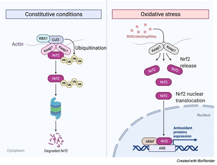
    ROS are profoundly reactive particles that begin primarily from the mitochondrial electron transport chain (ETC).

    Practically all cells and tissues constantly convert a little extent of atomic oxygen into superoxide anion by the equivalent decrease of sub-atomic oxygen in the ETC.

    The ROS are delivered by different pathways too, remembering the respiratory burst occurring for initiated phagocytes, ionizing radiation’s harming impact on parts of cell layers, and as results of a few cell catalysts including NADPH oxidases (Nox), xanthine oxidase (XO), and uncoupled endothelial nitric oxide synthase (eNOS).

    Because of the expected advantageous part of ROS showed by a few lines of exploration, going from their job as flagging particles to the more sudden function in the progress of certain malignancy, the expression “redox guideline” may end up being more precise than “redox stress”; there have been even a few circumstances where cancer prevention agents are portrayed to be “awful”.

    In any case, the expression “redox stress” is all the more generally utilized. Both the ER and the mitochondria take part in keeping up with typical cell homeostasis.

    It is through the ER job in keeping up with appropriate protein collapsing that this organelle is unpredictably associated with the general ROS guideline.

    The ER detects signs of changed cell redox states and afterward acts in like manner to reestablish and keep up with typical homeostasis. During the UPR of the ER, ROS will be grouped either because of genuine creation of ROS or because of utilization of the cell reinforcements like GSH.

    Since the ER can be a piece of an endless loop, where oxidative pressure prompts ER stress, and the last will additionally demolish the redox status, there are a few defensive systems to restrict the expected harm.

    A solid affiliation and a potential reason impact relationship exist between faulty mitochondria and metabolic illnesses. As in the ER case, a few defensive instruments exist to shield the mitochondria from oxidative harm. The cancer prevention agents, as superoxide dismutase, catalase, and glutathione peroxidase/reductase framework, are not in the extent of this survey.

    UCPs are normal controllers for mitochondrial ROS, reacting to and controlling the ROS production by decreasing the mitochondrial huge proton inclination. As of late, UCP2 has been connected to different capacities too.

    Reactive Oxygen Species vs Oxidative Stress

    Oxidative pressure suggests any divergence between reactive oxygen species and framework ability to detoxify these side-effects of digestion.

    Disappointment of ordinary rodox state prompts harmful impacts through creation of free revolutionaries and peroxides harming cell segments like lipids, proteins and DNA.

    Abiotic stresses like saltiness, dry season, and limits of temperature, UV radiations, synthetic poisonousness, and so forth are among the solid up-and-comers that hamper plant development and result in horticultural efficiency worldwide.

    This load of sort of stresses are joined by the overproduction of ROS. Microorganism attack is among one of the causes that makes ready for the age of exceptionally oxidizing ROS.

    Overproduction of ROS and its side-effects being toxic outcomes in oxidative pressure, it is, accordingly, the important to keep the amicability between the age and digestion of ROS and its results so that plant can play out its indispensable cell metabolic capacities easily.

    During the typical life cycle plants are consistently delivering Reactive oxygen species from different cell compartments with the serious paces of electron stream.

    It is a result of this reality that plants are completely outfitted with various enzymatic and non-enzymatic cancer prevention agent apparatus for searching ROS and to monitor them during ideal states of development.

    Then again during upsetting conditions, the unevenness between ROS creation and their searching may prompt oxidative pressure.

    Reactive Oxygen Species vs Cancer

    An alternate face of the ROS coin has been uncovered dependent on considering the impact of changes actuating the record atomic factor, nuclear factor-erythroid 2-related factor 2 (Nrf2).

    Nrf2 is a redox stress-delicate record factor that actuates a few cells reinforcement and detoxification qualities.

    Reactive Oxygen Species - research tweet 1

    Without redox stress states, Nrf2 is kept idle by restricting to another protein, Kelch-like ECH-related protein 1 or KEAP1, guaranteeing successful Nrf2 constraint.

    Substantial transformations in either Nrf2 or KEAP1 that forestall their limiting will result in constitutive Nrf2 enactment and record of Nrf2 target qualities.

    Such transformations have been segregated from patients with cellular breakdown in the lungs recommending a protumorigenic job of Nrf2.

    Moreover, drug obstruction in some antitumor treatment may happen because of such substantial transformations. All the more as of late it has been exhibited that in mice a few endogenous oncogenes, for example, Kras, Braf, and Myc effectively incite Nrf2 articulation, advancing a ROS detoxification program and subsequently making a more “decreased” intracellular climate, a program that the creators propose to be needed for tumor commencement.

    Thusly, the 10,000 foot view mirroring the commitments of different go amid in addition to nearby natural elements is by all accounts the real determinant for ROS-instigated results in both physiology and pathology, and subsequently it is fundamental to unwind the not-yet-surely knew portions of this complicated picture for better comprehension of the ROS incited modifications.

    There is developing proof that redox controllers, related dynamic intermediates, cell organelles works, and general conditions are altogether integrated in multifaceted organizations influencing the entire body, digestion, condition of wellbeing and illness and even life expectancy.

    Despite the fact that at present the utilization of cell reinforcements appears to be disillusioning in forestalling the movement of the ROS-related sicknesses, flow research discoveries have proposed novel focuses that may end up being more fitting cancer prevention agents.

    Further exploration is expected to examine the conceivable preventive or potentially restorative upsides of these particles.

    Reactive Oxygen Species Citations

    Share

    Similar Post:

    Continue Reading
  • Turner Syndrome: Symptoms, Causes, Diagnosis, and Treatment

    What is Turner Syndrome?

    Turner syndrome is one of the chromosomal conditions which affects only females and males are not affected mostly by this condition.

    It is considered as one of the genetic disorders which occurs due to the partial or complete loss of one of the X chromosomes.

    It is also referred to as monosomy of X.

    This condition leads to various developmental abnormalities and they are also suspected to many diseases.

    It was first observed by Henry. H. Turner, so this syndrome is commonly known as Turner syndrome.

    Turner Syndrome - research tweet 1

    Characteristics of Turner Syndrome

    Turner syndrome affects the primary and secondary sexual characteristics in females.

    The person with these conditions are usually sterile in condition, they are also short in stature and webbing of skin occurs and also webbing of neck is a common symptom.

    The person with this syndrome usually have other disorders such as hearing problems, cardiac abnormalities, vision loss, etc.

    There is no absolute cure for this disorder but they can be given speech and other intellectual trainings to improve their motor areas of the brain.

    There are usually short in stature and there will be inadequate progress in their sexual development.

    It is usually diagnosed by antenatal tests at the time of pregnancy.

    Types of Turner Syndrome

    Turner syndrome is one of the most occurring genetic disorder which is caused due to an aneuploidy of sex chromosomes.

    Where one X chromosome is found missing in all cells or few cells in our body. This condition occurs almost one in 2500 female births.

    They are classified into types depending on how they affect the body cells.

    I. Monosomy of X Chromosome

    This is the most common type of syndrome occurring in most of the cases.

    In this case there is a complete loss of one X chromosome. This occurs due to an abnormal cell division at the time of gamete formation.

    In this condition egg or sperm cell losses one chromosome which results in only 45 chromosomes in all the cells, this result in XO where there will be no bar bodies.

    Which means that X chromosome is not inactivated, but for the genes to get activated they must be present in pairs for the proper growth and development.

    II. Mosaicism

    The sex chromosomes are lost during mitosis after fertilization. During the development of zygote, it results in 45 chromosomes in some cells and where as the other cells have normal 46 number of chromosomes. This also results in Turner syndrome.

    Where as if the loss of chromosomes occurs late in the development, the number of aneuploidy cells are less and it results in reduced severity. When considering somatic mosaics, there will be a presence of 45X cells in few of the cells and in other cells it will be present in the extra X chromosome(47XXX).

    In this condition girls are taller. In some cases, the female with Turner Syndrome has an extra Y chromosome along with that 46X chromosome which leads to the condition of developing a cancer known as gonadoblastoma.

    III. X- Chromosome Abnormalities

    During this condition the 20% of Turner Syndrome cases results in two X chromosomes which are present in all the cells, but only one chromosome has an abnormal structure.

    This is usually due to the circular shaped chromosome which is present along the joint ends and forms a ring like structure which is known as ring chromosome.

    Sometimes it may also result in a condition where one of the X chromosomes has one long arm instead of the q arm. Which results in two long arms in a single chromosome. It is known as Isochromosome.

    Turner Syndrome Symptoms

    The genetic make up of an individual varies accordingly depending upon the traits present in the chromosome.

    Mostly females with this condition have rudimentary ovary and they are mostly sterile individuals along with short stature.

    The most common symptoms of turner syndrome are Abnormal facial features like drooping eyelids, narrow palate along with short jaw line and abnormal earlobes which are set low than normal ones.

    Growth is irregular among them in most of the cases.

    Puberty is delayed, but the females with this condition have good intelligence and reading skills along with good verbal communication. But few of these people face learning difficulties as a results in difficulty in memorizing and also in solving the mathematical skills.

    These people face difficulties in understanding the others emotion. These people suffer from undeveloped or poorly developed breasts and there will also be delay in menstrual cycles and in most of the cases they remain sterile.

    There will also be other disturbances like murmuring of heart, due to the narrowing of aorta and other abnormalities in heart, liver and kidney which results in other symptoms.

    Some symptoms also result in developing fetus, like Lymphedema which is characterized by swelling of muscles due to fluid leakage in the body.

    It also causes swelling or thickness of neck which results in lower weight than normal.

    Diagnosis of Turner Syndrome

    These conditions can be diagnosed at the time of pregnancy by the process of amniocentesis or other tests like ultra soundwave therapies which helps us to find the karyotype of the fetus.

    Turner syndrome can also be identified in infants by swelling of hands and feet and also other problems with kidney and heart and also webbed neck with broad chest and nipples are widely placed.

    In some cases, it cannot be diagnosed till puberty.

    Turner Syndrome Treatment Options

    Though there is no permanent cure for turner syndrome, the other severe symptoms arising along with this such as vision and hearing problems can be treated.

    We can also keep a regular check at heart discomforts and thyroid issues.

    Hormone therapy is followed now a days to prevent short stature, which also helps in inducing the sexual development.

    Invitro fertilization can be followed to get pregnancies among this kind of people.

    Other intellectual activities can be improved by giving proper therapies.

    Turner Syndrome Citations

    Share

    Similar Post:

    Continue Reading