Tag: Cytoskeleton

Tag: Cytoskeleton

  • Cytoskeleton: Description, Structure, and Function

    Cytoskeleton: Description, Structure, and Function

    Share

    Similar Post:

    Continue Reading
  • Cytoskeleton: Description, Structure, Types, and Function

    What Does the Cytoskeleton Do?

    We all know that cells form the structural and functional unit of the all the living organisms. Each and every cell in our body has its own structure. Have you all ever thinked how this stable structure is maintained.

    Cytoskeleton plays an important role in maintaining the structure and shape of the cell. Our cell organelles start moving here and there and loses its appropriate shape thus losing its functions, which makes various critical conditions in our body.

    If considering basically, what would happen if our internal organs move from its own places of origin, we find very difficult to move, speak and to do any kind of activities. In the same way, our cell also finds difficult to perform their activities.

    We all think that each and every cell are just a bilobed round structures which does not have its own properties, but the truth is that these cells have their magnifies appropriate structure which contains a network of filaments known as Cytoskeleton, which is literally explained as Cells skeleton”.

    It does not maintain the shape of the outer membranes like plasma membranes, it gives the overall structure of the cell and also plays an important role in placing the organelles in their correct positions and also aids in tracking the transport of the vesicles.

    What is Cytoskeleton?

    As mentioned above cytoskeleton is a group of fibers, which plays an important role in minting the structure and tracking the changes. Generally, in eukaryotes, there are about three types of protein fibers, which are named as follows:

    i. Microfilaments

    ii. Intermediate filaments

    iii. Microtubules

    i. Microfilaments

    Microfilaments are one of the three types of protein filaments that are present in our cytoskeletons. Of the three, microfilament is the narrowest one having a diameter of about 7 nm which are being made up of many linked monomers of the protein known ad actin, these actions are present as a monomer and combines together to form a double helical structure.

    Actin filaments usually have a directionality which means that they posses tow structurally different ends. Actin filaments usually consists of a variety of important functions in the cell.

    Actin filaments also consists of numerous important roles in the functioning of the cell. It also serves as a track for the movement of a motor protein which is known as myosin.

    As it has a relationship with the muscle protein myosin, actin is usually involved in performing many of the cellular functions. Microfilaments also aid in many functions of the cell like cytokinesis and in motility of the cell.

    ii. Intermediate Filaments

    These filaments are about 8 to 12 nm wide. They are called as intermediate filaments as their size is between the microfilaments and the microtubules. Intermediate filaments are generally made up of different kinds of proteins linked keratin, desmin, vimentin and lamin.

    All the intermediate filaments are found in the cytoplasm of the cell, except those of lamins, as they are found in the nucleus and they help in supporting the nuclear envelope. Intermediate filaments present in the cytoplasm maintains the shape of the cell, cell tension and also provides structural support to the cell.

    iii. Microtubules

    Among these three protein filaments, microtubules are considered as the largest cytoskeleton fibers which is about 23nm in size. These are hallow tubes that are made up of alpha and beta tubulin. Microtubules forms a structure like flagella, which are the tails that propels forward.

    Most of the microtubules in the cell usually comes from the cell organelles, the centrosome which is generally called as the organizing center of the cell.

    Cytoskeleton Function

    As mentioned above, cytoskeleton consists of several functions. The first and fore most function of the cytoskeleton is to give its cytoskeleton the appropriate shape.

    Cytoskeleton is very much important in the cells without cell walls, as such animal cells do not get their shape from the thick outer layer. It provides the cell a good movement.

    he microfilaments and the microtubules help in reassembling and disassembling the cells. It also helps the cell to crawl and migrate. The cytoskeleton also plays an important role in keeping the cell organelles in the place and it also helps in movement of the organelles throughout the cell.

    Cytoskeleton Citations

    Share

    Similar Post:

    Continue Reading
  • Cytoskeleton: Description, Structure, and Function

    Cytoskeleton

    o The cytoskeleton is a cellular “scaffolding” or “skeleton” contained within the cytoplasm.

    o The cytoskeleton is present in all cells; it was once thought this structure was unique to eukaryotes, but recent research has identified the prokaryotic cytoskeleton.

    o It is a dynamic structure that maintains cell shape, protects the cell, enables cellular motion (using structures such as flagella, cilia and lamellipodia), and plays important roles in both intracellular transport (the movement of vesicles and organelles, for example) and cellular division.

    Cytoskeleton Component

    Cytoskeleton, Cytoskeleton Description, Cytoskeleton Structure, Cytoskeleton Function 1

    Types of Cytoskeleton

    There are two types of filaments in the cytoskeleton;

    I. Microtubules

    o Microtubules are built from subunits-molecules of tubulin-each one of which is itself a dimer composed of two very similar globular proteins called a-tubulin and b-tubulin bound tightly together by noncovalent bonds.

    o Although tubulin is globular it can polymerize into long straight filaments.

    o Thirteen of these filaments lie alongside each other to form the microtubule.

    o In nine triplet sets (star-shaped), they form the centrioles.

    o Centrioles function in the production of flagella and cilia, but are NOT necessary for microtubule production.

    o The major portion of each flagellum and cilium, called the axoneme, contains nine pairs of microtubules forming a circle around two lone microtubules.

    o The latter formation is commonly referred to as a “9+2” arrangement, wherein each doublet is connected to another by the protein dynein.

    o The mitotic spindle is made from microtubules.

    o Mitotic spindle provides the machinery that will segregate the chromosomes equally into the two daughter cells.

    o Microtubules have a + and a – end.

    o This is because of the polarity of the a and b tubulin, thus making the microtubule polar.

    o The – end attaches to a microtubule-organizing center (MTOC) in the cell.

    o The major MTOC in animals is the centrosome.

    o A microtubule grows away from an MTOC at its + end.

    o Centrosomes are composed of two orthogonally arranged centrioles Centrosomes are often associated with the nuclear membrane during interphase of the cell cycle.

    o In mitosis the nuclear membrane breaks down and the centrosome nucleated microtubules can interact with the chromosomes to build the mitotic spindle.

    o The centrosome is copied only once per cell cycle so that each daughter cell inherits one centrosome, containing two centrioles.

    o The centrosome replicates during the S phase of the cell cycle.

    II. Microfilaments

    o Microfilaments are the thinnest filaments of the cytoskeleton found in the cytoplasm of all eukaryotic cells.

    o These linear polymers of made of actin subunits are flexible and relatively strong.

    o Microfilaments produce the contracting force in muscle as well as being active in cytoplasmic streaming, phagocytosis, and microvilli movement, cytokinesis, and muscle movement.

    o In fungi, the Septa is usually perforated to allow exchange of cytoplasm between cells, called cytoplasmic streaming.

    o Both microtubules and microfilaments are involved in intracellular movements in eukaryotic cells.

    o In both cases the movements are generated by motor proteins, which bind to microfilaments and microtubules and use the energy derived from repeated cycles of ATP hydrolysis to travel steadily along the microtubule or microfilaments in a single direction.

    o The motor proteins that move along cytoplasmic mircrotubules are: kinesins ( – ⇒ + ) and dyneins ( + ⇒ – ).

    o Actin uses myosin as its motor protein.

    Eukaryotic Flagella and Cilia

    o Eukaryotic flagella and cilia are specialized structures also made from microtubules.

    o The major portion of each flagellum and cilium, called the axoneme, is arranged in nine doublets oriented about two additional microtubules (wheel-shaped) they form cilia and flagella.

    o This formation is commonly referred to as a “9+2” arrangement, wherein each doublet is connected, via a crossbridge, to another by the protein dynein.

    o The cross bridges cause the microtubule pairs to slide along their neighbors creating a whip action in cilia causing fluid to move laterally, or a wiggle action in flagella causing fluid to move directly away from the cell.

    o Both cilia and flagella grow from a basal body.

    o In humans cilia are only found in the fallopian tubes and the respiratory tract and in cerebrospinal fluid in ependymal cells.

    o Bacterial flagella are long, hollow, rigid, helical cylinders made from a globular protein called flagellin, these shouldn’t be confused with eukaryotic flagella which are made up of microtubules.

    o They rotate counterclockwise. When they are rotated clockwise, the bacterium tumbles.

    o This tumbling acts to change the orientation of the bacterium allowing it to move forward in a new direction.

    o A basal body is an organelle formed from a centriole, a short cylindrical array of microtubules.

    o It is found at the base of a eukaryotic cilium or flagellum and serves as a nucleation site for the growth of the axoneme microtubules.

    Cytoskeleton Citations

    Share

    Similar Post:

    Continue Reading
  • Cytoskeleton: Definition, Importance, and Functions

    What are Cytoskeleton?

    The presence of cytoskeleton in the design of the cellular material was proposed by Koltzoff in 1928. They are intertwining organization of protein fibers stretched out all through the cytoplasm and lattice of various proteins in cells.

    As its name infers it assists with keeping up with cell shape and is significant in cell motility.

    It is a unique three-dimensional construction that fills the cytoplasm. This construction goes about as both muscle and skeleton, for development, locomotion, and strength.

    Features of Cytoskeleton

    The interior movement of cell organelles, velocity, and muscle fiber withdrawal can’t happen without the cytoskeleton.

    It is accepted that cytoskeleton is the trademark highlight of eukaryotic cells however late examination demonstrated that prokaryotic cells have proteins that structure a cytoskeleton.

    Over the span of the human genome project more than 800 presumably cytoskele­ton related qualities are found.

    Based on three kinds of protein fibers, cytoskeletons are of three sorts like microtubules, halfway fibers and microfilaments.

    Microfilaments

    Microfilaments are fine, string-like protein strands, 5-7 nm in breadth, address the dynamic or motile piece of the cytoskeleton.

    They seem to assume a significant part in cyclosis and amoeboid movement. They are made overwhelmingly out of a contractile protein called actin, which is the most plentiful cell protein.

    These fibers are cross connected into organizations or groups. The semi adaptable microfilaments make cells versatile, to isolate in mito­sis (cytokinesis) and are answerable for strong compression. The adaptable transitional fila­ments fortify the cell furthermore.

    As a rule a shell of microfilaments upholds the plasma layer. Microfilaments are a polymer of actin protein subunits in addition to appended proteins like cross-linkers.

    Most multi-cell creatures have a few actin iso-structures. People have six actin qualities; four encode alpha-actin, one beta-and one gamma-actin.

    Alpha-actin is found in muscle cells where it assumes a significant part in getting the cell, though beta-actin is limited toward the front of moving cells and gamma-actin structures pressure strands.

    Actin protein as a polymer without connected proteins is called filamentous actin (F-actin), while the globular actin monomers are called G-actin.

    Actin Filaments

    Actin fibers are 8 nm in distance across and comprise of two strands of the protein actin that are bound around one another.

    They are particularly unmistakable in muscle cells, where they accommodate the withdrawal of muscle tissue, monomers of the protein actin polymerize to frame long, slight filaments.

    Cytoskeleton - Research Tweet 1

    A few elements of actin fibers structure a band just underneath the plasma layer that gives mechanical solidarity to the phone, joins trans-film proteins (e.g., cell surface receptors) to cytoplasmic proteins, secures the centrosomes at inverse posts of the cells during mitosis, and squeezes partitioning creature cells during cytokinesis.

    It produces cyto­plasmic gushing in certain cells, velocity in cells, for example, white platelets and the one-celled critter, and connect with myosin (“thick”) fibers in skeletal muscle strands to give the power of solid constriction.

    Microtubules

    Microtubules are profoundly unique protein polymers that structure an essential piece of the cytoskeleton in every single eukaryotic cell.

    Robertis and Franchi (1953) noticed the first time in the axoplasm of the myelinated nerve filaments, which they called neurotubules.

    Microtubules were first depicted exhaustively by Ledbetter and Porter (1963).

    Microtubules are transport lines inside the cells.

    Microtubules are round and hollow cylinders, 20-25 nm in measurement, and made out of protein tubulin subunits.

    Cytoskeleton - Research Tweet 2

    These subunits are named alpha and beta. Each miniature tubule is com­posed of eleven sets of these tubulin subunits masterminded in a ring.

    A significant part of microtubules is giving a pathway to intracellular developments of organelles and proteins.

    This is finished by engine proteins (kinesins and dyneins) under the utilization of ATP.

    The centrosome is situated in the cytoplasm appended to the outside of the core. It is dupli­cated during the S phase of the cell cycle. Not long before mitosis, the two centrosomes move separated until they are on inverse sides of the core.

    As mitosis continues, microtubules develop out from every centrosome with their in addition to closes developing toward the metaphase plate. These clusters of microtubules are called axle strands.

    Importance of Cytoskeleton: Cell Movement

    Cell movement is achieved by cilia and flagella. Cilia are hair-like constructions that can beat in synchrony causing the movement of unicellular Paramecium.

    The two cilia and fla­gella are built from microtubules, and both give either motion to the cells (e.g., sperm) or move liquid (e.g., ciliated epithelial cells that line our air entries and move a film of bodily fluid towards the throat).

    Every cilium or flagellum is made of a round and hollow exhibit of 9 equally divided microtubules, each with a halfway microtubule connected to it. 2 single micro­tubules run up through the focal point of the pack, finishing the supposed “9+2” design.

    The whole get together is sheathed in a layer that is just an expansion of the plasma mem­brane.

    Some eukaryotic cells move about through microtubules connected to the outside of the plasma layer. These microtubules are called flagella and cilia.

    Flagella and cilia both have a similar construction: a ring of nine tubulin trios orchestrated around two tubulin sub-units.

    The distinction among flagella and cilia lies in their movement and numbers. Flagella are appended to the cell by a “wrench” Mike mechanical assembly that permits the flagella to turn. Cilia, then again, are not joined with a “wrench,” and beat to and fro to give move­ment.

    Ciliated cells generally have many these projections that cover their surfaces.

    For instance, the protist Paramecium moves through a solitary flagellum, while the protist Didinium is covered with various cilia.

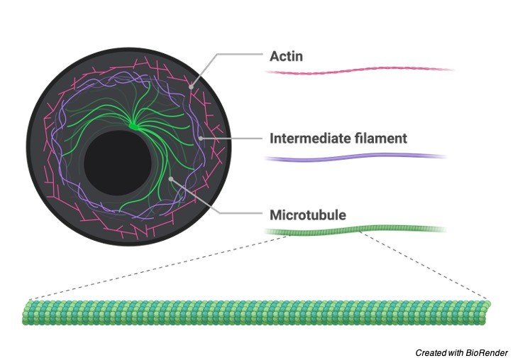
    In microtubules one alpha-and one beta-tubulin structure a hetero-dimer. Long chains of these hetero-dimers made out of proto-fibers, wherein consistently an alpha-tubulin is fol­lowed by a beta-tubulin.

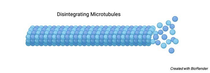
    Each microtubule has a (- ) and a (+) end. At the (+) or beta-tubulin end new heterodimers are added quicker and at lower tubulin fixations than at the (- ) or alpha-tubulin end.

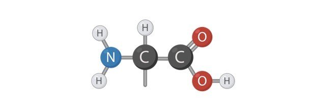
    The alpha-tubulin just as the beta-tubulin subunit ties a little guanosine tri-phosphate (GTP). Cells have protein engines that tight spot two particles, and utilizing ATP as energy, influence one atom to move in relationship to the next.

    Two sorts of these protein engines are myosin and actin, and dynein or kinesin and microtubules. These groups of proteins all have a mo­tor end, however, may have a few sorts of various atomic designs on the limiting end.

    At the point when these proteins tie the atoms they are moved to various organelles. When connected to different microtubules, protein engines can cause movement if the finishes are fixed or broaden the lengths of the fiber groups if the closures are free.

    Cytoskeleton Citations

    Share

    Similar Post:

    Continue Reading