Author: Admin
-

Amphibolic Pathway: Definition and Example I Research...
Continue ReadingWhat is Amphibolic Pathway?
Survival of the organism depends on the nutrient uptake from the environment and complete utilization of nutrients.
This process is vital and forms a dynamic nature of the cell. The conversion of external nutrient to an internal biomolecule is a main function of any biosystem present and is achieved through the process of metabolism.
Metabolism is the process by which nutrient are converted to desirable products for synthesis and breakdown of biochemical components
"A pathway which participates in Catabolism and anabolism is termed as amphibolic pathway"
Metabolism is otherwise termed as Catabolism + Anabolism. Catabolism is a process of breaking down of Complex food molecules to simpler compounds.
Anabolism is mainly concerned with the biosynthesis of Chemical compounds which are essential for storage and other functions from smaller molecules.
Synthesis (Anabolism) and degradation (Catabolism) of any biomolecule takes place in a series of process which are distinct in its own mechanism and have specific pathway.
But there are certain pathways which can act both catabolic an anabolic under appropriate circumstances.
A pathway which participates in Catabolism and anabolism is termed as amphibolic pathway.
Amphibolic Pathway involves many intermediates which forms new compounds (biosynthesis) such as amino acid, fats, glucose etc., or they get oxidized to produce energy.
An ideal example for Amphibolic Pathway is the CITRIC ACID CYCLE. The citric acid cycle is Amphibolic because it oxidizes Carbohydrates, Fats, Proteins to yield energy and it is the common oxidative pathway for any biochemical compound.
Any biochemical degradation either directly yields acetyl Co – A or through pyruvate and other components of citric acid cycle.
Similarly, the intermediates from the citric acid cycle becomes precursor for the synthesis of Glucose, fats, Amino Acid etc.,
The catabolism and anabolism of the intermediates present in the citric cycle depends upon the internal cues of the body.
Internal cues can be the requirement of synthesis or breakdown of a compound and is dependent on many other factors.
Amphibolic Pathway Characteristics
The main characteristics of amphibolic pathway is the involvement of both catabolic and anabolic pathway and Citric Acid Cycle becomes an ideal cycle to understand the Amphibolic Pathway

Catabolic Nature of Citric Acid Cycle
In cellular respiration, citric acid cycle is the common oxidative pathway where the Acetyl Co – A breakdown yielding NADH, FADH and ATP in plants GTP in Animals.
The basic biological compound namely Proteins, Fats and Glucose oxidizes to form Acetyl Co – A directly or through Pyruvate enters the common pathway of oxidation.
From the basic cellular respiration, we know that the substrates of Citric Acid Cycle are not completely derived from glycolysis but the amino acids and lipids also contribute to the substrate.
Catabolic activities of citric acid cycle are,
1. Oxidation of Glucose
2. Transamination of Amino Acids
3. Oxidation of Fatty acids
Glucose is broken down to pyruvate by Glycolysis.
Fatty Acids on Beta – Oxidation provides Acetyl Co – A directly
Amino Acid Catabolism
Essential and non – essential amino acids are present in body for the proper functioning of the living system.
Among all of them each amino acids do not form acetyl Co – A directly.
Few forms other intermediates of the Citric Acid Cycle by the process of Transamination or Aminotransferase. The Aminotransferase reaction involves the transfer of alpha – amino group from the carbon skeleton and transforming the skeleton to Amphibolic intermediate to produce energy.
Few amino acids oxidize to become pyruvate and Acetyl Co -A also.
For example:
I. Glutamine is transformed to Glutamate with enzyme involving Glutaminase. Glutamate then by transamination becomes alpha – ketoglutarate.
II. Similarly, Asparagine becomes Aspartate on transamination’s yields oxaloacetate and Certain Amino acids like alanine, Serine etc., directly oxidizes to produce pyruvate
Anabolic Nature of Citric Acid Cycle
Biosynthesis of glucose, fatty Acids, Amino Acids, Nucleic Acids are produced from the intermediates of the Krebs cycle.
The intermediates form many precursors corresponding to their respective substrates from which the biosynthesis takes place.
Anabolic activities are;
I. Citrate from mitochondria enters cytosol to oxidize to form Acetyl Co – A, this initiates the Biosynthesis of Fatty Acids.
II. α-ketoglutarate is the precursor for Amino Acid Glutamate which on Transamination provides Glutamine, Arginine and Proline and provides Purines.
III. Succinyl Co – A becomes a precursor for Porphyrin and Heme forming oxygen carriers in Blood.
IV. Oxaloacetate is the precursor for Aspartate and they form Asparagine to produce pyrimidines and other Amino acids that acts as an intermediate for Gluconeogenesis
Amphibolic Pathway Citations
Share
Similar Post:
-

What is Lactic Acid? Where Does it...
Continue ReadingWhat is Lactic acid?
Do you know what type of acid is present in all the milk products including cheese, milk, curd and butter.
Yeah, absolutely it’s a Lactic acid.
Lactic acid is generally considered as an organic acid and it is produced by a fermentation process which occurs in milk sugar.
It was first identified by Carl Wilhelm in 1780 in milk. It is a colorless water-soluble substance which has been widely used in food and pharmaceuticals and it has also been synthesized by our body in many metabolic conditions.
The First Demonstration of Lactic Acid
Lactic acid was first identified by Scheele in sour milk in the year 1780. Further when Pasteur was undergoing research on pasteurization, he found the microorganism which is necessary for lactic acid production.
Lactic acid is also being commercially sold for many purposes.
How Lactic Acid is Utilized by Human Body?
Lactic acid is used for the process of conversion of glucose into energy when our body has very low oxygen content.
We often release a painful sensation when we exercise or do heavy activities suddenly or after a long interval, it is due to the accumulation of lactic acid in the muscles which results in muscle cramps at a certain area in our body.
It does not cause any serious issues as liver has the capability to breakdown the extra lactate.
How Lactic Acid Build up in Our Body?
In some cases, our muscles do not have enough oxygen to breakdown glycogen into glucose for the synthesis of energy molecules which is known as ATP, at this time lactic acid gets accumulated in a certain area where enough oxygen is not available which is often referred to as anaerobic metabolic process.
When lactate gets accumulated it leaves to formation of lactic acidosis.
It may also upgrade at the times of heart attack, sudden shock, and at the cases of lung diseases.
Lactic acid forms in many varieties at each area according to their nature.
When they are being accumulated at muscles and blood tissues they are known as dextrorotary form.
The lactate which is obtained as a result of sucrose formation is known as levorotary form, when the same lactate if produced by bacteria during fermentation they are known as racemic form.
Thus, each of its form has its origin from the process of their accumulation or synthesis.
To explain in detail about how our body produces lactic acid, as said earlier lactic acid gets accumulate in our body when we perform heavy works or exercises during a sudden period of time.
This because when we perform powerful activities, we take a long and faster breathes which leads to the usage of more oxygen by the muscles, which tend to perform that action, which results in synthesizing energy anaerobically.
We generally get energy by the process of glycolysis, where glucose is reduced to pyruvic acid and release of ATP molecules occurs through further process which is important for the energy source.
This is a general process, but when our muscles are in a state with absence or lack of oxygen the pyruvate which is produced through a process of glycolysis convert itself into a substance namely lactate and supplies energy in the anaerobic form which leads to the accumulation of lactate at higher levels.
But this is performed by our body, for a longer time it leads to side effects by the accumulation of lactate at higher levels in the tissues which also disrupts the other metabolic activities of the muscle and creates an acidic environment around the muscle cells.
So, it results in degrading a working capability of the muscle and cannot perform any heavy or powerful activities further.
But on considering an immune response of our body, natural immune barriers prevent the muscle tissues from permanent damage and initiates the cells to work normally by upcoming the situation and by performing regular muscle contraction improves the lactic acid accumulation.
This is because our body gets slow during these conditions and allows the reverse mechanism of conversion of lactate into pyruvate and normal aerobic respiration is followed by the tissues.
Fermentation of Lactic Acid
Lactic acids are also synthesized in industries for various purposes through fermentation.
Several microorganisms are involved in the production of lactic acid, each of it has the mechanism of producing either D (-) lactic acid or L (+) lactic acid depending on their nature.
The racemic form is formed due to the presence or production of an enzyme known as racemase, the lactic acid usually be in an active form unless it is deactivated by any other enzymes.
The two most important steps involved in the production of lactic acid are as follows,
1. Homo-fermentative Process
This type of process involves the utilization of certain bacteria such as Lactobacillus delbruckii, L.casei, L.leichmanii, streptococcus lactis etc. which utilizes its EMP pathway to synthesis pyruvic acid which helps in the production of lactic acid by reducing lactate dehydrogenase.
Though these microbes can serve under anaerobic conditions they are known an anaerobic microbe.
2. Heterofermentative Process
This process involves the synthesis of lactic acid, ethanol, acetic acid, carbon dioxide and water with the help of Leuconostoc mesenteroids.
Manufacturing of Lactic Acid
The medium should be prepared such that it contains masses of molasses, maltose, lactose, sucrose, calcium carbonate, hydrogen and phosphate which are kept at a pH of 5.5 to 6.5.
As lactic acid is soft and delicate in nature the mixture of metals is avoided in the culture medium and fermenters are nature based and mostly wood fermenters are used.
The use of Thermophilic clostridia in the culture results in the formation of major and important constituents of lactic acid namely butanol and butyric acid.
Uses of Lactic Acid
Considering lactic acid as a commercial one it can also be used in polymer production since it has a property of weak acid.
It is also being used in food and other industries such as production of beverages in preservation as it has an acidic property which is due to the anaerobic property of that which prevents the food from spoilage.
It is also being used in leather industries for de-lining and also in laundry and textiles for the treatment of fabric.
Calcium lactate is being used as a source of calcium in pharmaceuticals and also in baking units.
Lactic Acid Citations
- Lactic acid and exercise performance : culprit or friend? Sports Med . 2006;36(4):279-91.
- Lactic acid fermentation of human excreta for agricultural application. J Environ Manage . 2018 Jan 15;206:890-900.
- Biosynthesis of D-lactic acid from lignocellulosic biomass. Biotechnol Lett . 2018 Aug;40(8):1167-1179.
Share
Similar Research Position:
-

Pathogenesis and Transmission of Mycobacterium tuberculosis
Continue ReadingMycobacterium tuberculosis quickly after infection encounters with alveolar resident macrophages, after which dendritic cells and monocyte-derived macrophages also take part in the phagocytic process.
Transmission of Mycobacterium tuberculosis
Patients, suffering from active pulmonary TB are the principal source of TB transmission. These patients expel aerosolized tubercle bacilli by the respiratory route and may infect any individual, unfortunate enough to inhale the aerosolized bacteria.
Respiratory droplets are generated by human’s coughs and sneezes and they remain airborne for about 6-10 hours.
Thus, approximately one third of the world population, i.e. ~2 billion peoples are latently infected with this etiological pathogen.
Mycobacterium tuberculosis quickly after infection encounters with alveolar resident macrophages, after which dendritic cells and monocyte-derived macrophages also take part in the phagocytic process.
Although, both pathogenic and non¬pathogenic mycobacteria can enter in macrophages with similar facility, but only the pathogenic species can survive therein.
Replication and dissemination of the pathogen are restricted by mononuclear phagocytes and control the infection by cell-mediated immunity (CMI).
Mycobacterium tuberculosis remains dormant until the balance between bacillary persistence and the immune response gets disturbed.
The infected alveolar macrophages containing the pathogen either destroy their predators (a mechanism that has not yet been proven, but probably accounts for a small proportion), or they fail to contain the pathogen and die.
Immune response and virulence of M. tuberculosis are balanced, intracellular bacteria are contained by the macrophages, and the immune system isolates the primary site of infection by granuloma formation (primary lesion).
Mycobacterium tuberculosis can persist in a dormant state for long periods of time even sometimes lifelong.
"Mycobacterium tuberculosis remains dormant until the balance between bacillary persistence and the immune response gets disturbed"
Any disturbance of the balance between host and pathogen after weakening of the cellular immune response (immuno-suppression) causes endogenous exacerbation which leads to active (post primary) TB.
An impaired host response due to various reasons including aging, malnutrition, steroids or HIV allows reactivation of the bacilli resulting in clinical manifestation of disease.
Understanding the patho-mechanisms of latent persistence of Mycobacterium tuberculosis will therefore facilitate novel approaches towards prevention and control of infection, reactivation and re-infection.
Intracellular multiplication of Mycobacterium tuberculosis in alveolar macropbages
Most alveolar macrophages are highly activated cells capable of destroying or inhibiting the growth of inhaled bacilli, especially if these bacilli are not fully virulent.
However, some alveolar macrophages are poorly activated and allow ingested tubercle bacilli to multiply intracellularly.
Virulent strains of Mycobacterium tuberculosis also uses a variety of strategies to avoid phagosome-lysosome fusion in macrophage and multiply continuously, eventually lead to the lysis of the infected cell.

Once the bacteria are transported into the deeper tissues by macrophage and perhaps other phagocytic cells, additional macrophages gather at individual infected cell to form granuloma.
The extracellular bacilli are then taken up by other macrophages and by blood monocytes that are attracted to the focus and then develop into immature macrophages.
Thus, the bacillary multiplication cycle is repeated within immature macrophages, which lead to the spread of mycobacteria to deeper tissues and other organs including lymph nodes, where they multiply.
Once the bacteria are transported into the deeper tissues by macrophage and perhaps other phagocytic cells, additional macrophages gather at individual infected cell to form granuloma.
The tuberculous granulomas in humans and mice have a large complement of T lymphocytes, some B lymphocytes, dendritic cells, neutrophils, fibroblasts, and extracellular matrix components.
Although the role of all the accessory cells in the granuloma formation has not yet been clarified, certain T lymphocytes subsets play an unequivocal role in the maintenance of the granuloma and in restriction of the bacterial growth in human infection.
Granuloma formation: Host versus Mycobacteria
Inside the necrotic lesion the tubercle bacilli survive in the solid caseous lesions, but fail to multiply there because of anoxic conditions, reduced pH and the presence of inhibitory fatty acids.
At this stage, the cell mediated immune response gets activated, which initiates the proliferation of T- lymphocytes and macro phages that accumulate around caseous centre to prevent the extension of lesion. Depending on the host, an appropriate immune response can control mycobacterial growth.
Thus, it is of prime importance to define differences in architecture and functional properties of the granuloma. Several studies have shown that the cytokines interferon-gamma (IFN-γ) and tumor necrosis factor-alpha (TNF-α) play a key role during the latent phase of infection.
The TNF-α and IL-10 seems to play a role in containing persistent M. tuberculosis and preventing them from reaching other region of the lung or other organs.
Both CD4 and CD8 positive T cells are found mainly in the periphery of intact granulomas, and their total number correlates with the structure integrity of granuloma.
Production of limphotoxin-alpha3 (LT-α3) by CD4 T -cells seems to mediate granuloma formation and maintenance. Reduced numbers of CD4 T -cells in HIV -positive patients are therefore a major risk factor for reactivation of persistent M. tuberculosis.
"Depending on the host, an appropriate immune response can control mycobacterial growth"
The massive activation of macrophages that occurs within tubercles often results in the concentrated release of lytic enzymes.
These enzymes destroy nearby healthy cells; resulting circular regions of necrotic tissue from a necrotic lesion with a caseous.
As these caseous lesions heal, they become calcified and are readily visible on X-rays, where they are called Ghon complexes. The caseous necrosis is the basic process of tuberculosis disease in human.
Protection against Mycobacterium tuberculosis
Elimination of Mycobacterium tuberculosis infection mainly depends on the success of the interaction between infected macrophages and T-Iymphocytes. Primary as well as acquired immunodeficiencies, especially human immunodeficiency virus infection, have dramatically shown the importance of cellular immunity in TB.
CD4+T cells exert their protective effect by the production of cytokines, primarily IFN-γ, after stimulation with mycobacterial antigens.
Other T -cell subsets, like CD8+ T cells, are likely to contribute as well by lysing infected cells.
The acquired T-cell response develops in the context of the major histocompatibility comples (MHC), which may contribute to differences in disease susceptibility or outcome.
"Elimination of M. tuberculosis infection mainly depends on the success of the interaction between infected macrophages and T-Iymphocytes"
In mycobacterial infection, Th I-type cytokines seem to be essential for protective immunity. Indeed. IFN-γ gene knockout (KO) mice are highly susceptible to M.tuberculosis and individuals lacking receptors for IFN-γ suffer from recurrent, sometimes lethal mycobacterial infections.
Th2-type cytokines inhibit the in vitro production of IFN-γ, as well as the activation of macrophages, and may therefore weaken host defense. It has shown an increase in Th2-type cytokines in TB patients.
However, this is not a consistent finding, and the relevance of the Th1-Th2 concept in disease susceptibility or presentation remains uncertain.

Phagocytic cells play a key role in the initiation and direction of adaptive T-cell immunity by presentation of mycobacterial antigens and expression of costimulatory signals and cytokines.
Activated T cells migrate via the bloodstream to the site(s) of infection, emigrate from the intravascular space into the tissue and deliver macrophage-activating cytokines.
This result in the formation of granuloma and effective cell recruitment must be sustained for the life of the host in order to maintain control of the infection.
More recently it was found that acquired T-cell immunity in vaccinated mice effectively protects them from disseminated tuberculosis but does not prevent the initial pulmonary infection. In human disease, the same holds true.
"Phagocytic cells play a key role in the initiation and direction of adaptive T-cell immunity by presentation of mycobacterial antigens "
Acquired T-cell immunity after vaccination with Mycobacterium bovis BCG is more effective against disseminated infection than against pulmonary disease.
Similarly, naturally acquired T-cell immunity does not prevent exogenous re-infection of the lung. Thus, local, T -cell-independent host defense mechanisms clearly are involved in protection against pulmonary infection.
Immune Response in Tuberculosis
Recognition of Mycobacterium tuberculosis by phagocytic cells leads to cell activation and production of cytokines, which in itself induces further activation and cytokine production in a complex process of regulation and cross-regulation.
This cytokine network plays a crucial role in the inflammatory response and the outcome of mycobacterial infections.
"The IgM antibodies appear in the initial stages followed by rise and persistence of IgG antibodies"
Mycobacterium tuberculosis infection induces humoral response (antibodies) in infected host that are capable of binding to various mycobacterial antigens, majority of antibodies have been found to be directed towards the cell wall antigens.
B cells are recruited to the lungs of mice infected with Mycobacterium tuberculosis and contribute to granuloma formation, yet mice that lack mature B cells are able to control the growth of the bacteria in the lungs.
The observation that B-cell deficient mice recruit fewer neutrophils, macrophages and CD8+ T lymphocytes to their lungs implies a role for B cells in the regulation of chemokine and/or adhesion protein expression after infection with Mycobacterium tuberculosis.
Grange 1984 reported the induction of various classes of immunoglobulins in TB patients to mycobacterial antigens.
The IgM antibodies appear in the initial stages followed by rise and persistence of IgG antibodies.
But their role in providing immunity against Mycobacterium tuberculosis infections suggests that antibodies do not protect the host from TB.
It may be concluded that contribution of antibodies in inducing immunity against Mycobacterium tuberculosis if any is not clear.
Share
-

What is Sex Determination System? Definition and...
Continue ReadingSex Determination Overview
By nature, we all look every living organism in this environment as male or female, considering from a plant, pets and we the humans; because these characters are externally shown and they can be easily differentiated into separate individuals.
We will be discussing here about how each sexes are determined, either through gene or through other external features.
Usually, all organisms in this living environment are classified as two sexes namely male and females depending upon their gametes and sexual characters and differentiation among them.
Accordingly, the Latin term sex means separation or section. Some organisms of lowest forms contain more than two sexes, where the species of protozoa which is known as Paramecium bursaria has eight sexes.
The organisms which usually posses two sex organs in a same individual are referred as hermaphrodite individuals.
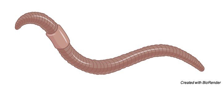
Earthworms are hermaphrodites: each carries male and female sex organs.
Where as in flowers the staminate or anther is seen in males and pistillate is being seen in females but they are present in the same plant hence the term monoecious is used here.
But most of the flowers has both male and female organs in a same plant and it is called as the original or a perfect flower.
As such for animals the organisms which produce male and female gametes individually are referred by the term dioecious.
However, these sex cells and reproductive organs and their respective hormones play an important role in determining their primary and secondary sexual characters.
Mechanism of Sex Determination
Mostly our sexes are determined in a genetical manner and they are classified into four categories as follows,
1. Heterogamesis or sex chromosome mechanism
2. Genic balance mechanism
3. Male haploidy or haplodiploidy mechanism
4. Single gene effects.
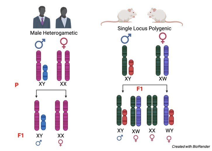
1. Heterogamesis
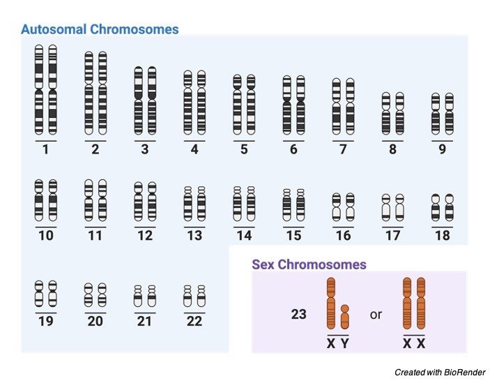
Generally, in dioecious organisms two types of chromosomes are present such as autosomes and sex chromosomes.
The sex chromosomes form two types of sex determination depending on two systems namely heterogametic females and heterogametic males.
In heterogametic sex, females have two x chromosomes where as the males have one x and one y chromosomes with 50 percent of the gametes.
The heterogametic male contains XX-XO type in plants and XX-XY type in mammals.
Where as the heterogametic female contains ZZ-ZO types in moths and ZW-ZZ type in fishes and certain insects
2. Genic Balance
This mechanism explains that some genes are being carried by sex chromosome which is responsible for the determination of sex in individuals.
There will be mostly an inherited mechanism of sexes in both individuals considering from masculine features in male to feminine characters in female.
This was discovered by Wilson, Bridges and Goldschmidt in different organisms.
If an individual is found intermediate between both sexes they are referred to as intersexes.
Sex Determination in Drosophila
The Y chromosomes present in a drosophila are very much important for maintaining the fertility in male flies but they do not have much importance in determining the sex.
The sex of the flies are determined polygenically, where the genes are situated in a distributive manner such that autosomes determine the male character and where as in female it is determined by X chromosome.
Bridges crossed the polyploid flies of experimentally produced female drosophila which has triploid set of chromosomes (3AA:3XX) to that of the diploid male chromosomes.
Which resulted in the formation of intersexes, superfemales and super males and also a normal diploid male along with the triploid females.
From this experiment he concluded that autosomes also carry the genes which are responsible for sex determination.
The intersexes obtained were sterile with both male and female phenotypic characters. This proves the genic balance mechanism.
Sex Determination in Human
Like Drosophila, sex determination can also be seen in humans, where male has XX-XY pair of chromosomes, here Y trait has a potential to bring all male determining characters, where as X chromosome is considered as a feminine character determiner.
Whereas the male having one X and one Y chromosomes can be a normal male by overcoming the feminine characters of X chromosome until there is no chances of chromosomal abnormalities or abbretions, which results in syndromes such as Turner and Klinefelter’s which causes feminine characters in male; like inhibiting sperm production, lack of masculine features; such as bread growth and body hairs, etc. which leads to many physical and mental abnormalities.
Where in Turners syndrome the lack of female characters tends to occur, such as shield chest, short stature, congenital malformations and webbing of neck etc,.
The individual who has addition of both type of chromosomes that is extra X and Y chromosomes, shows true hermaphroditism which have both ovarian and testicular tissues and genitalia appears to be intersexual.
3. Male Haploidy or Haplodiploidy
This is considered as one of the types of parthenogenesis mechanism which is commonly seen in hymenopterous insects.
These types of insects include ants, bees, wasps and sawflies.
Where fertilise eggs develop into diploid females and also as unfertilised eggs are developed into haploid males.
So, this is also considered as a form of sex determination. Considering an example of honey bee, where they can lay two varieties of eggs.
Such that it produces a fertilised egg by controlling a splinter of her sperm receptacle and an unfertilized one. Here the fertilised egg has diploid zygote and develops into a female and an unfertilised egg has only 16 zygotes and develops into a male.
Later the females are grown up as a honey bee or worker bee depending upon the food they consume or diet they undertake and the one which is developed from unfertilised eggs are males.
Single Gene Control of Sex
In certain organisms like Neurospora, Chlamydomonas, asparagus, yeast, maize etc, the single gene plays a very important role in determination of sexes and also in expressing the genes hence it is known as single gene control of sex.
Sex Determination System Citations
- Putting the heat on sex determination. Genetica . 1992;87(1):1-6.
- Prenatal sex determination and selection. Hum Reprod . 1993 Oct;8(10):1545-9.
- Genetic sex determination of mice by simplex PCR. Biol Sex Differ . 2017 Oct 17;8(1):31.
- Sonographic fetal sex determination. Obstet Gynecol Surv . 2009 Jan;64(1):50-7.
- Polygenic sex determination. Experientia . 1964 Apr 15;20(4):190-9.
- Sex Determination and Sex Chromosomes in Amphibia. Sex Dev . 2017;11(5-6):298-306.
Share
Similar Post:
-

In vitro Cell Migration, Wound Healing Full...
Continue ReadingWhat is Cell Migration?
Cell migration, movement of cells from one area to another generally in response to a chemical stimulus.
Migration is a central to achieving functions such as wound repair, cell differentiation and embryonic development, immune response, and disease processes such as cancer metastasis and inflammation.
Methods to examine cell migration are very useful and important for a wide range of biomedical research such as cancer biology, immunology, vascular biology, cell biology and developmental biology
Principle of Cell Migration
Migration assay is commonly used test to study the migratory response of endothelial cells or tumor cells to tumor promoters or inhibitors.
In migration assay the cells are seeded and when it reaches a confluency of 90% a scratch is made to mimic a wound.
This wounded cells are exposed to different concentration of test compounds to check its effect on the migratory property of cells.
Cell Migration Assay Requirements
1. Cancer cell line
2. Complete growth media
3. 1X PBS (pH-7.2)
4. 6-well plate
5. Micro-tips
6. Pipettes
7. Light Microscope
8. CO2 incubator
9. Biosafety cabinet level II
Procedure of Cell Migration Assay
1. Seed appropriate number of cells (e.g. for MDA-MB-231 seed approx. 2.5×10^5 cells per well) in 6 well plates.
Make duplicates for each treatment and control group. Incubate in 5% CO2 incubator for 24-36 hours until they reach ̴ 90% confluence.
2. When the cells are ̴ 90% confluence, make a scratch with 200 μL tip in the middle of each well and flush the scratch area with 1 ml pipette gently, so as any of adhered cells from the scratch area get detached.
3. Discard the media wash with 1 ml PBS and add freshly prepared treatment media.
4. Take the images at 0, 12, 24 and 48h, as per the requirement of your experiment (Observe the cells before taking images each time, sometimes some percentage of cells at higher doses get died or undergo apoptosis after 12h or 24h.
These cells come out in the scratch area and we get blurred image of the scratch area with undefined boundaries.
In such case, first collect the treatment media in separate tubes, wash with PBS and keep PBS in wells while taking images.
After taking images, replace PBS with treatment media).
5. Arrange the representative images for each treatment group and perform ImageJ analysis to measure the scratch area and represent your results as percent migration area or wound closure area.
Cell Migration Assay Precautions
Do not use non sterile tips to make the scratch.
Make the scratch straight with steady hands otherwise the wound will not be clear and the cells will be disturbed which will result in insignificant data.
Always take the image of area which shows least dead or floating cells to produce significant and publishable data.
Cell Migration Citations:
Share
Similar Post:
-

Cover Letter Template For PhD Program /...
Continue Reading(1) Cover Letter Template
Dear Dr. XXX,
I am a graduate student in the laboratory of Dr. xxxx at xxxx University, India (Ph.D. Awarded). This letter is in reference to any postdoc position available in your lab. Having been familiar with your research and recent publications, I’m interested in joining your lab, with particular interest in the area of epigenetic modifications of tumor suppressive non-coding RNAs and cancer manifestation. I believe that your laboratory offers the perfect opportunity for me to apply all that I have learned in graduate school and further my scientific endeavor.
My graduate research was focused on ameliorating potential of thymoquinone against the toxic effects of iron oxide nanoparticles both in vitro (primary splenocytes) and in vivo model system (albino rats). Superparamagnetic iron oxide nanoparticles (SPIONs) offer a broad spectrum of application in the various field of biomedical sciences including magnetic resonance imaging (MRI), drug delivery, cell labeling, hyperthermia etc. (xxx et al., xxx. 2020). SPIONs are known to induce cytotoxicity in various cancer cell lines through the generation of reactive oxygen species (ROS). However, the studies on its potential to induce toxicity in normal cell lines and in vivo system are limited and ambiguity still exists. Additionally, small molecules are known to interact with the DNA and cause damage to the DNA (xxx et al., 2019). We found that thymoquinone (natural antioxidants) have potential to ameliorate the cytotoxic and genotoxic effects induced by SPIONs both in vitro and in vivo (xxx et al., xx 2020). I am familiar to various techniques such as cell culture, cell viability assays, ROS estimations, ELISA, gene amplification (PCR), gene manipulation, microbial culture, chromosomal aberration assay, micronucleus test, single cell gel electrophoresis (comet assay), UV-Vis spectroscopy, Spectrofluorometry, histopathology, animal handling etc. Now, I’m keen to learn and embrace additional techniques being used in your lab and whatever new will be required to execute research plans.
Since my graduate schooling, I am always very interested in epigenetic phenomenon, specially related to cancer and its progression. What I can understand from your work and that of others are that epigenetic factors play an important role in tumorigenesis. Since its very well known that many oncogenic miRNAs contributes to cancer development, my interest is to identify that factors contributing to epigenomic modifications during oncogenesis. For instance, mutations in chromatin modifiers and its relation to cancer related process. The other aspects that I would like to address is genomic loss of genes via karyotyping (band analysis), for instance, epigenome related to tumor suppressive miRNAs as well as its chemical modifications (methylation). The gene and drug delivery using ligand conjugated nanoparticles will also be one of my priorities. Given an opportunity, I would like to work in your laboratory with my full efforts and technical expertise. However, I’m open to shape and adapt the research plan according to your precise vision and scientific goals. I’m hopeful that you will extend your guidance to me. I can prepare a more focused and concise plan of work after getting your consent and current research work going on in your lab. Please find enclosed my curriculum vitae for your review and let me know if you have any query.
Thank you for your time and consideration.
I look forward for your response.
With kind regards
Sincerely yours
xxx
(2) Cover Letter Template
Dear Dr..,
This is regarding to my interest for the Postdoctoral position in your laboratory. My background is biotechnology with extensive experience in plant metabolite production, cell biology, cell culture, protein expression, purification and characterization, bioinformatics tool docking and simulation.
Currently, I have completed my PhD in November 2020 under supervision of Prof xxx, Department of Bioengineering, xxx University. My PhD research is based on “Study of anticancer potential of xxx sprout and its nanoformulation on liver carcinoma cell line, since Dec xx.
During Ph.D, I worked as a JRF in the project entitled “In vitro and in vivo study of hepatoprotective activity of xxx extracts in various germination stages”. In the study, the mechanism of the cytotoxicity of N. sativa and its sprouts on liver cancer cell line using in silico, in vitro and in vivo approaches was elaborated. The work has been published in high quality journals in herbal drug/ plant drug analysis.
I have working experience in few formulations for skin cancer and liver cancer performed short term animal study with Swiss albino mice model and analysed the skin tissue sample for western blotting and PCR at protein and mRNA level and also done the project on expression, purification and characterization of protein using Pichia pastoris as an expression system. And I have some working experience on technique like HPLC, LC-MS, biological activities such as extraction and purification of secondary metabolites from plant extract, establishment and maintenance of animal tissue culture laboratory, handling & culturing mammalian cell lines, media preparation for mammalian cell lines, DNA & RNA isolation, PCR,qRT PCR, fluorescence microscopy, cell viability analysis, cytotoxic analysis, genotoxic analysis, reactive species generation monitoring, TLC, SDS-PAGE and Agarose Gel Electrophoresis and bioinformatics.
I am highly interested in exploring the bioactive natural product and therapeutic intervention thereafter. I have experience in preparation of different nanoformulation from plant extract apart from other in vitro and in silico studies. Further, I am prone to learn new techniques and implement them precisely in my studies. Please find my other credentials and references in the resume attached herewith. If you require any further information I shall be happy to furnish the same. Hope, I will be a significant addition to your lab.
Sincerely,
Dr. xxx
Integral xxxx
(3) Cover Letter Template
Dear Prof. xxxx
I, Dr. xxxx, wish to express my interest for a postdoctoral position available in your esteemed laboratory. I am highly motivated by your research theme and feel great pleasure to write you about the possible opportunity. In December 2016, I have been awarded with Ph.D. degree in Science on the topic entitled “xxxxxx of Metals and Carbon-based Nanomaterials (CNMs) induced toxicity” from xxx.
During my doctoral research, I get acquainted with nitty-gritty of nanoscience and nano-bio interface, particularly the synthesis and interaction of cerium and carbon family nanomaterials (carbon nanotubes, graphene oxide and graphite carbon nanofibers) with human lung cells as well as Swiss albino mice lungs. We explored that cerium oxide, carbon nanotubes and graphene oxide causes cell death in lung cells (cancer and normal) via oxidative stress induced apoptosis and necroptosis. These cell death pathways were closely associated with the unique physicochemical properties of NM and cellular phenotypes (xxxx et. al., one manuscript under preparation). Moreover, graphite carbon nanofibers induced cell death in human lung cells was assessed which describes the underlying role of autophagy-apoptosis-oxidative stress axis (xxx et al., revision submitted in xxx). Furthermore, the adverse effect associated with the in vivo exposure of CNM in mice lungs was explored which showed the role of frustrated phagocytosis, fibrosis and TGF β/Smad pathway underlying the toxicity of CNM (Manuscripts under review/preparation).
My doctoral research has provided me the opportunity to gain experience in in-vivo and in-vitro experimentation, genotoxicity, biochemical and molecular biology techniques, Flow-cytometry, animal handling and exposure, cell signaling, independent planning and execution of experiments and data analysis.
I am highly motivated and independent research scholar having a strong interest in the field of nano – biointerface particularly the interaction of nanomaterials with proteins and their implication in nanomedicine and targeted drug delivery. Presently, we are studying the anti-cancer effect of graphene oxide – chloroquine nanoconjugate in human lung cells. We identified the role of impaired autophagic flux mediated necroptosis and trying to understand the underlying molecular mechanism. Also, we are working on to assess the combinatorial effect of Gold-Zinc nanoparticles with X-ray to improve the anti-cancer potential of current radiotherapy techniques.
It would be a great pleasure for me to be a part of your lab. Provided an opportunity, I firmly believe that with my research experience and skills, I can significantly contribute to the ongoing research themes in your lab. Kindly find enclosed my CV including list of publications and referee details for your kind consideration. I will feel happy to provide any other information, if required.
Thanking you for your time and kind consideration. I look forward for a positive reply from your end.
xxxxx
(4) Cover Letter Template
Dear Dr. xxxx,
I am writing this letter to express my interest for the “Postdoctoral Research Opportunity floated by your group in the field of Theoretical/computational biophysics, computational biology/ medicine, molecular & structural biology. I find the floated job profile is very much similar to my research interest and I strongly feel that I could be one of the potential candidate for this job and possibly I can prove my suitability if I get chance.
In my Postdoctoral research I am working on Membrane simulations specific to understanding the “Membrane disruption Mechanism of Antimicrobial Peptides. In my 1.5 year postdoctoral experienced I have already published two papers and we are working on 3 more manuscripts that can be possibly get published before the end of this year. The membrane protein simulation specifically understanding the membrane disruption mechanism of Antimicrobial peptides (AMP) are longer time scale biological events. We have generated about 200 μs cumulative all atoms MD trajectory for several AMP. We have observed several spontaneous events that are new for scientific community and preparing our manuscripts for targeting high impact journals. My postdoctoral group is well known for publishing high impact papers in this field that can be seen by visiting our group web. I would be happy to discuss my research in details if I get opportunity. In PhD, I get experienced in computational Biophysics specific to “Molecular Modeling and Simulation of Proteins and Peptides. I worked on certain protein and peptides to explore the role of Dynamics in protein/peptides function and applications like drug design (See the publications for details).
Recently, I have submitted a proposal entitled “Understanding the Mechanism of Aggregation and Non-gated Ion-channel Formation of Aβ-Peptides on/in Membrane models: A Molecular Dynamics Simulation Study” to DST INDIA, and this proposal is on final stage of selection. Thus, I have already been very much aware about the recent literatures on Aβ-Peptides. I am sending my project summary with this covering letter for your consideration and I would be happy to discuss in details if you like.
With my expertise and experience in Science I firmly believe that I am a potential candidate for this position. Please see my resume for additional information.
Thank you for your time and consideration. I look forward to speaking with you about this employment opportunity.
Sincerely yours,
xxxx
(5) Cover Letter Template
Dear Professor xxxx,
I am writing this letter in pursuance of availability of a Postdoctoral Fellow position in your laboratory. I earned my PhD degree in Biotechnology and Bioinformatics in 2014 from the University of Hyderabad, India. Subsequently, I pursued as postdoctoral research fellow (May 2015- April 2018) in the Laboratory of System Biology of Cancer at the Department of Molecular Biology and Genetics, xxxx.
My PhD research work mainly concerned with the implementation of various in silico and in vitro approaches to understand the regulatory roles of microbial oncogenes in the context of chronic inflammation, apoptosis and cancer. During this period, I had extensively contributed towards standardizing the methods for bacterial gene cloning, protein expression, purification and characterization, flow cytometry analysis and cell-based immune signaling. My recent research experience as postdoctoral scientist at xxxx University relates with system level analysis of mechanisms of therapy resistance and metastatic progression in breast cancer models. I therefore gained expertise in using the computational approaches (RNA-seq and proteomics) and clinical data analysis to identify novel druggable targets (both coding and non-coding RNA drivers) for cancer prevention. Moreover, I have gained exposure in applying the cytobiology, biochemistry and functional genomics tools in in vitro and in vivo settings. Of particular interest, I have also been experienced in siRNAs, shRNAs and sgRNAs/ CRISPR/ Cas9 based screening of targets genes/factors in primary and resistant cancer cell line models, 3D culture and xenograft mouse tumor models for in depth understanding of signaling mechanisms involved in therapy-resistant cancer development. In this context, my current focus of research is to explore the transcription/ post-transcriptional regulatory activities of mediator complex proteins (CDK8-module) in development of hormonal-therapy resistance in ER+ breast cancer. I am confident that my research and technical backgrounds will greatly help me in impactful accomplishment of the project in your lab.
As you can see in my curriculum vitae (CV) that I have authored 8 scientific papers in peer-reviewed international journals and currently two articles are in the process of submissions (including my recent research work as first author). Besides, participation in a number of national and international level scientific programs enabled me to develop strong communication and problem solving skills.
I would highly appreciate you for considering my application. Please also find attached copy of my CV which provides more details of my experiences, research and skills to meet the requirement of this position. Please feel free to contact me to further explore my suitability.
Sincerely,
Dr. xxx
Share
Similar Research Position:
-

Introduction to Cell Culture: SubCulture, Media, Protocol
Continue ReadingWhat is Cell Culture?
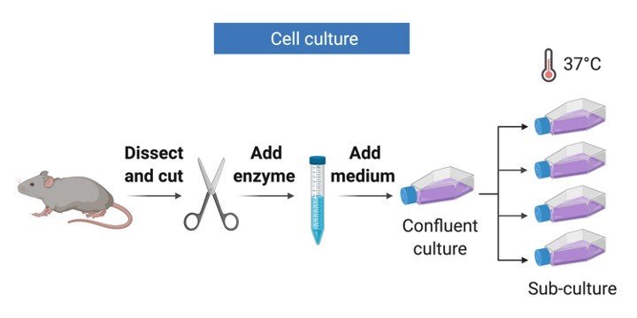
Cell culture refers to the removal of cells from an animal or plant and their subsequent growth in a favourable artificial environment.
The cells may be removed from the tissue directly and disaggregated by enzymatic or mechanical means before cultivation, or they may be derived from a cell line or cell strain that has already been established.
Mammalian cell culture is used widely in academic, medical and industrial settings.
It allows the study of physiology and biochemistry of the cell and its development.
It has also widen the scope and its application in the field of cell and molecular biology where the use of reproducible model systems is attained by cultured cell lines.
For medical use, cell culture provides test systems to assess the efficacy and toxicology of potential new drugs.
Large- scale mammalian cell culture has allowed production of biologically active proteins, initially production of vaccines and then recombinant proteins and monoclonal antibodies; recent innovative uses of cell culture include tissue engineering to generate tissue substitutes.
Basic Requirements for Successful Cell Culture
The first necessity is a well-established and properly equipped cell culture facility.
The level of bio containment required (Levels 1–4) is dependent on the type of cells cultured and the risk that these cells might contain, and transmit, infectious agents.
All facilities should be equipped with the following: a certified biological safety cabinet a centrifuge, preferably capable of refrigeration and equipped with appropriate containment holders that is dedicated for cell culture use; a microscope for examination of cell cultures and for counting cells; and a humidified incubator set at 37°C with 5% CO2 in air.
The second requirement for successful cell culture is the practice of sterile technique prior to beginning any work, the biological safety cabinet should be turned on and allowed to run for at least 15 minute to purge the contaminated air.
All work surfaces within the cabinet should be decontaminated with an appropriate solution; 70% ethanol or isopropanol are routinely used for this purpose.
A third necessity for successful cell culture is appropriate, quality controlled sterile reagents, culture media and required sterile plastic wares.
Adopted from BioRender
Types of Cell Culture
There are two types of cell culture system, Primary and continuous culture.
1. Primary Culture: Primary cultures are derived directly from excised, normal animal tissue and cultures either as an explant culture or following dissociation into a single cell suspension by enzyme digestion.
The preparation of primary cultures is labour intensive and they can be maintained in vitro only for a limited period of time.
During their relatively limited lifespan primary cells usually retain many of the differentiated characteristics of the cell in vivo.
2. Continuous Culture: Continuous cultures are comprised of a single cell type that can be serially propagated in culture either for a limited number of cell divisions (approximately thirty) or otherwise indefinitely.
Cell lines of a finite life are usually diploid and maintain some degree of differentiation.
The fact that such cell lines senesce after approximately thirty cycles of division means it is essential to establish a system of Master and Working banks in order to maintain such lines for long periods.
Continuous cell lines that can be propagated indefinitely generally have this ability because they have been transformed into tumour cells.
Tumour cell lines are often derived from actual clinical tumours, but transformation may also be induced using viral oncogenes or by chemical treatments.
Transformed cell lines present the advantage of almost limitless availability, but the disadvantage of having retained very little of the original in vivo characteristics.
Cell Culture Morphology
In terms of growth mode cell cultures take one of two forms, growing either in suspension (as single cells or small free floating clumps) or as a monolayer that is attached to the tissue culture flask.
The form taken by a cell line reflects the tissue from which it was derived e.g. cell lines derived from blood (leukaemia, lymphoma) tend to grow in suspension whereas cells derived from solid tissue (lungs, kidney) tend to grow as monolayers.
Attached cell lines can be classified as endothelial, epithelial, neuronal or fibroblasts and their morphology reflects the area within the tissue of origin.
There are some instances when cell cultures may grow as semi-adherent cells, ( e.g. marmoset B-lymphoblastoid cell line), where there appears to be a mixed population of attached and suspension cells.
For these cell lines it is essential that both cell types are subcultured to maintain the heterogeneous nature of the culture.
In vitro age of cell culture Two terms are predominantly used to define the age of a cell culture:
1. Passage number indicates the number of times the cell line has been sub-cultured.
2. The population doubling (pd) number indicates the number of cell generations the cell line has undergone i.e. the number of times the cell population has doubled.
Cell Culture Maintenance
In culturing mammalian cells in vitro, one attempts to reproduce in a culture vessel the physiological environment and characteristic responses of individual cell types.
At a minimum, the fluid medium in which cells are cultured must provide for their nutritional requirements, provide an energy source, maintain pH, and provide a level of osmolarity compatible with cell viability.
Culture media commonly used today consist of two parts: a basal nutrient medium and supplements.
The basal nutrient medium, such as Dulbecco’s modified Eagle’s medium (DMEM; also known as Dulbecco’s minimal Eagle’s medium), RPMI 1640, or Ham’s F-12, is a buffered aqueous solution of inorganic salts, vitamins, amino acids and other anabolic precursors, energy sources such as glucose and glutamine, and trace elements.
Supplements are either undefined, such as fetal bovine serum (FBS), tissue extracts, and conditioned medium, or defined, such as hormones and growth factors, transport proteins, and attachment factors.
The compositions of basal nutrient media and medium supplements may vary considerably; however, both components of the complete medium are necessary for support of cell viability and proliferation.
There are two formats of media available i.e. dehydrated and liquid media.
Preparation of Cell Culture Media
Requirements:
1. Dehydrated medium (i.e. RPMI or DMEM)
2. Double autoclaved water
3. Autoclaved bottles for storing medium
4. 0.22 micron filtration unit (capacity 1 litre)
5. Antibiotics (Penicillin, Streptomycin, Amphotericin B)
6. Additives for the medium as required (like sodium bicarbonate, sodium pyruvate, glutamate etc.)
7. 1N NaOH and HEPES
8. Conical Flask (1L or 2L for dissolving the dehydrated medium)
9. Measuring Cylinder
10. Discard beaker
Cell Culture Procedure
1. In a sterile biosafety cabinet dissolve powdered medium with constant stirring in a 0.8× to 0.9 × volume of water. (If a commercially prepared liquid medium is being used, add penicillin and streptomycin from commercial stock solutions and proceed to step 8)
2. Add an amount of HEPES that yields a concentration of 15 mM in the final volume of medium. (Omit this step if the powdered medium is formulated with HEPES.)
3. Add the amount of sodium bicarbonate recommended by the medium supplier for use in a CO2-controlled atmosphere (e.g., 14 to 36 mM in 5% CO2 atmosphere). (Omit this step if the powdered medium contains sodium bicarbonate.)
4. Add glutamine to give a final concentration of 2 mM and pyruvic acid to give a final concentration of 0.01% (w/v). (Omit this step if the powdered medium contains glutamine & pyruvic acid.)
5. Add penicillin G to give a final concentration of 100 IU/ml and streptomycin to give a final concentration of 50 μg/ml.
6. (Other antibacterial agents or antifungal agents should not be routinely included in culture medium. Gentamicin at a final concentration of 50 μg/ml or kanamycin at 100 μg/ml may be useful in eliminating gram-positive and gram-negative bacteria from primary cultures or from irreplaceable cultures, but it is best to discard any cultures that are contaminated with bacteria, yeast, or fungi.)
7. Adjust the pH of the medium to 7.4 with 1 N NaOH, and add water to achieve the final (1×) volume. Readjust the pH of the medium to 7.4, if necessary.
8. Sterilize the medium by filtration through a 0.2-μm filter. Store the medium at 4°C in the dark. Vacuum-operated filtering units or bottle-top filters are useful for small volumes of medium (0.1 to 2 liters), whereas filter capsules (2 to 5 liters) or filter stands (>10 liters) that are used under positive pressure are more suitable for larger volumes.
9. Make an aliquote of prepared media and keep at 37°C to check for any contamination.
10. Add serum (5-20%) to the desired final concentration at the time of use.
Cell Culture Precautions
Basal nutrient medium and the serum supplement should be stored individually at 4°C, and the complete medium should be made up at the time of use and only in the volume necessary.
Working volumes of serum should be stored at 4°C and used within several weeks.
Serum should not be subjected to repeated freezing and thawing, but it can be stored for at least 2 years at −20°C with little deterioration in growth-promoting activity.
In this way, medium components are not wasted, and the chances of detecting, isolating and eliminating contamination with minimal losses are increased.
Cell Culture Citations:
Share
Similar Post:
-

Cytoplasm: Definition, Function, and Example
Continue ReadingWhat is Cytoplasm?
We all know that cells are the basic, structural and functional units of all living organisms.
Cells are made up of many organelles and they are bound embedded in the cytoplasm.
It plays a very important role in functioning of the cell and also contains the genetic information of the cell, as it contains chromosomes in it.
The term cytoplasm was first coined by Rudolf Von Kolliker, a Swiss biologist.

"Cytoplasm is contained within cells in the space between the cell membrane and the nuclear membrane"
Cytoplasm is considered as the fluid compartment of the cell, which contains cytosolic filaments, ion substances, proteins and other macromolecular substances. Along which the other cell organelles are found suspended.
The Eukaryotic cytoplasm contains all the cell organelles found embedded in it except the nucleus.
The prokaryotic cytoplasm is found throughout the cell carrying its genetic material as the prokaryotic organism does not have its well- defined nucleus.
Does Cytoplasm Have Any Structure?
Cytoplasm doesn’t have a specific structure its just a jelly or glassy liquid like substance which fills up the space in each cells volume and make the other cell organelles being embedded in it and provides a better environment for the organelles and genetic materials to carry out their function.
Cytoplasm Vs Cytosol
In biology, Cytoplasm is often confused with cytosol due to their similarity in understanding of these words.
Cytoplasm is the liquid part of the cell which is found outside or around the nucleus.
Where as Cytosol is denoted as the substances or proteins which are found in the cytoplasm apart from other cell organelles.
Protoplasm Vs Cytoplasm
The protoplasm is jelly like substance, which is composed of macromolecules and water.
It is actually a colorless living part of the cell. It can be generally known as the organic or inorganic substance which is made up of cytoplasm, nucleus, mitochondria and plastids of a cell, which play a major role in functioning of the cell.
Thus, cytoplasm is a part of a protoplasm which is placed in between the nucleus and the cell membrane in all eukaryotic cells. Where the organelles are embedded in it.
Function of Cytoplasm
Cytoplasm provides a suitable environment for all other cell organelles to maintain their turgidity which helps them to maintain their shape.
The fluid form of the cytoplasm is made up of salts and water which form a jelly like structure and helps the other cell organelles to embed themselves in that space.
The Cytoplasm also acts as a center for other cell organelles to perform their function, as it contains many enzymes and molecules which helps them to perform their metabolic reactions.
As cytoplasm helps the cell organelles to fit it into, so that they cannot move here and there and fix themselves in their origins, if not there is a high chance of mixing up of molecules of one organelle into the other which results in change in body functioning.
The fluid matrix of the cytoplasm which is known as cytosol has no organelle, so it fills up the space which is left out by the organelles.
Cytoplasm also paves the role for many cellular functions to occur in it where the ribosomes synthesize their proteins and the process of cellular respiration takes by initiating glycolysis and reproduction of cell phases like mitosis and meiosis occurs.
Cytoplasmic Streaming
Cytoplasmic streaming is often referred to as cyclosis by which the molecules or substances present in the cell are circulating within the cell.
Cyclosis mostly occur in all types of cells such as Plant cells, in bacteria and fungi.
But these movements change accordingly with the type of chemicals and hormones present in it and also according to the temperature and humidity of the cell.
Plants perform cyclosis to settle the chloroplast in the area, where we can get suitable sunlight.
Whereas chloroplast is a very important and essential component in performing the photosynthesis which help the plant to stay alive.
Whereas in protists such as slime molds and amoeba the cyclosis movement is used for the locomotion.
These temporary movement of cells is termed as pseudopodia which helps the cell in capturing the food.
Cyclosis also helps the cell in cell division processes where the daughter cells are divided by the process of mitosis and meiosis.
Other Features of Cytoplasm
As discussed above cytoplasm is a glassy liquid substance which contains all the cell organelles and other substances except the nucleus.
The eukaryotic cytoplasm is present between the cell membrane and the nucleus of the cell, where as in prokaryotes cytoplasm just acts as a covering to all the substances in a cell membrane.
However, in both the cases the role of cytoplasm is to carry all the metabolism and growth and functions of all other organelles present in a cell.
The organelles present in the cytoplasm perform various functions, where as the nucleus contains the genetic information and helps in caring our hereditary characters throughout the generations.
Mitochondria which is found embedded in the cytoplasm acts as a powerhouse of the cell producing Adenosine tri phosphate.
The chloroplast present in plants cell helps in performing photosynthesis.
Likewise, each cell organelles perform various functions, but without the help of cytoplasm which provides them a good outer condition by providing proper humidity and other conditions, these cells cannot undergo their functions.
The cytosol which acts as a fluid part of the cell, consisting of 70% of water and other molecules including ion particles such as chloride, potassium, bicarbonate magnesium and calcium serves as a site for many chemical reactions.
Cytosol also plays an important role in cell signaling and osmoregulation by inducing action potentials such that of nerve and muscle cells.
Cytoplasmic Biochemical Reactions
Basically, cytoplasm acts as a center for many biochemical reactions, the biochemical reactions are thereby classified as anabolic and catabolic reactions, where catabolism refer to a breakdown of complex molecules and anabolism refers to a synthesis of biomolecular substances with the help of ATP (Adenosine Triphosphate) to form a complex substance.
Biological Functions of Cytoplasm
Cytoplasm acts as a center for both growth and metabolism, as said earlier it helps in both generation and degradation processes.
Whereas glycolysis, which is the first process in the cellular respiration occurs at a cytosol, the fluid component of the cell and this process for further followed by oxidative decarboxylation reaction and Krebs cycle or citric acid cycle and followed by electron transport chain.
Citations
Share
Similar Post:
-

Transfer RNA (tRNA): Definition, Structure, and, Function
Continue ReadingThe role of RNA in protein synthesis
1. mRNA: This is a messenger RNA, transfer the genetic detail duplicated from DNA in fashion of a sequence of 3-base code ‘term’, every term states a certain amino acid.
2. Ribosomal RNA (rRNA): It links with a batch of proteins to configure ribosomes. It is the complicated form, moves physically along with a mRNA molecule, facilitates the fabrication of various amino acids into protein chains. They also link tRNAs and different supplementary molecules necessary for protein synthesis. Ribosomes are made up of a large and small subunit, each of which having their unique rRNA molecule or molecules.
3. Transfer RNA (tRNA): It is pivotal in decrypting the key codons in mRNA. Every amino acid possesses their individual kind of tRNA, which attach it and transfer it to the expanding end of a polypeptide chain when the subsequent codon on mRNA needs it. The correct tRNA with its attached amino acid is selected at each step because each specific tRNA molecule contains a 3-base sequence that can pair with base of its reciprocal codon in the mRNA.

About tRNA
Every tRNA carry a set of 3 nucleotides known an anticodon. The anticodon of a said tRNA can attach to one or more particular mRNA codons.
The codons are predetermined for the tRNA thus, transferring the amino acid to that specific location.
Variety of tRNA are streaming within a cell, possessing their own anticodon along with complimentary amino acid and they attach to codons inside of the ribosome, where they remit amino acids for incorporation into the protein chain.
Thus, proteins are constructed from miniature units known as amino acids, which are described by 3-nucleotide mRNA sequences called codons.
The 3D Structure of tRNA
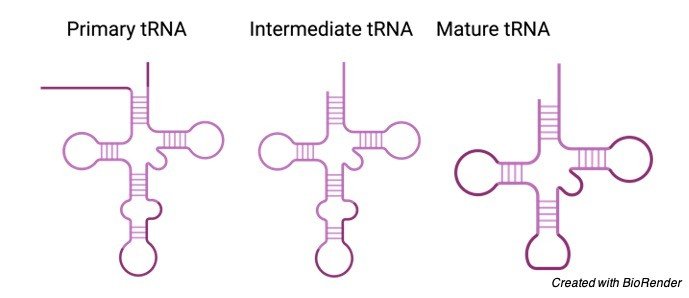
A tRNA is formed from a single strand of RNA in a similar fashion mRNA is formed.
However, the strand takes on a complex three dimensional structure since base pairs form between nucleotides in different parts of the molecule.
This creates double-stranded regions and loops, overlapping the tRNA into an L shape.
The tRNA molecule has a noticeable folded structure with 3 hairpin loops that form the structure of a 3-leafed clover.
One of these hairpin loops contains a sequence known as the anticodon, which can admit and decode an mRNA codon.
Every tRNA have their correlated amino acid attached to its end.
When a tRNA identifies and attaches to its corresponding codon in the ribosome, the tRNA shifts the suitable amino acid to the tip of the lengthening amino acid chain.
Then the tRNAs and ribosome carry on to decipher the mRNA molecule until the entire series is translated into a protein.
tRNA and its Decoding Role
The genetic information passed from DNA to protein via mRNA in which the nucleotide sequence of mRNA is converted into chain of the amino acid to form protein.
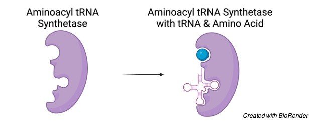
However, this deciphering procedure is carried out with the help of two kinds of adapter molecules: tRNAs and an enzymes known as aminoacyl-tRNA synthetases.
There are 2 functions which are performed by all tRNAs:
1. To get chemically connected to a specific amino acid and to base-pair with a codon in mRNA so as to put in the amino acid in the lengthening peptide chain.
Every tRNA molecule is exquisitely identified by the 20 aminoacyl-tRNA synthetases.
In a similar fashion, every enzyme molecules inimitably attaches the 20 amino acids to a specific tRNA, forming an aminoacyl-tRNA.
2. When the accurate amino acid is linked, then a tRNA identifies a codon in mRNA, thereby bringing its amino acid to the expanding polypeptide.
How Synthetases Recognize tRNAs.
After further studies on tRNA, 30 – 40 variety of tRNAs were recognized in bacterial cells while about 50 – 100 in animal and plant cells.
Therefore, the count of tRNAs in almost all cells is exceeding the number of amino acids observed in proteins.
Moreover, they differ from the number of codons in the genetic code.
Accordingly, several amino acids possess more than one tRNA to which they can link.
Furthermore, several tRNAs can attach to more than one codon. Aforementioned, the majority of amino acids are encoded by more than one codon, needing some tRNAs to identify more than one codon.
Function of tRNA Molecules
The function of 70 – 80 nucleotides long tRNA is based on their accurate 3D structures.
In solution, all tRNA molecules overlap into a same stem-loop setting that mimic a cloverleaf which when drawn in two dimensions.
The 4 stems are small double helices fixed by Watson-Crick base pairing; 3 out of 4 stems have loops containing 7 or 8 bases at their tail end, whereas the rest, unloop stem embody the free 3′ and 5′ ends of the chain.
3 nucleotides entitled the anticodon, located at the middle of one loop, can form base pairs with the 3 corresponding nucleotides forming a codon in mRNA.
As delineated earlier, determined aminoacyl-tRNA synthetases identifies the surface structure of every tRNA for a particular amino acid and covalently bind the specific amino acid to the unloop amino acid acceptor stem.
The 3′ terminal end of each tRNAs has the sequence CCA, which in most instance adjoins when synthesis and processing of the tRNA are finish.
Observed in 3 dimensions, the overlapped tRNA molecule has an L shape with the anticodon loop and acceptor stem forming the ends of the two arms.
Loading tRNA With an Amino Acid
Enzymes known as aminoacyl-tRNA synthetases have this pivotal role.
There is an individual synthetase enzyme for each amino acid, wherein an enzyme recognizes only particular amino acid with its respective tRNAs.
Whenever the amino acid and its tRNA binds its respective enzyme, the enzyme blends them together.
The above reaction has been powered by the “energy currency” molecule adenosine triphosphate (ATP).
Sometimes, an aminoacyl-tRNA synthetase miscalculate, wherein it attaches to the incorrect amino acid.
For instance, the threonine synthetase occasionally seize serine by coincidence and binds it to the threonine tRNA.
Fortunately, the threonine synthetase has a proofreading site, which dislodges the amino acid from the tRNA.
tRNA Summary
Genetic information is transmitted into mRNA in the form of a triplet code.
Every amino acid is encoded by surplus of 3 – base sequences, or codons, in mRNA.
Each codon identifies one amino acid, however, majority of amino acids are encoded by multiple codons.
All tRNAs have a same 3 – D structure that comprises an acceptor arm that binds a particular amino acid and a stem-loop with a 3- base anticodon sequence at its ends.
The anticodon can base-pair with its complimentary codon or codons in mRNA.
Since it is a nonstandard interplay, a tRNA may base-pair with more than one mRNA codon, and in contrast, a specific codon may base-pair with several tRNAs.
Each of the 20 aminoacyl-tRNA synthetases identifies a single amino acid and covalently links it to an associated tRNA, producing an aminoacyl-tRNA.
This reaction triggers the amino acid, so it can involve in peptide-bond development.
Transfer RNA (tRNA) Citations
- tRNA Modifications: Impact on Structure and Thermal Adaptation. Biomolecules . 2017 Apr 4;7(2):35.
- tRNA biology charges to the front. Genes Dev . 2010 Sep 1;24(17):1832-60.
- Transfer RNA: From pioneering crystallographic studies to contemporary tRNA biology. Arch Biochem Biophys . 2016 Jul 15;602:95-105.
Share
Similar Post:
-

Green Revolution: Definition, Advantages, Importance, & Facts
Continue ReadingWhat is Green Revolution?
Green Revolution is really the way toward expanding agrarian creation by utilizing present day machines and procedures.
It was a logical exploration-based innovation drive performed among 1950 and the last part of the 1960s, that expanded horticultural creation around the world, especially in the creating scene, starting most uniquely in the last part of the 1960s.
It utilized HYV seeds, expanded utilization of manure and more specialized techniques for water system to build the creation of food grains.
Green Revolution in India
In India Green Revolution started in the mid 1960s that prompted an expansion in food grain creation, particularly in Punjab, Haryana, and Uttar Pradesh.
Significant achievements in this endeavor were the advancement of high-yielding assortments of wheat.
The Green revolution is revolutionary in character because of the presentation of new innovation, groundbreaking thoughts, the new use of data sources like HYV seeds, composts, water system water, pesticides, and so on.
As every one of these were brought out of nowhere and spread rapidly to accomplish sensational outcomes subsequently it is named as a revolution in green agribusiness.
Reason Behind Green Revolution
The world’s most exceedingly awful recorded food debacle occurred in 1943 in British governed India known as the Bengal Famine.
An expected 4,000,000 individuals died of yearning that year alone in Eastern India (that incorporated the present Bangladesh).
The underlying hypothesis set forward to clarify that disaster was that there was an intense shortage in food creation nearby.
Notwithstanding, Indian financial analyst Amartya Sen (beneficiary of the Nobel Prize for Economics, 1998) has set up that while food lack was a supporter of the issue, a more strong factor was the aftereffect of madness identified with World War II which focused on food supply for the British rulers.
The insanity was additionally misused by Indian merchants who accumulated food to sell at more exorbitant costs.
By and by when the British left India four years after the fact in 1947, India kept on being spooky by recollections of the Bengal Famine.
It was in this way normal that food security was a vital thing on free India’s plan. This mindfulness drove, on one hand to the Green Revolution in India and on the other, administrative measures to guarantee that financial specialists could always again be unable to store nourishment for reasons of benefit.
Notwithstanding, the expression “Green Revolution” is applied to the period from 1967 to 1978. Somewhere in the range of 1947 and 1967, endeavors at accomplishing food independence’s were not totally fruitful.
Endeavors until 1967 to a great extent focused on growing the cultivating regions. Be that as it may, starvation passings were all the while being accounted for in the papers.
In an ideal instance of Malthusian financial matters, populace was developing at a lot quicker rate than food creation. This called for extreme activity to expand yield. The activity came as Green Revolution.
The expression “Green Revolution” is an overall one that is applied to fruitful rural analyses in numerous Third world nations. It’s anything but explicit to India. However, It was best in India.
Factual Results of Green Revolution
A record grain yield in 1978-79 around 131 million tons happened because of the Green Revolution. Consequently, it made India as one of the world’s greatest agrarian maker.
In India Green Revolution recorded a significant degree of accomplishment. India likewise turned into an exporter of food grains around that time.
Monetary Results of Green Revolution
Yield regions under this venture required more water, more composts, more pesticides, and certain different synthetic substances.
This expanded the development of the nearby assembling area. Expanded modern development made new positions and added to the nation’s GDP.
The expansion in water system made the requirement for new dams to saddle storm water. The put away water was utilized to make hydro-electric force.
The entirety of this brought about modern development, made positions and worked on the personal satisfaction of individuals in towns.
Sociological Results of Green Revolution
This new innovation utilized regular use of water, composts, bug sprays, bigger volumes of transportation, power, and so on The agrarian specialists as well as modern laborers landed a lot of positions on account of the production of offices like processing plants, hydro-electric force stations, and so on to back up the revolution.
Political Results of Green Revolution
Quite possibly the main factors that made Mrs. Indira Gandhi (1917-1984) and her gathering the Indian National Congress, an extremely incredible political power in India is this Green Revolution. India changed itself from a destitute country to an exporter of food.
This gave India adoration and appreciation from everywhere the world, particularly from the Third world country.
Disservices of the Green Revolution
The negative social impact of the Revolution was additionally soon noticeable. Differences in pay have been augmented by these advancements in horticulture.
Rich landowners have power over the horticultural info and worked on synthetic composts.
The most noticeably terrible part is that the helpless ranchers wound up impeded by little homesteads of land and insufficient water supply. With complete horticultural methods and data sources, the Green revaluation would in general have its most focused application on enormous homesteads.
As a convergence of the new innovation to enormous homesteads, the Inequalities have additionally Increased.
The helpless ranchers have been unfavorably influenced by a developing propensity among the rich ranchers to recover land recently rented out under tenure understanding, which has been made beneficial by better yields from new innovation.
Poor people and in reverse class of ranchers has been progressively driven into the position of the landless worker.
An intense expansion in a more elevated level of lease with land esteem taking off.
Likewise in light of unnecessary utilization of manures soil began to become soluble or acidic relying on the idea of the compost utilized.
Conclusion
India has made a colossal accomplishment in term of the Green Revolution, as it’s anything but an extraordinary degree of food security.
It’s anything but countless needy individuals out of destitution and aided numerous non-needy individuals keep away from the neediness and yearning they would have encountered had it not occurred.
This revolution has saved over a billion group everywhere on the world from starvation.
Green Revolution Citations
- The green revolution. Nutr Rev . 1969 May;27(5):133-6.
- From green to gold: agricultural revolution for food security. J Exp Bot . 2020 Apr 6;71(7):2211-2215.
- A Strigolactone Biosynthesis Gene Contributed to the Green Revolution in Rice. Mol Plant . 2020 Jun 1;13(6):923-932.
- Toward a “Green Revolution” for Soybean. Mol Plant . 2020 May 4;13(5):688-697.
- Green revolution: impacts, limits, and the path ahead. Proc Natl Acad Sci U S A . 2012 Jul 31;109(31):12302-8.
Share
Similar Post: