-

Chromatin: Definition, Characteristic, and Examples
Continue ReadingChromatin Definition
Chromatin is a nucleic acid (DNA or RNA) and protein combination (e.g. histones). It was discovered in 1882. Originally considered to be merely a colourful material in the nucleus, chromatin was subsequently shown to be proteins linked to DNA, and DNA was recognised as the bearer of genetic information. As a result, we may describe chromatin as a material made up of DNA and proteins (known as histones).
Histones
Histones are basic proteins with a positive charge that attach to DNA’s negatively charged phosphate units. Chromatin consists of two primary components: the cell’s DNA and related proteins. Histones are the proteins that are involved. Or, to put it another way, chromatin is made up of proteins known as histones.

Chromatin is a kind of DNA packing. It can wrap around itself or get harmed during cell division if it isn’t packed correctly. Cells are measured in micrometres, while DNA may be up to 3 metres long. Tight packing is necessary to accommodate such a long structure inside a micrometre cell.
Nucleosomes
Nucleosomes are formed when a DNA molecule wraps around histone proteins to form tight loops.
The nucleosomes coil and bundle together to create chromatin fibre, which is a kind of fibre. These chromatins, in turn, loop and fold around to form a chromosome with the aid of proteins. That is why a chromosome is said to carry a portion or all of an organism’s genetic material. Because of its tightly coiled shape, DNA is now protected after it is condensed into a chromosome.
Chromatin is also important for controlling the transmission of genetic information. A material composed of DNA or RNA and proteins, such as histones. During cell division, it condenses to create a chromosome.
Where is Chromatin Found?
Chromatin is present in the nucleus of eukaryotic cells. Here’s a diagram to help you visualise where it’s located within the cell nucleus.
Genes in Chromatin
The genes that are present in chromatin can be switched on or off. It indicates that in certain cells, one half of the gene is active (“switched on”) and the other is inactive (“switched off”). It’s chromatin, of course.
The researchers confirmed this by studying the on and off states of genes in chromatin using a fruit fly as a model organism. Their research revealed five distinct kinds of chromatin based on the presence of certain proteins.
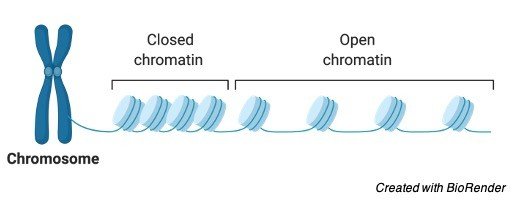
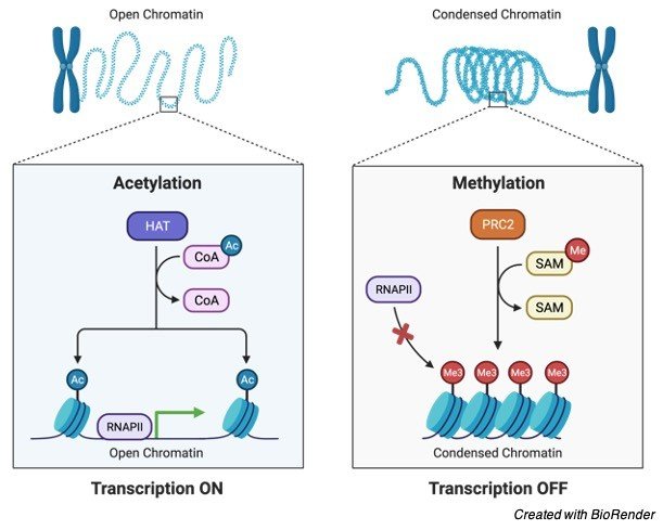
Chromatin Structure
In eukaryotic chromatin, the histone proteins and DNA have almost the same mass (although there are also some cells with non-histone proteins instead). The nucleosome is a structural unit of chromatin, which is made up of DNA and proteins (histone and non-histone). This structure can be found throughout an organism’s genetic material. Below is a diagram showing chromatin packing into a higher-order structure.
"Beads on a String" Model of Chromatin
The initial degree of compaction for DNA inside the nucleus is provided by DNA and histone proteins. The nucleosome is chromatin’s fundamental structural unit. When DNA is wrapped around histones (the protein core) to create a “bead-like” structure, it is called a nucleosome. A nucleosome is the name of this bead-like structure. The “beads-on-a-string” type of chromatin is shown in figure 3 as the second from the top. The nucleosome is a 146-base-pair DNA molecule that is wrapped around eight proteins called histones. When DNA is wrapped around histones, a nucleosome is produced.
H1, H2A, H2B, H3, and H4 are the five distinct kinds of histones. When two H2A and H2B proteins join with H3 and H4 proteins, a histone core is formed. A nucleosome is formed by wrapping 145 base pairs of DNA twice around this protein structure. The length of linker DNA varies based on the gene activity of the species and can range from 10 to 95 base pairs. Every 200 base pairs, a nucleosome with a length of 10 nm appears.
When seen under a microscope, chromatin resembles beads strung on a string. Nucleosomes are the name of these beads. The nucleosome is made up of eight proteins called histones. The nucleosomes wrap themselves into a 30nm spiral to produce a solenoid. Additional histone proteins aid in the formation of the chromatin structure in this solenoid. Due to its more compact shape, chromatin condenses into chromosomes.
Chromatin and DNA
DNA is packaged in chromatin. To fit inside a nucleus, DNA and related proteins are compacted into chromatin.
How DNA is Assembled in Chromatin Structure?
The process of assembling DNA into chromatin involves multiple stages. H3e and H4 proteins are the first to bind to DNA, followed by H2A and H2B. A sub-nucleosomal particle with 146 base pairs of DNA is produced. The second phase is maturation, which involves ATP establishing a constant nucleosome core spacing. Folding of linker histones into a nucleofilament of 30 nm structure is the next stage. Further folding happens in the final phase, resulting in a greater amount of packing. The packing ratio is estimated to be around 7000.
Euchromatin vs Heterochromatin
Heterochromatin and euchromatin are the two types of chromatin. Heterochromatin is extremely condensed and can not generally be transcribed, whereas euchromatin is less condensed and can be transcribed. Constitutive heterochromatin and facultative heterochromatin are two types of heterochromatin. Constitutive heterochromatin refers to the DNA sequences found in all of an organism’s cells. Highly repetitive DNA is linked to constitutive heterochromatin.
Similarly, not all cells have facultative heterochromatin. The gene that codes for beta-globin in animals, for example, is found in certain cells but not in blood cells. In eukaryotic cells, chromatin is a combination of proteins and DNA, as previously stated. Nuclear DNA is tightly compacted and wrapped around nuclear proteins to fit inside the nucleus, rather than existing as linear strands.
In the interphase nucleus, chromatin is divided into two types: euchromatin and heterochromatin. Euchromatin refers to a kind of chromatin that is structurally loose. In terms of transcription and replication, it is generally active. Allowing RNA and DNA polymerases to transcribe and replicate DNA, respectively, is permissive. Heterochromatin is the chromatin that is less active. It contains dormant genes and is smaller in size.
Chromatin Function
Initially, chromatin was thought to be the material that gave the cell nucleus its colour. It was later discovered that it is more than simply a colouring agent; it is also one of the most significant DNA expression controllers. The structure of chromatin is also crucial for DNA replication. DNA is packaged in chromatin and nucleosomes, resulting in a tightly closed structure that enzymes essential for transcription, replication, and repair of DNA can not access.
DNA structural packing is transcriptionally restrictive, allowing only a modest amount of gene expression. DNA may be copied and transcribed more easily when nucleosome structures are open or broken.
Some repressors and activators interact with RNA to regulate gene activity and modify the chromatin structure during the transcription process. Activators alter the structure of nucleosomes, causing RNA polymerase assembly to be stimulated. A comparable control of chromatin shape happens during replication, allowing the replication mechanism to be in place at the replication origin.
The control of gene expression is another function of chromatin. The genes can be made transcriptionally inactive by placing them near quiet heterochromatic chromatins using the location effect variegation method. Silent heterochromatin chromatins and genes can be separated by up to 1000 kilobase pairs. Because it causes phenotypic diversity, this phenomenon is referred to as epigenetic.
The extremely condensed structure of heterochromatin, according to scientists, inhibits DNA transcription. However, how the surrounding non-heterochromatic areas are impacted is still unknown. Proteins in chromatin can travel to nearby areas, causing a similar restrictive effect, according to the researchers. The researchers also speculated that heterochromatin may be found in compartments of the nucleus that are not accessible to transcription factors. As a result, transcription factors may not have direct access to chromatin in the nucleus.
DNA replication is influenced by chromatin structure. The euchromatin and other active regions of the genome, for example, replicate faster. The replication process is also sluggish in the heterochromatin and quiet regions surrounding it. Below are some of chromatin’s other key activities.
i. DNA Packaging
The primary purpose of chromatin is to compact lengthy DNA strands into a much smaller area. In comparison to where it resides, DNA has a relatively long linear length. DNA must be compressed in some way in order to fit comfortably and securely without tangling or harm. Condensing is the process of compacting DNA into the nucleus. The packing ratio refers to how tightly DNA is packed within a body. DNA has a packing ratio of around 7000. DNA is not packed directly into the structure of chromatin because of the high amount of compaction. Rather, there are numerous organisational hierarchies.
The nucleosome is initially packed by wrapping DNA around it. A packing ratio of 6 is obtained as a result of this. Both heterochromatin and euchromatin have the same packing. Wrapping the beads in a 30 nm fibre, which is also present in mitotic chromosomes and interphase chromatin, achieves the second degree of compaction. The packing ratio is increased from 6 to 40 with this wrapping. Compaction is done in the third stage by injuring the fibre into loops, domains, and scaffolds. In mitotic chromosomes, the packing ratio climbed to 10,000, whereas interphase chromatins had a packing ratio of 1000.
During metaphase, chromosomes are most compact. The DNA of eukaryotic cells must be evenly split into two daughter cells during cell division. The DNA is severely compressed during this phase, and the chromosome uncoils after the cell division is completed. When linear DNA is compared to the length of metaphase chromosomes, the packing ratio can be as high as 10,000:1. Phosphorylation of histone H1 results in this high amount of compaction.
ii. Transcription Regulation
Transcription is the process of genetic information being transferred from DNA to proteins. After that, the information is translated into RNA. The translation of RNA into functional proteins is the final step.
Chromatin regulates the transcription process. The transcription will halt if chromatin is reinforced and access to read the proteins is restricted. Heterochromatin is a densely packed form of chromatin in which proteins are unable to read the DNA. While euchromatin is not as densely packed, proteins can carry out the DNA description process. Similar to active and inactive chromatins, there are active and inactive chromatins that can contribute to transcriptional bursts or discontinuity.
The attachment and dissociation of transcription factor complexes in chromatin are other factors in transcription. This phenomenon is thought to be the cause of the significant diversity in gene expression among cells in an isogenic population.
iii. Chromatin and DNA Repair
The packaging of DNA into chromatin is essential for all DNA-based activities. Because of the dynamic organisation of proteins, chromatin can change its form and structure. Chromatin relaxation happens when DNA is damaged. This relaxes the proteins, allowing them to attach to DNA and repair it.
iv. Chromatin in Mitosis
Mitosis is a cell division process in which the resultant two cells (daughter cells) have the same chromosomal type and number as the parent nucleus. During the four stages of mitosis, chromatin plays a crucial role.
The chromatin fibres wrap around to create the chromosomes during this phase. At the centromere, two chromatids are joined to form a replicated chromosome.
Metaphase: During this phase, chromatin condenses to an excessive degree.
Anaphase: The spindle microtubules bring the two identical chromosomes to the cell’s end and separate them during this phase.
Telophase: The new chromosomes are split into their own nucleus during this phase. Uncoiling causes chromatin fibres to become less condensed at this stage. Two identical cells with the same number of chromosomes are generated.
Structure and Function of Chromatin
Chromatin is a macromolecule composed of DNA, RNA, and proteins. Its name, which literally means colourful substance, comes from the fact that it can be easily identified by staining. The nucleosome is chromatin’s fundamental structural unit. A DNA segment is wrapped around the histone protein cores of each nucleosome in chromatin. When this nucleic acid and protein combination becomes compact during cell division, it becomes a chromosome. Its responsibilities include packing DNA into a smaller volume so that it can fit within the cell, strengthening DNA to aid mitosis and meiosis, and regulating expression. In eukaryotic cells, chromatin is located in the nucleus, whereas in prokaryotic cells, it is found in the cytoplasm.
Chromatin, Chromosome, and Chromatid
Despite the fact that all three structures, chromatin, chromosomes, and chromatids, are made up of DNA and are found in the nucleus of the cell, they are each uniquely designated as follows:
Chromatin vs Chromosome
The primary distinction between chromatin and chromosomes is that chromatin is made up of DNA and histones packed into a fibre, whereas chromosomes are single-stranded forms of condensed chromatin. The fine fibre of chromatin serves as the foundation for the chromosomal structure. While the activities of chromatin have been defined, the function of chromosomes is critical for mutation, regeneration, cell division, variation, and inheritance. Furthermore, chromatin condenses to form a chromosome during cell division, and the chromosome is double-stranded with an X shape. The centromere is a region that connects the two strands to the centre of the cell.
Chromosomes Location in Cells
A eukaryotic cell’s chromosomes are found in the nucleus. In prokaryotes, such as bacteria, the chromosome is usually a single loop of stable chromosomal DNA in the nucleoid. Non-histone proteins are linked to prokaryotic DNA. There is no nucleus in viruses, so the chromosome appears as a small linear or circular DNA or RNA molecule, typically without any structural proteins, wrapped in an envelope or capsid around its head.
Chromatin vs Chromatid
Chromosomes are divided into two strands. A chromatid is the solitary strand of the chromosome. At the end of cell division, these chromatids split to form daughter chromosomes. Thus, chromatin is distinct from chromatid in that chromatin is made up mostly of DNA and associated proteins in the form of fibre, whereas chromatid is a chromosomal component. Yes, chromatin is present in the chromatid.
Chromatin vs Nucleosome
The nucleosome is a piece of DNA that wraps around the protein’s core. Chromatin is a DNA-protein complex that aids in the condensation of DNA for packing into the nucleus.
Chromatin Citations
- Local Chromatin Motion and Transcription. J Mol Biol . 2020 Feb 7;432(3):694-700.
- Formation of Chromatin Subcompartments by Phase Separation. Biophys J . 2018 May 22;114(10):2262-2270.
- Chromatin remodelling comes into focus. F1000Res . 2020 Aug 20;9:F1000 Faculty Rev-1011.
- Chromatin’s physical properties shape the nucleus and its functions. Curr Opin Cell Biol . 2019 Jun;58:76-84.
- Chromatin and nucleosome dynamics in DNA damage and repair. Genes Dev . 2017 Nov 15;31(22):2204-2221.
Share
Similar Post:
-

Coenzyme: Definition, Characteristic, and Examples
Continue ReadingCoenzyme Definition
Coenzyme is a molecule necessary for catalysis of a chemical reaction by a specific enzyme. Many are produced from vitamins, particularly water-soluble vitamins that have been phosphorylated. When coenzymes attach to the active site of an enzyme (called an apoenzyme) and then create the active enzyme, they engage in catalysis (called holoenzyme). Despite the fact that coenzymes activate enzymes, they are not considered reaction substrates.
The main role of a coenzyme in a reaction is to act as an intermediary carrier of transferred electrons or functional groups. Nicotinamide adenine dinucleotide (NAD), nicotineamide adenine dinucleotide phosphate (NADP), and flavin adenine dinucleotide constitute examples of coenzymes (FAD). These three coenzymes are involved in hydrogen transport or oxidation. Another is coenzyme A (CoA), which is involved in acyl group transfer.
What is Coenzyme?
Enzymes have the ability to break down complex large molecules into smaller ones, as well as combine tiny molecules or atoms to produce massive metabolites. Enzymes, as a result, play a crucial role in biochemical and cellular order. Enzymes are comparable to catalysts in that they have the chemical capacity to speed up processes without changing or consuming themselves. Carboxyl group transfer, peptide linkage hydrolysis, carbon bond breaking, and the conversion of compounds to their optical isomers are examples of biological processes.
Enzymes may or may not work alone in certain processes, and they may require the aid of a cofactor. A holoenzyme, also known as an active enzyme, is a two-part complex that includes the protein component (apoenzyme) and the cofactor part. The protein component, or apoenzyme, can not operate without the help of the cofactor. An activator, which is generally a cation, might be a cofactor. It might also be a coenzyme, which is an organic molecule with a complex structure. The presence of non-protein molecules termed coenzymes is critical for enzyme catalytic function. Because cofactors are tightly linked to apoenzymes, it is impossible to separate coenzymes from apoenzymes without denaturing the enzyme proteins.
Coenzyme Characteristics
Coenzymes are involved in a variety of biochemical processes, including the breakdown of macronutrients into smaller molecules (catabolism) and the synthesis of new biological substances in the body (anabolism).
Because it binds to the enzyme together with the substrate at the start of a chemical process and leaves the enzyme changed at the conclusion, a coenzyme is also referred to as a co-substrate. Coenzymes, on the other hand, are named after the fact that they attach to the enzyme before other substrates do. Furthermore, other enzymes in the cell convert coenzymes back to their original form, allowing them to be reused. A coenzyme is a type of activated vitamin that is required for metabolic processes to function. Coenzymes and enzymes create complexes. These complexes turn nutrients into energy that may be used. They create biomolecules, which are the building blocks of life.
Cofactors and coenzymes are nutrients that function as cofactors and coenzymes. Others are broken down with the assistance of coenzymes. As a result, it is critical to maintain trace element consumption in the diet in order to create the energy necessary for survival.
Enzymes that rely on coenzymes to operate will be unable to sustain normal metabolic activities or the activity of natural biochemical processes that keep the cell’s normal functions active, such as cell growth, differentiation, division, and repair.
Coenzymes also help to maintain the structural integrity of several regulatory proteins and hormones.
Some vitamins function as coenzymes in biochemical processes such as catabolism, anabolism, and energy generation. Vitamins A and K are fat-soluble vitamins that function as coenzymes or cofactors, although all water-soluble enzymes may do the same. Vitamins play an important part in the synthesis of hormones, the integrity of collagen in bones, blood clotting, and correct eyesight, in addition to their function as cofactors.
Coenzyme Examples
Coenzymes are not substrate-specific; rather, they serve as a carrier for the reaction products. Coenzymes are regenerated so that they can be utilised again. The nicotinamide adenine dinucleotide (NAD) needed to activate the lactic dehydrogenase enzyme is an example of a coenzyme.
NAD is reduced by absorbing hydrogen atoms for catalytic processes in the dehydrogenation of pyruvate to lactate, but certain enzymes require the nicotinamide adenine dinucleotide phosphate (NADP) phosphate, which is also reduced.
NADP coenzyme is necessary for the production of steroids. The reduced enzyme is then re-oxidized by transferring hydrogen through a hydrogen acceptor chain, where it is mixed with molecular oxygen to create a water molecule.
The molecule NAD+ is the first to bind to the enzyme and the last to dissociate from the complex. As a result, it is the biochemical reaction’s rate-limiting phase. As a result, it is classified as a coenzyme rather than a substrate.
Nicotinamide adenine dinucleotide (NAD) and nicotinamide adenine dinucleotide phosphate (NADP) aid in the catabolic process of amino acids, lipids, and carbohydrates, as well as enzymes involved in the production of steroids, fats, and other metabolites.
Types of Coenzyme
Several enzymes, such as flavoproteins and some pyridoxine-and biotin-containing enzymes, have a built-in cofactor termed prosthetic groups. Metal-containing enzymes are known as flavoproteins. They transfer hydrogen atoms from their coenzymes, such as reduced NAD, to their prosthetic group. When absorbing hydrogen, the flavin adenine dinucleotide (FAD), which is a derivative of riboflavin, functions as a prosthetic group. The flavin is then re-oxidized by coenzyme Q, which continues the electron transport cycle to generate a water molecule. Because biotin is involved in the production of fatty acids, it is predicted to have a role in fatty acid-derived hormones like prostaglandin.
There are several additional biological processes in which coenzymes are involved. Another example is coenzymes that help in the breakdown of carbohydrates for energy generation by removing carbon dioxide (decarboxylation) from a molecule, such as the active form of vitamin B1, thiamin. Others transport hydrogen in order to participate in oxidation processes that generate energy from high-energy foods. About 120 enzymes, including synthetases, racemases, cleavage enzymes, decarboxylases, and transaminases, require the cofactors pyridoxal phosphate (PLP) and pyridoxamine phosphate (PMP). PLP and PMP are involved in distinct metabolic pathways using amino acids.
Coenzyme A is required for fatty acid, amino acid, carbohydrate, and other biological substances metabolism. It includes pantothenic acid (PA), a vitamin B derivative. As an acyl-carrier protein cofactor, PA also plays a role in fatty acid production. The coenzyme forms of vitamin B12 are involved in the production of methionine (amino acid).
Biotin’s coenzyme is biocytin. It helps to improve the metabolism of fatty acids and amino acids by assisting in various carboxylation processes. Biocytin is also involved in the production of urea. The coenzyme form of folate has one carbon unit, which is required for the conversion of amino acids to the pyrimidine and purine bases required for DNA and RNA synthesis.
Hydroxylases require ascorbic acid as a cofactor. They hydroxylate lysine and proline to maintain collagen structure, as well as cholesterol for the production of bile acids and tyrosine hydroxylation for the creation of the hormone noradrenaline.
Retinol, a vitamin A aldehyde form, is a cofactor for apoproteins present in the eye. Apoproteins are responsible for eyesight in low-light situations. They are also important for the retina’s strong light and colour vision.
Function of Coenzyme
As coenzymes or cofactors, minerals and vitamins play a critical part in the anabolic and catabolic processes that lead to the production of biomolecules, including lipids, nucleic acids, proteins, and carbohydrates.
Vitamins as Coenzymes: Because the metabolite form of vitamin A, retinoic acid, serves as a gene regulator, it is critical for cell growth. Vitamin K is a coenzyme that helps enzymes transport CO2 groups around (g-carboxylases). The carboxylic group released binds to calcium, which is necessary for the production of osteocalcin, a key protein in bone remodelling. It is also necessary for the production of prothrombin, which is essential for blood coagulation.
Minerals as Cofactors and Catalysts: Minerals can act as cofactors and catalysts in biological processes. Minerals that function as catalysts do not bind to an enzyme or its substrate. They do, however, speed up the enzyme’s biological interaction with its substrate. Minerals that function as cofactors, on the other hand, become a component of the enzyme or protein structure that is required for the biochemical reaction to take place. Manganese, selenium, magnesium, and molybdenum are minerals that serve as cofactors. Cobalt, iodine, calcium, and phosphorus are minerals that function as cofactors for non-enzymatic proteins. Non-enzymatic and enzymatic proteins, for example, need copper, zinc, and iron as cofactors.
Vitamin Deficiency: The rate of reaction is related to the concentration of enzyme in normal circumstances. As a result of the high concentration of substrate and enzyme, there is a fast rate of product turnover; enzymatic reactions, like catalysed chemical processes, are reversible. However, under normal circumstances, enzymatic reactions only go in one direction since the products are periodically absorbed by the next enzyme in the biochemical reaction chain. Because coenzymes necessary for metabolic processes are lacking in vitamin deficiency, the products of reactions pile up in the body, potentially reversing the reaction.
Coenzyme Citations
Share
Similar Post:
-

Dominant: Definition, Characteristic, and Examples
Continue ReadingDominant Definition
In biology, the terms dominant or dominant characteristic are frequently used. The broad definition of dominant is “in command.” Other definitions include commanding conduct, being powerful, and having the upper hand over others. It may also refer to having power or influence over anything else. The term “dominant” is most commonly used in genetics and ecology, two disciplines of biology.
In genetics, the term “dominant” refers to an allele or a gene that is expressed in an organism’s phenotype, concealing the influence of the recessive allele or gene when it is present; it may also refer to a feature or character that is expressed over one that is not. In ecology, it refers to an individual or group of species that has the most impact or power over other organisms in its ecological community. It refers to the typical propensity for one side of the body or one of a pair of organs to dominate or be utilised more consistently than the other in anatomy and physiology.
Dominant Etymology
Dominant derived from the French dominant, which is derived from the Latin dominant-domin (us), which means “master” +-ant.
What is Dominant?
The features that arise in the following generation of a species are known as dominant traits in genetics. A dominant allele is passed down from one generation to the next. The phenotype refers to the observable features or traits, whereas the genotype refers to the genetic component that determines the trait. A gene is defined as dominant when it is prevalent in a population, meaning it is commonly passed down from generation to generation. The term dominant, on the other hand, refers to the connection between alleles in this context.
Alleles are gene variations by definition. G.J. Mendel was the first to establish the notion of dominance in 1860. He established the ideas that subsequently became the rules of Mendelian Inheritance, albeit without using the names gene, allele, phenotype, and so on. The Law of Dominance is one of these laws.
In essence, one allele is dominant and the other is recessive when the gene determining a trait is heterozygous, meaning the alleles are not the same. The dominant gene variation (allele) is more highly expressed than the recessive gene variant (allele). As a result, the dominant allele is considered the dominant allele, whereas the recessive allele is considered the recessive allele. A capital letter represents the dominant allele, whereas a lowercase letter represents the recessive allele.
Mechanism of Dominance
Sexually reproducing animals contain two sets of chromosomes. Humans have 46 chromosomes in total, with 23 pairs on each chromosome. An elephant has 56 chromosomes, while a monkey has 48. Thousands of genes are found on these chromosomes. The genes contain the instructions for making proteins.
The code contains all of the information about how the protein should evolve, as well as the traits and qualities that it should have. As a result, liver protein is distinguished from muscle and kidney proteins. Proteins differ not just on a molecular level, but also in structural features such as structure, shape, and size, as well as physiological qualities.
On a single chromosome, there are thousands of genes. In most cases, a chromosome contains at least two copies of each gene. The positions of these gene copies (alleles) on a chromosome are identical (called locus). They are placed together because they are in the same position.
Take the ear lobe characteristic, for example. There are two gene variations for it: one that causes a detached earlobe trait and the other that causes an attached earlobe trait. These alleles are found in pairs. One allele is inherited from the mother, while the other is inherited from the father. When we state that a gene is dominant, we are referring to a certain allele. The dominant allele is dominant, whereas the recessive allele is recessive.
As previously stated, the dominant allele is denoted by a capital letter, therefore in this case, we’ll use the letter “F” to denote the dominant allele for the dominant characteristic, which is the free-hanging earlobe. The recessive allele responsible for the connected earlobe characteristic (recessive trait) will be indicated by a tiny letter, which in this case is “f”. When one of the parents has a pair of free-hanging earlobes, and the child has the same earlobe feature, the parent’s allele was dominant.
A specific phenotype is produced by the dominant allele (observable changes in physical characteristics). Protein functions are best expressed by a dominant allele. Because the dominant copy of the gene generates enough enzymes to provide a cell with the necessary material and code, this is the case. As a result, the dominant allele-controlled cell has the same characteristics as the dominant allele-controlled cell.
Furthermore, the dominant and recessive behavior of genes is only a description of the interaction between the two alleles; in fact, the interactions are far more complicated. A dominant allele might be dominant over one allele while being recessive for others. It is dependent on the type of protein and how they interact.
For example, a polygenic characteristic is regulated by multiple genes that, when turned on, are produced as a unit, rather than merely two alleles of a single gene (monogenic). Polygenes are many genes that regulate a single characteristic; polygenic traits in humans include eye colour, hair colour, and skin colour. Polygenic characteristics are more frequent in people than monogenic features. Other monogenic characteristics in humans include widow’s peak, hitchhiker’s thumb, and colour blindness, in addition to the earlobe trait.
When an allele is entirely dominant, the consequences of the recessive gene are usually completely hidden. Complete dominance can be found in heterozygous individuals in particular (those that have two different alleles at a locus, e.g. sickle cell trait). The two alleles that make up a pair in a homozygous organism are identical. As a result, a homozygous dominant for a characteristic would have the same coding, resulting in the same protein. Both alleles are recessive in a homozygous recessive creature, and because there is no dominant gene to conceal their impact, the recessive characteristic will appear.
There are several instances where dominance is insufficient. The dominant alleles generate distinct enzymes in such instances. However, none of the genes outweigh the natural characteristics. Physical effects are produced in equal amounts by both alleles. The Punnett square below, for example, depicts a cross of two red and white flowers. Pink is the resulting colour. It’s important to note that incomplete dominance impacts a wide range of enzymes, not only colours.
A codominance of alleles can occur in various circumstances. However, in codominance, both alleles are expressed in their separate regions. A red cow, for example, has red coat alleles, whereas a white bovine has white coat alleles. When both alleles are present in the same cow, the outcome is a coat with red and white spots. Allele codominance has been seen in dogs and cats, resulting in a broad range of coat and colour patterns.
In genetics, the dominant allele is the allele that defines an organism’s phenotype. Its impacts are more easily detected than recessive influences. The dominant allele in a monogenic trait is indicated by a capital letter, whereas the recessive allele is represented by a small letter, such as Aa (where “A” refers to the dominant allele and “a” to the recessive allele).
Dominant in Ecology
In ecology, the term dominant refers to the degree to which one species outnumbers its competitors in terms of population, production, and size in the ecosystem. Temperature, humidity, groundwater conditions, and the existence of animals that feed on selected plants all have a role in species dominance in the ecological environment.
To ensure that the species’ generation survives and reproduces, they seek a suitable habitat. To maintain their dominance, certain species can alter their surroundings in a variety of ways. During the winter, many birds and animals move from colder climates to warmer ones. This habit also allows them to maintain their ecological dominance over other species.
A dominant in ecology is an entity, such as an animal, that rules over a social hierarchy and is generally the top predator with the best access to food, territory, and mates.
Dominant in Other Areas
The term dominating is sometimes used to describe one person’s behavioural domination over another. In ethology (the science of animal behaviour), dominance is characterized as one individual having preferred access to resources over another. In anthropology, dominance refers to possessing a greater social standing than others, with other individuals responding to the dominating species in a subservient manner.
The advantage of this type of domination is that the dominating individual has access to food or prospective partners without having to engage in combat with another person. Dominance in anatomy and physiology refers to the natural propensity for one side of the body or one of a pair of organs to dominate or be utilised more consistently than the other, such as right-handedness vs. left-handedness.
Dominant Examples
Below are some instances of dominant characteristics, genetics, and ecology.
i. Dominant Examples in Genetics
There are a number of characteristics in human genetics that are governed by dominant alleles.
When compared to other eye forms, such as round eyes, almond-shaped eyes are a dominating feature in humans.
The majority of people have detachable earlobes, whereas just a handful have connected earlobes. As a result, the allele for detached earlobe is dominant.
The allele that causes people to be right-handed is dominant over the one that causes them to be left-handed. Furthermore, right-handed people outnumber left-handed people in the population.
A dominant allele results in the development of six fingers rather than five. Polydactyly is the condition of having an additional finger (or toe). Mutations in the GLI3 gene on chromosome 7 have been linked to a variety of diseases in humans, one of which is polydactyly (e.g., preaxial polydactyly IV, postaxial A1, etc.)
The brown hue of the eyes takes precedence over the blue. However, it’s important to note that eye colour is a polygenic characteristic, not one regulated by a single gene.
Selective breeding and genetic engineering approaches have been used to promote desirable dominant characteristics, such as crops with dominant alleles that give superior disease and drought tolerance. When using these approaches, however, caution should be exercised since they may result in the transfer of characteristics to other species, such as weeds obtaining the desired feature and outgrowing essential crops. As a result, scientists must investigate the good and negative effects of selective breeding.
ii. Dominant Examples in Ecology
The following are some instances of dominance in ecology:
1. White polar bears dominate the Arctic because they can withstand extreme cold, whereas brown bears dominate Asia, Europe, and North America since their habitats are comparably warmer.
2. In temperate bogs, sphagnum (a moss) is the dominating plant.
3. Tidal wetlands in tropical areas are dominated by mangrove species.
Dominant Citations
Share
Similar Post:
-

Growth: Definition, Characteristic, and Examples
Continue ReadingGrowth Definition
Growth (biology definition) is the unstoppable rise in the size of an organism through time. Development and maturity are synonyms.
What is Growth?
Growth is the unstoppable rise in the size of an organism through time. It may also be described as one of a living thing’s qualities. The phrase “biological growth” is used in biology to characterise an organism’s development. The growth of an organism might continue throughout its life or stop when it reaches maturity. A plant seed that grows into a fully grown tree is an example of biological growth. Cell growth refers to a rise in the size or number of cells at the cellular level. Growth has been linked to pathology in medical research (disease). Pathological growth, for example, might suggest the presence of a tumour, which is an abnormal mass of cells or tissue.
Growth Process
Throughout their lives, living creatures tend to develop. It is, however, a procedure that occurs on a regular basis and in phases. The existence of a single cell is generally the starting point for growth. This cell can either proliferate and become larger in particular places, as in plants, or it can diversify and grow in other sections of the organism, as in animals. The rate at which an organism grows is determined by a variety of variables, both external and internal. Changes in these elements throughout centuries have had an impact on how living things develop and flourish to this day.
Types of Growth
In biology, there are three (3) different forms of growth to consider. They are as follows:
i. Growth in cells
ii. Growth in plants
iii. Growth in animals
i. Growth in Cells
When cells increase in bulk and physical size, they are said to be growing. Because all living things are made up of cells, cell development is the foundation of all creatures’ growth. Mitosis, a kind of cell development that divides the parent cell into two genetically identical daughter cells, is a common way for cells to proliferate. Mitosis is a natural process that happens in nearly all cells. It replaces old and sick cells with new ones that are healthier.
Mitosis is the process by which multicellular eukaryotes reproduce new cells that can specialise and take on particular functions in the body. Monerans, also known as prokaryotes, do not have this sort of cell proliferation since they are unicellular creatures. Instead, due to the cell’s size, it must replicate by simply splitting or binary fission. Because these cells lack organelles, their development pattern is less intricate, and their cellular organisation is less complex than that of eukaryotic cells.
ii. Growth in Plants
In mitotically active areas, growth occurs. This is when the stem and roots grow longer, or new plant components emerge. Plants expand in size as a result of both cell division and growth. Cell division will increase the number of cells in the plant, whereas cell expansion will increase the size of the plant. Cell differentiation is the process through which some cells in a plant specialise and are employed for specific activities inside the plant. The cells that stay undifferentiated, on the other hand, play a bigger part in the development of the plant, allowing it to continue to divide.
Meristems are where these undifferentiated cells may be discovered. The meristem allows the organism to expand by allowing the stem and roots to continue to extend. This is referred to as primary growth. Secondary growth occurs when lateral cell division occurs in the meristem, resulting in an increase in the plant’s girth and thickness.
iii. Growth Animals
The growth process for animals differs from that of plants. Cell division and growth, for example, are not concentrated in animals, as they are in plants, but are distributed throughout the body. When it comes to animals, the rate of cell division and growth vary as well. The proliferation of these cells promotes tissue development, which leads to the creation of organs.
Organs build organ systems, which in turn build an organism. Animals typically go through three phases of development: embryonic, juvenile, and adult. The adult stage of the life cycle has little to no growth, whereas the embryo stage has the highest development. Most animals go through this basic development phase.
Factors Regulating Growth
Growth, like all other traits of living organisms, needs control. The process of growth is enabled by a variety of variables. For example, if something in the environment isn’t working properly, an organism’s growth will be hampered or accelerated abnormally. These physical variables might also result in various structures taking diverse shapes.
Plants that are subjected to significant physical stress, such as drought, will wither as a result of a lack of water. For the buttercup plant, however, leaves produced in the air have a distinct morphological form than those developed in water. All living creatures require the proper temperature, since most animals will succumb to hypothermia if the temperature is too low.
Internal influences can encourage or impede the development of organisms. This is frequently found in an organism’s gene pool, and it can cause either normal or aberrant growth in a living thing. Hormones, which are regulatory stimulants that cause the body to execute certain activities, control the rate, size, and physical traits that an organism develops in its system. These can even influence whether or not an organism will survive and how long it will live.
The pituitary growth hormone, for example, has a significant impact on the rate at which animals develop. A pig given growth hormone will not only grow faster than a normal pig, but it will also be leaner and require less food.
Growth Dynamics
Because no two organisms develop in the same way, measuring growth has proven to be a difficult undertaking throughout biology’s history. The methods used to assess the growth of living organisms are mass and volume. Although mass is easier to measure than volume, volume provides a more precise assessment of growth. Cellular growth can be measured in terms of mass or dry mass, although volume is preferred.
Dry mass, on the other hand, is the best method for determining the size of tiny herbaceous plants. Scientists must, however, exercise caution when using mass for tiny plants, since their water content varies greatly. The metric of volume will be used to measure more large vegetation, such as trees. The average animal is typically measured by mass since it is a straightforward technique for these frequently mobile and restless animals.
The study of how organisms grow and develop throughout their lives is known as developmental biology. This discipline continues to investigate the many ways in which organisms grow and evolve, as well as to investigate new concepts and findings. The research of hox genes in Drosophila, or fruit flies, has lately assisted with the mislocation problem in vertebrates’ organs. This and a slew of other examples demonstrate how studying bodily growth may aid in biological research and discoveries.
Growth Examples
Cells can multiply in a variety of ways. The cell size will rise if there is no cell division but the cell continues to duplicate DNA. Endoreplication is a type of cell proliferation that occurs in the platelet-producing cells of the bone marrow (megabaryoblasts). Their cells, on the other hand, expand without producing new DNA molecules. These cells mature to the point where they can carry out their specialised functions and seldom divide. To do so, these cells must continually manage their ambient variables in order to avoid growing in size and frequency. Neurons and heart muscle cells are frequently affected by this.
A plant’s life cycle of growth is fascinating, from seed germination through senescence. The “Christmas” conical tree form is an excellent illustration of initial development in plants. The shoots and root terminals of these trees usually divide quickly. When the tree’s tips (apical buds) are removed, the tree grows outwards, giving it a “fuller” appearance.
Secondary growth is typically less visible in plants, especially trees, because it takes such a long time for them to mature in this way. A simple examination of the rings on the trunks of trees, on the other hand, reveals years of secondary development within the organism. Each circular ring on many trees represents a year of growth for that particular plant.
Animal growth can be measured in terms of the organism’s overall physical size or the number of specialised cells. Animals’ muscle, nerve, and reproductive cells all develop and differentiate to perform their respective roles.
If a structure is fully formed, the cells within it will stop developing until it is damaged. The ears of a cat, for example, will not change in size as the animal grows older. That ear will only grow if it is somewhat injured; an ear that has been completely cut will not regenerate.
In general, all animals will grow in size throughout the course of their lives. Hormones generated by hormone-synthesizing glands in the body typically promote these changes. These are released into the circulation and carried to the body’s vital organs.
T3, a hormone found in metamorphic vertebrates, is responsible for the formation of structures, organ rearrangement, and other processes that occur during the metamorphosis stage. Amphibians go through this process of development.
Growth Citations
- Light-Mediated Hormonal Regulation of Plant Growth and Development. Annu Rev Plant Biol . 2016 Apr 29;67:513-37.
- Long-Term Adverse Effects of Early Growth Acceleration or Catch-Up Growth. Ann Nutr Metab . 2017;70(3):236-240.
- A systematic review and meta-analysis to revise the Fenton growth chart for preterm infants. BMC Pediatr . 2013 Apr 20;13:59.
- Physical Growth, Body Scale, and Perceptual-Motor Development. Adv Child Dev Behav . 2018;55:205-243.
Share
Similar Post:
-

Life: Definition, Characteristic, and Examples
Continue ReadingLife Definition
The ability of a living creature to distinguish itself from a dead organism or a non-living entity, as evidenced by its ability to grow, metabolise, respond (to stimuli), adapt, and reproduce. The flora and fauna of a certain area.
What is Life?
When it comes to the topic of when life begins, there is no universal agreement. The origin of life is likewise a subject of debate. Despite the ambiguous answers to concerns regarding life, the following are the essential qualities of a living thing:
Organization: Living things have a well-organized structure that allows them to carry out a certain purpose. A living item, in particular, is made up of a single or a group of cells (s). Any organism’s basic structural and functional unit is the cell. A living form would be able to maintain its existence by controlling its internal environment to maintain a constant or favourable condition, for example.
Metabolism: Through anabolic processes, a living creature would be able to transfer energy from molecules into cellular components. It would also be capable of catabolism, which would allow it to decompose organic materials.
Growth: is the process through which a living thing expands in size or quantity.
Response: An organism’s capacity to respond to stimuli or its environment, generally through a sequence of metabolic processes, is known as response.
Reproduction: The ability to reproduce, or the ability to create a new of its kind, is one of life’s characteristics.
Adaptation: An organism has the ability to change throughout time in order to adapt to its surroundings.
Evolutionary History of Life
Evolution is essential in biology because it promotes biodiversity. Over time, certain qualities will become more prevalent, while others will become rare. Life may not be as we know it if evolution does not occur. It will be less varied than it is now.
The Earth undergoes a sequence of transformations. At one time, the Earth was a livable world. The Earth’s primordial state was inhospitable to life. Life was thought to have begun around one billion years after the Earth initially came into existence. All living organisms are thought to have descended from RNA-based, self-replicating creatures. These living forms developed into single-celled creatures over a long period of time. Then emerged multicellular creatures. About 600 million years ago, they initially appeared.
Several major extinctions may be found in between the bursts of life throughout the history of life during distinct geologic epochs. For example, the Earth had a supercontinent named Pangaea surrounded by the Panthalassa ocean during the Permian epoch of the Paleozoic era. The interior became extremely dry and arid as a result of this. Reptiles thrived as a result of their ability to thrive in such environments. Dimetrodon is a reptile group that developed into therapsids. Therapsids developed into cynodonts, which were the first animal predecessors. Archosaurs, the first dinosaur ancestors, arose at this time.
According to legend, a catastrophic extinction known as “the Great Dying” happened, wiping out around 90% of life on Earth. The next epoch (the Mesozoic epoch) is known as “the Age of the Dinosaurs.” These creatures ruled the Earth’s land, oceans, and atmosphere. However, a catastrophic extinction occurred, resulting in the loss of dinosaurs and other big creatures. Mammals, on the other hand, seized the opportunity and expanded.
Evolution is necessary for life to survive on an ever-changing planet. Adaptability, both genetically and phenotypically, is required of organisms. It’s also possible that forming symbiotic partnerships with other creatures might help you survive and grow. Speciation happened in tandem with evolution. Throughout evolution, species have been divided into two or more descendent species. Unfortunately, most of the creatures that formerly thrived on Earth have already perished. Almost all of the Earth’s species have become extinct. These creatures died, and their species vanished completely. As a result, it appears that the extinction of species is unavoidable.
Last Universal Common Ancestor (LUCA)
The evolutionary connections between species are depicted in an evolutionary tree diagram. The classification is based on genetic and physical features that are similar and different. The branching pattern depicts how species or things descend from a common ancestor. Following the path of development of all living creatures on Earth would eventually lead to LUCA, the common ancestor (last universal common ancestor). The putative progenitor of all living creatures, LUCA, is thought to have appeared between 3.5 and 3.8 billion years ago.
There is still no consensus on how life began on Earth. Many others, on the other hand, thought that RNA-based, self-replicating organisms were the ancestor of all living beings. Single-celled creatures with cytoplasmic structures but no internal compartmentalization developed from these entities. Prokaryotes are single-celled creatures that lack membrane-bound organelles.
Endosymbiotic Theory
The prokaryotes appeared first, followed by the eukaryotes. They were able to withstand the Earth’s initial harsh environment. Around 1.6 to 2.7 billion years ago, single-celled eukaryotes emerged. The endosymbiotic theory suggests that bigger organisms, such as bacteria and cyanobacteria, take in smaller cells for a cooperative relationship (endosymbiosis). They went through coevolution as a group. Smaller prokaryotes eventually developed into semi-autonomous organelles. Bacteria developed into mitochondria, whereas cyanobacteria developed into chloroplasts. Eukaryotes emerged as a result of the existence of membrane-bound organelles inside the cell.
Multicellularity
The earliest multicellular organisms appeared during the Neoproterozoic era, namely during the Ediacaran epoch (about 600 million years ago). Until now, the origins of multicellularity have remained a mystery. In this sense, Haeckel’s hypothesis is the most popular. Multicellularity, according to his Gastraea Theory, arose when cells of the same species gathered in a blastula-like colony, and certain cells in the colony underwent cell differentiation over time. Based on Ediacaran biota fossils, sponge-like creatures emerged during this time period. They were thought to be the earliest creatures.
Cambrian Explosion
The Paleozoic era follows, with geologic eras ranging from the Cambrian to the Permian, each marked by important evolutionary events. A rapid explosion of life happened during the Cambrian epoch (about 541 million years ago). The Cambrian explosion was the name given to this geologic event. Plants and animals of all kinds arose. Plants and fungus began to colonise the land. Arthropods and other creatures came ashore soon after, most likely to mate and lay eggs.
Rise of Invertebrates
Invertebrates were the main creatures throughout the Ordovician epoch (485 to 440 million years ago). Primitive fish continued to evolve, and a mass evolution of fish occurred in the Silurian geologic epoch. Arachnids and arthropods began to occupy the land, not simply venture it, during the Silurian (440 to 415 million years ago). Internal gas exchange systems, waterproof exterior layers, skeletal structures (endo-or exoskeletons), and a method of reproduction that did not require water were developed, making life on land a possibility.
Age of Fish
The Age of the Fish refers to the Devonian period (415 to 360 million years ago). The fish has become the most common marine vertebrate. Plants developed on land, creating new habitats in the form of primitive plants, trees, and shrub-like forests. Animals developed and diversified in tandem with the emergence of terrestrial plants. The earliest tetrapods to arise were amphibians. They first appeared around 364 million years ago.
Emergence of Amniotes
A significant evolutionary event happened during the Carboniferous epoch (360 to 300 million years ago). Amniotic eggs-laying tetrapods appeared. Tetrapod amniotes were able to migrate further away from the waterside and therefore dominate further inland by depositing amniotic eggs in a drier environment. As a result, towards the end of the era, these early amniotes had become very diverse.
Permian Reptiles
Reptiles and synapsids flourished throughout the Permian epoch (300 to 250 million years ago). Soon after, a significant evolutionary event took place, resulting in the appearance of beast-faced therapsids. The cynodonts evolved from these therapsids (the early ancestors of mammals). In the Permian epoch, the first archosaurs (early dinosaur ancestors) emerged.
Age of the Dinosaurs
The Mesozoic period (252 to 66 million years ago) follows the Paleozoic era and is known as “the Age of the Dinosaurs.” Dinosaurs inhabited the Earth and ruled it. However, there was a huge extinction catastrophe. By the end of the period, they had died along with the other huge animals (weighing more than 25 kg).
New Life
The next epoch, the Cenozoic (66 million years ago to the present), is known as the “New Life.” Mammals have grown in number and diversity. The evolution of giant apes led to the evolution of hominids, which led to the emergence of the Homo species. Homo sapiens is the sole living member of the genus Homo (anatomically modern humans).
Life Citations
Share
Similar Post:
-

Enzyme: Definition, Characteristic, and Examples
Continue ReadingEnzyme Definition
A biomolecule that speeds up particular chemical processes by acting as a catalyst. Proteins and RNA (ribozymes) molecules are both examples of enzymes.
What is Enzyme?
An enzyme is a type of biomolecule that can be made biologically (naturally) or by other methods (synthetically). Its primary purpose is to act as a catalyst, speeding up a chemical reaction while remaining unchanged in the process. Enzymes are protein molecules with a particular amino acid sequence that folds to form a three-dimensional structure that provides the molecule with its unique characteristics.
Proteins are one of the main macromolecules, with carbohydrates (particularly polysaccharides), lipids, and nucleic acids rounding out the list. In nature, enzymes are proteins made up of polymers of amino acids. Peptide bonds connect the amino acids together. The DNA in the cell that generates the enzyme protein encodes the kind and sequence of amino acids in the enzyme protein. Proteins aren’t all enzymes, and enzymes aren’t all proteins.
Ribozymes are examples of enzymes that aren’t proteinaceous in nature. A ribozyme is an RNA-based enzyme rather than a protein-based enzyme. The ribosome, which is a combination of protein and catalytic RNA molecules, is an example of a ribozyme.
Enzyme Characteristics
Enzymes are frequently spherical in shape. They can be found alone or as part of a complex. They are frequently bigger than the substrates on which they grow. Even while enzymes are enormous in comparison to their substrates, only a tiny fraction of them are actually engaged in catalysis. The catalytic site is the location where catalysis takes place. The binding site is another place in an enzyme structure where the substrate interacts or binds.
The active site of an enzyme is made up of the catalytic and binding sites. The enzyme’s allosteric site refers to the location where another molecule can attach and cause the enzyme to alter its shape, resulting in an increase or decrease in activity. Inhibitors and activators are the two main categories of chemicals that influence enzyme activity. Molecules that limit or reduce enzyme activity are known as inhibitors.
Activators are substances that stimulate or boost the activity of enzymes. Denaturants are another factor that can influence enzyme function (chaotropic agents). Denaturation of the enzyme occurs when it is exposed to temperatures and pH levels that are outside of its optimum range. The enzyme’s structure and catalytic capabilities are lost after denaturation. The structure of an enzyme regulates its function. An enzyme’s specificity is determined by its 3D structure.
Types of Enzyme
The processes that enzymes catalyse are generally categorised and named after them. The EC numbers were created by the International Union of Biochemistry and Molecular Biology as a nomenclature for enzymes. The specifics are as follows:
1. Oxidoreductases: catalyse the processes of oxidation and reduction.
2. Transferases: a functional group is transferred (e.g. a methyl or phosphate group)
3. Hydrolases: these enzymes catalyse the hydrolysis of a variety of bonds.
4. Lyases: cleave bonds in a variety of ways other than hydrolysis and oxidation.
5. Isomerases: catalyse changes in isomerization within a single molecule.
6. Ligases: covalent bonds are used to connect two molecules.
Examples of Enzymatic Reaction
Transcription and translation are involved in the creation of enzymes through protein synthesis. Transcription and translation mechanisms within the cells produce an enzyme. Transcription is the process of creating an mRNA template from DNA, which encodes the sequence of amino acids in the form of a trinucleotide code and serves as a template for translation. Translation is the process by which amino acids are joined together in a precise order based on the genetic code’s instructions.
It is divided into four phases:
(1) activation (the amino acid is covalently bonded to the tRNA),
(2) initiation (the small subunit of the ribosome binds to the 5′ end of mRNA with the help of initiation factors),
(3) elongation (the next aminoacyl-tRNA in line binds to the ribosome with the help of GTP and an elongation factor), and
(4) termination (the ATG is released from the rib
On the newly produced proteinaceous structure, additional processes like as post-translational modification and folding will be conducted.
An enzyme, like any other catalyst, would be able to speed up a chemical reaction without disrupting its equilibrium. It denotes the absence of a catalyst in a reaction. An enzyme, on the other hand, is far more selective than non-biological catalysts. An enzyme must first attach to its substrate before it can catalyse a process. According to Daniel Koshland’s induced fit model, the enzyme reshapes as it interacts with its substrate, and the substrate may also change form somewhat, such that they finally fit into one another. By decreasing the activation energy, the enzyme accelerates a biological process.
It does this by either
(1) stabilising the transition state,
(2) offering an alternate route, or
(3) destabilising the substrate ground state.
For their catalytic activity, certain enzymes require non-protein substances known as cofactors. Organic or inorganic cofactors are also possible. Cofactors generally attach to the enzyme’s active site. The enzyme is called an apoenzyme when the cofactor is free, and a holoenzyme when the cofactor is bonded (however, holoenzyme also refers to an enzyme containing multiple protein subunits).
A coenzyme is a molecule that transfers chemical groups from one enzyme to another, such as hydride ions, phosphate groups, acetyl groups, and methyl groups. Coenzymes encompass NADH, NADPH, ATP, FMN (flavin mononucleotide), FAD (flavin adenine dinucleotide), TPP (thiamine pyrophosphate), and THF (tetrahydrofolate).
Biological Function of Enzyme
Enzymes are enzymes that act as biological catalysts. Chemical reactions are sped up by them. Enzymes are used in almost all metabolic processes to speed the conversion of substrates to products. When compared to a process without an enzyme, enzymes speed up the reaction rate by a million times.
Enzyme Citations
Share
Similar Post:
-

Genetic Engineering: Definition, Characteristic, and Examples
Continue ReadingGenetic Engineering Definition
Genetic engineering is a technique for selecting and implanting a specific gene into a recipient organism. As a result, the cell that got such an implant can begin generating molecules that perform the required activities. Recombinant DNA, molecular cloning, and transformation are all used in genetic engineering.
What is Genetic Engineering?
Scientists can shift desired genes from one plant or animal to another, or from a plant to an animal, or vice versa, using genetic engineering. desirable, we imply that it can generate a result that is usually considered “helpful” or “useful.” A “genetically modified organism,” or GMO, is an organism that has undergone such genetic alteration.
Because of the numerous benefits, genetic engineering has become a commonplace aspect of our lives.
Genetic Engineering Examples
• Crop types that are considered to be “more beneficial” have been created thanks to genetic engineering. Modern genetic engineering, unlike selective breeding, is more gene-specific. One disadvantage of selective breeding is the risk of creating undesirable characteristics. Modern genetic engineering, which adds particular genes, prevents this. Because the method is very simple, it is faster than selective breeding in terms of producing crops with the desired characteristics. Drought-resistant plants, disease-resistant crops, plants that develop quicker, and plants fortified with more nutrients (e.g. legumes) are all examples of genetically modified plants with more desirable characteristics. The latter can be accomplished by adding genes that code for (1) trace-element-binding proteins, (2) overexpression of already-existing storage proteins, and/or (3) enhanced expression of proteins involved in trace element absorption in plants.
• Organisms can be ‘tailor-made’ to exhibit desired traits. Trees, for example, may have their genes altered to absorb more CO2 and lessen the threat of global warming. Genetic diseases can also be treated through genetic engineering by replacing the defective gene with a functioning one. Disease-carrying insects, such as mosquitoes, might be genetically modified to become infertile. This will aid in the prevention of illnesses such as malaria and dengue fever.
• Genetic engineering has the potential to expand genetic diversity and create more variant alleles that can be crossed across and implanted in other species. It is conceivable, for example, to change the genetics of wheat plants to make them produce insulin. Every coin, however, has two sides to it. While genetic engineering is helpful in the manner stated above, it is also linked to several “unpleasant” or “disadvantageous” outcomes.
• Inadvertent consequences, such as the development of food that might induce an allergic reaction, GMOs that can cause severe genetic effects, and genes transferred from one species to another that is not genetically modified, are all causes for concern. GMO agricultural plants have been demonstrated to transfer beneficial genes to wild populations. Sunflowers that have been genetically modified to repel specific insects are an example. It was discovered that they had passed the gene on to their weedy relatives. Nature is a complicated web of interconnections. Some experts fear that adding genetically modified genes might have irreversible repercussions that are yet to be determined.
• Many moral and ethical concerns are teetering on the edge of genetic engineering. One of the most important concerns posed is whether people have the authority to influence natural laws and processes.
Importance of Genetic Engineering
Along with the discovery of the atom and space flight, genetic engineering may be one of the most significant discoveries in modern history. There are, however, possible dangers. As a result, countries have enacted legislation to regulate the kinds of genetic engineering studies that are conducted. Genetic engineering has developed despite the tight regulations.
Over the years, it has resulted in several experimental achievements.
• In July 1996, scientists at the Roslin Institute in Scotland succeeded in cloning an identical replica of a sheep dubbed “Dolly.” This was the first successful mammalian artificial cloning.
• A rat’s genetic code was successfully altered to develop a human ear on its back by scientists.
These methods are basically “therapeutic cloning” in nature. Cloning of embryonic cells is now possible. They’re cultivated for medical reasons, such as obtaining biological organs for transplantation. In the laboratory, cells are also cloned for scientific objectives. Cloning a human being is not possible at the moment. The genotype, but not the phenotype, may be cloned.
The revelation of the intricate and microscopic nature of DNA and its component nucleotides paved the way for genetic engineering. It is possible to map chromosomes and DNA for future reference in order to better comprehend them. Because of their simplicity, species like the fruit fly (Drosophila) have had their chromosomes mapped. They will be able to function with fewer genes.
Splicing a region of a chromosome, a gene, that regulates a certain bodily feature is the process of genetic engineering. Endonuclease is an enzyme that splits DNA sequences and separates genes from the rest of the chromosome. This gene, for example, may be designed to generate an antiviral protein. This gene has been deleted and may now be transplanted into a different creature. It can, for example, be put in a bacterial cell and sealed into the DNA chain by ligase.
The bacterial cell is successfully re-programmed to replicate this new antiviral protein once the chromosome is sealed once more. While human intervention has changed the bacterium’s genetic code to make the protein, it can continue to live a healthy life.
Genetic Engineering Citations
Share
Similar Post:
-

Genetic Material: Definition, Characteristic, and Examples
Continue ReadingGenetic Material Definition
Cellular material capable of self-propagation and variation that plays a key role in shaping the structure and type of cell substances. A gene, a portion of a gene, a set of genes, a DNA (or RNA) molecule, a fragment of DNA (or RNA), a group of DNA molecules (or a group of RNA molecules), or an organism’s whole genome can constitute the genetic material of a cell. Depending on whether the organism is a prokaryote or a eukaryote, it can be located in the nucleus, mitochondria, or cytoplasm.
What is Genetic Material?
Genetic material is the cell’s hereditary component. It houses all of an organism’s distinct data. It’s also known as DNA (deoxyribonucleic acid) or RNA (ribonucleic acid) (ribonucleic acid). Where can you find genetic material? DNA is found in the cytoplasm of prokaryotes such as bacteria. DNA is present in the nucleus of eukaryotes such as plants and animals (nuclear DNA) and, to a lesser extent, in extranuclear locations such as mitochondria (containing mtDNA) and chloroplasts (containing chloroplast DNA) (containing cpDNA).
In the somatic cells of a multicellular creature, genetic material regulates the organism’s makeup and is similar. The genetic material can multiply within the cell, resulting in new cells with the same genetic material as the parent. Plasmids are plasmids, which are genetic materials.
Plasmids are pieces of genetic material located outside of bacteria’s chromosomes. They’re distinct, circular, supercoiled, and a fraction of the size of chromosomal genetic data. Non-essential characteristics like antibiotic resistance and the generation of toxins are frequently encoded in plasmids. Plasmids may multiply without the help of the cell. Conjugative plasmids can be passed across bacteria, causing new characteristics to emerge in the recipient bacterial cell.
A gene, a segment or set of genes, or even the whole genome might be found in genetic material. The three forms of genetic material are DNA, RNA, and genes.
Genetic information is transmitted from one generation to the next during reproduction. It might be done in a sexual or asexual way. The “clone” obtains genetic information that is identical to its parent in asexual reproduction. In sexual reproduction, on the other hand, the “offspring” inherits genetic material from both their father and mother. As a result, the offspring’s genetic material is not a carbon duplicate of its parents’.
Genetic Material Structure
Scientists investigated the structure and function of genetic material and discovered that it is stored in chromosomes. Because chromosomes include both proteins and DNA, it was unclear whether the genetic information was carried by the proteins or the DNA at first. Because proteins lack the most crucial characteristic of genetic material, replication, Hershey and Chase’s studies demonstrated that DNA, not proteins, constituted the genetic material.
The genetic material of a human cell is in the form of double-stranded DNA molecules that create a double helix structure. It is made up of two DNA strands formed by a sequence of nucleotides.
The two strands split during cell replication, resulting in the formation of two new DNA molecules. The original DNA molecule and the freshly duplicated DNA molecule are identical.
Guanine (G), cytosine (C), adenine (A), and thymine (T) are the four nucleotides that make up DNA.
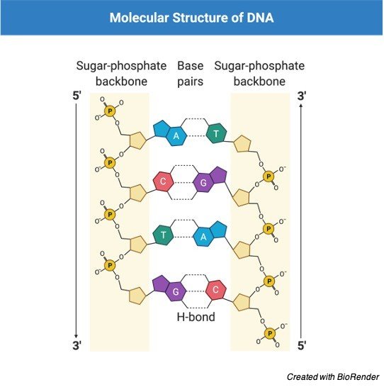
On the other hand, C is coupled with G, while A is paired with T, creating a base. A nucleotide is formed when a base is joined to a phosphate molecule and a ribose sugar.
In humans, DNA is present in the nucleus as chromosomal linear strands. In bacteria, on the other hand, it exists as a solitary genophore. Structural proteins bind to chromosomal DNA to create chromatins which compress, organise, and regulate DNA strand accessibility.
In eukaryotes, chromatin is typically made up of nucleosomes containing DNA segments coiled around histone proteins. A genome is an organism’s whole set of genetic material.
Some viruses have RNA as their genetic material. It is made up of a single strand with a phosphate group, sugar, and nucleotides on one end. Furthermore, the RNA strand’s bases are as follows:
Guanine (G), cytosine (C), adenine (A), and uracil (U), with uracil replacing thymine in DNA. The base pairs of RNA are identical to those of DNA, with the exception that adenine and uracil are coupled.
Genetic Material Function
The genetic material is crucial since it contains all of the information about the creature. Individuals’ genetic composition differs due to variations in the sequence and order of the nucleotides that make up DNA.
In order to keep genetic material secure, DNA strands are extremely condensed and ordered in the cell. It should, however, be accessible to the cell for use as a template in biological activities like protein synthesis.
The cell uses the DNA strand as a guide to synthesizing proteins, since DNA is utilised to make messenger RNA, which is complementary to one of the DNA strands.
Ribosomal RNA then translates the messenger RNA into proteins made up of different amino acids. The genetic code is the sequence of amino acids.
Characteristic of Genetic Material
Every cell has genetic information. It contains all of the organism’s data. It changes depending on the organism. It regulates several activities within the cell, as well as cell replication and the creation of new cells. It also takes into account the diversity of organisms.
DNA is the genetic substance found in all cells. It is handed down through the generations via DNA. Previous generations are linked to an individual through genetic information. Individual traits like hair and skin colour, as well as other invisible or recessive qualities, are inherited by genes.
Some genetic alterations (mutations) may impact an individual’s genetic material and may be passed down to the next generation. Other alterations may not impact genetic material and, as a result, are not passed down to kids. The DNA sequence is currently being utilised to develop new treatments for hereditary disorders that are difficult to treat.
Genetic Material Citations
Share
Similar Post:
-

Limiting Factor: Definition, Characteristic, and Examples
Continue ReadingLimiting Factor Definition
In biology, a limiting factor is any element in the environment that might restrict a process, such as the development, abundance, or dispersion of a population of organisms in an ecosystem. Examples of ideas or laws that may be used to explain limiting variables in an ecosystem include Liebig’s law of the minimum, Blackman’s law of limiting factors, and Shelford’s rule of tolerance. The population increase might be limited by the scarcest resources, not the abundant ones, under the law of minimums.
Limiting Factor Etymology
The phrase limiting factor is derived from the Latin words limitare, which means “to bind,” and factor, which means “a doer,” “performer,” and factus, which means “done” or “made.” Limiting resources; ecological factors; and constraining factors are synonyms.
What is Limiting Factor?
A biological or ecological process that is dependent on several elements will likely have a pace restricted by the slowest factor, according to the law of limiting factors. According to the rule of tolerance, an organism’s survival success is said to be determined by a complicated combination of environmental variables.
Density-dependent or density-independent limiting factors are possible. Those that are density-dependent tend to limit population growth, abundance, or dispersion based on population density.
A density-independent limiting factor, on the other hand, can control population growth, abundance, or dispersion regardless of population density. Limiting factors can also be single-limiting, indicating that only one component limits the system. A co-limiting factor is a factor that has an indirect restricting impact or amplifies the effect of a direct limiting factor.
Food, nutrition, shelter, and mate are examples of limiting variables that might restrict the expansion of a population. Because these resources are scarce in the environment, they may force living creatures to compete for them.
Limiting Factor Limitations
The ecosystem is influenced by a variety of limiting forces.
(1) keystone species,
(2) predators,
(3) energy,
(4) accessible area, and
(5) food supply are the factors to consider.
In biology, a limiting factor is a component or variable in the environment that has the ability to limit the development, abundance, or dispersion of a population in an ecosystem. These ingredients are in short supply. As a result, species fight for limited resources in the environment.
Limiting Factor Principle
The concept of limiting factors is described as the notion that a factor in low supply will restrict an organism’s or community’s growth and development. The laws that describe the concepts of limiting factors include Liebig’s law of the minimum, Blackman’s law of limiting factors, and Shelford’s law of tolerance.
i. Liebig’s Law of the Minimum
Carl Sprengel created the law of the minimum, which was subsequently popularised by Justus von Liebig. This rule asserts that rather than the total resources available, a limiting factor, i.e., the scarcest resource, controls growth. In biology and ecology, this indicates that the variables that are scarcest limit the expansion of a population, not the factors that are numerous. This was determined by observing crop growth. As a consequence, adding an excess of nutrients did not result in enhanced growth.
Conversely, adding limited nutrients, which is the limiting factor in this situation, resulted in enhanced crop growth. This indicates that even if some nutrients in the soil are abundant, crop development will be limited or limited if other nutrients are limited or scarce. When this concept is applied to other biological groups, it implies that growth occurs only in response to the most limiting constraint. William Cumming Rose utilised this technique to determine the amino acids that were designated as necessary.
ii. Blackman’s Law of Limiting Factor
Frederick Frost Blackman, a British plant scientist, introduced the limiting factor law in 1905. According to this law, the rate of a process that is dependent on several elements is restricted by the slowest factor’s rate. Photosynthesis, for example, is a biological process that is influenced by a number of variables.
6CO2 + 12H2O + energy = C6H12O6 + 6O2 + 6H2O is the general chemical process of photosynthesis. CO2, H2O, and light energy (sunlight) are the reaction’s limiting variables, according to this equation. If any of them becomes available at a slower or lower rate than usual, photosynthesis is predicted to slow down depending on the slowest factor’s rate. Even if H2O and light energy levels are abundant, the rate of photosynthesis slows if CO2 concentrations become limited (e.g., owing to the closing of stomatal openings in response to increased temperatures in the environment).
The same thing will happen if light energy becomes less accessible or strong; photosynthesis will slow down despite the availability of CO2 and H2O. When a plant is unable to capture light, such as owing to shadows caused by a dense population of plants, light becomes a limiting factor in photosynthesis.
iii. Shelford’s Law of Tolerance
Victor Ernest Shelford, an American naturalist, created the law of tolerance in 1913. It asserts that an organism’s success is determined by a complicated collection of environmental variables (environmental factors). And that organism’s performance would be determined by a set of minimal, maximum, and optimum environmental variables. These represent the organism’s tolerance limit. Within the same organism, tolerance ranges might differ, for example, depending on life stage (larval vs. adult).
Types of Limiting Factors
i. Density Dependent Limiting Factor
Density-dependent limiting factor is the density-based element that limits the growth of a population. A big, dense population is more influenced than a small, sparsely populated area. A dense population, for example, would have higher food and water demands than a small population. The limiting element in this situation is food and water availability, which is dependent on density. Disease is also a density-dependent component. It spreads quicker in densely populated areas than in sparsely populated areas.
ii. Density Independent Limiting Factor
The limiting factor that is not reliant on density is known as a density-independent limiting factor. The limiting factor can limit population size regardless of population density. For example, regardless of population density, a catastrophic event such as an earthquake or a volcanic eruption might induce a population drop.
iii. Single Limiting and Co-limiting
When there is just one limiting element in a system, it is called a single-limiting factor. When a factor indirectly impacts the population of species in an environment but enhances the limitation of the factor directly impacting the population, it is referred to as a co-limiting factor.
Limiting Factor Examples
A population will expand exponentially as long as the environment to which all people in that population are exposed remains constant, according to the law of population growth. As a result, assuming environmental circumstances remain unchanged, the population is anticipated to increase. However, there will come a moment when the population outnumbers the environment’s capacity to support it. This is referred to as the carrying capacity, or the environment’s maximum load.
The carrying capacity of an ecosystem refers to the number of people it can support without causing harm or devastation to the organisms or the environment. As a result, population growth may continue until carrying capacity is reached. The population will ultimately shrink if it exceeds this limit. Carrying capacity is determined by limiting variables. Food, water, habitat, and mate are all typical limiting variables in ecosystems. The presence of these elements will have an impact on the environment’s carrying capacity.
As the world’s population grows, so does the need for food. Due to the scarcity of food, organisms will begin to compete for it. The same may be said for nutrients, space, and partners. Because these resources are only accessible for a short time, residents of a given ecosystem will compete, perhaps against other species of the same species (intraspecific competition), or against other groups of species (extraspecific competition) (interspecific competition).
Another common symbiosis in the environment is the predator-prey interaction. If predation is excessive, deer numbers, for example, may suffer. If the number of wolves is larger than the number of deer that they hunt, the deer population may decline. However, as the quantity of deer decreases, the number of wolves may also decrease. In an ecosystem, this predator-prey relationship is an example of a biotic factor.
Abiotic factors comprise the different physico-chemical elements in an ecosystem, and biotic factors include the actions of a biological component of an ecosystem. Sunlight, humidity, temperature, the atmosphere, soil, land geology, and water resources are all examples of physico-chemical variables. Temperature, for example, is a key limiting factor because it influences the efficacy of enzymes and catalysts, which are critical components of an efficient biological and chemical system.
Limiting Factor Citations
- The agony of choice: how plants balance growth and survival under water-limiting conditions. Plant Physiol . 2013 Aug;162(4):1768-79.
- Limiting factors to oxygen transport on Mount Everest 30 years after: a critique of Paolo Cerretelli’s contribution to the study of altitude physiology. Eur J Appl Physiol . 2003 Oct;90(3-4):344-50.
- Limiting factors for maximum oxygen uptake and determinants of endurance performance. Med Sci Sports Exerc . 2000 Jan;32(1):70-84.
Share
Similar Post:
-

Passive Transport: Definition, Characteristic, and Examples
Continue ReadingPassive Transport Definition
An instance of passive transport in action. A channel protein transports water-soluble compounds along the concentration gradient across the membrane (an example of facilitated diffusion). Fat-soluble compounds pass through the lipid bilayer membrane with ease (an example of unassisted diffusion).
Passive transport is a method of transporting substances from a high-concentration to a low-concentration region. As a result, chemicals travel in a concentration gradient.
What is Passive Transport?
Passive transport refers to the movement of things such as ions and molecules along concentration gradients. It indicates that the drug prefers to migrate from a high-concentration location to a low-concentration area. Substances gravitate to areas where there are fewer of them.
Active transport, another kind of cellular transport that fundamentally requires ATP to move substances against their concentration gradient, does not require metabolic energy (e.g. ATP) since its movement is downhill or along its concentration gradient. Simple diffusion, assisted diffusion, filtration, and osmosis are the four primary forms of passive transport.
Passive Transport vs Active Transport
Passive and active transport are two types of cellular transport processes that a cell uses to transfer things across a biological membrane. Passive transport, on the other hand, differs from active transport in that the chemicals travel in the direction of their respective concentration gradients, whereas active transport moves substances against their gradients. In passive transport, substances travel in the opposite direction as they do in active transport.
In passive transport, the concentration is transferred from one location to another with a lower concentration. The direction of travel is described as downward. As a result, unlike active transport, cellular energy (e.g. ATP) is not required. Passive transportation, on the other hand, is propelled by kinetic and natural energy. Active transportation, on the other hand, moves people uphill, from a less densely populated region to one where it is already densely populated. As a result, ATP is commonly used as the driving force in the process.
Types of Passive Transport
Passive transport may or may not require the aid of a membrane protein when transporting molecules across a biological membrane.
(1) simple diffusion,
(2) assisted diffusion,
(3) filtration, and
(4) osmosis are the four primary forms of passive transport.
The net migration of molecules from higher to lower concentrations is referred to as simple and assisted diffusion. The migration of a solvent (typically water molecules) from lower to higher solute concentrations via a semipermeable membrane is referred to as osmosis. Filtration is the process of moving water and solute molecules across a cell membrane, which is aided by the circulatory system’s hydrostatic pressure.
Passive Transport Examples
Plants and animals require passive transport in order to operate properly. Gases such as carbon dioxide and oxygen, for example, diffuse into and out of plant cells through stomatal openings based on concentration gradients. Plants require carbon dioxide because it is one of the primary reactants in photosynthesis. Animals that emit carbon dioxide during expiration are one of the sources of the gas. Passive transport is responsible for the emission of carbon dioxide into the atmosphere.
Carbon dioxide diffusion occurs between the blood and the tissue fluid, in particular at the capillary beds. It travels from the tissues to the blood, then to the lungs, where it diffuses from the blood into the alveoli before being exhaled. As carbon dioxide leaves the body, oxygen enters the lungs and is delivered into the body’s tissues by simple diffusion.
Passive Transport Citations
- Passive water transport in biological pores. Int Rev Cytol . 2002;215:203-30.
- Cell Membrane Transport Mechanisms: Ion Channels and Electrical Properties of Cell Membrane. Adv Anat Embryol Cell Biol . 2017;227:39-58.
- Vitamin E intestinal absorption: Regulation of membrane transport across the enterocyt. IUBMB Life . 2019 Apr;71(4):416-423.
Share
Similar Post: