-

Phenotype: Definition, Characteristic, and Examples
Continue ReadingPhenotype Definition
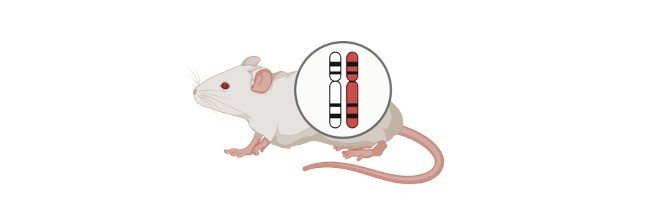
The word “phenotype” is defined in biology as an organism’s observable and quantifiable traits as a result of the interplay between the organism’s genes, environmental variables, and random variation. The phenotype of an organism includes not only observable traits like appearance, but also molecules and structures like RNA and proteins generated as coded by genes; this is referred to as “molecular phenotype.”
What is Phenotype?
The visible features of an organism as a multifactorial result of hereditary traits and environmental effects are referred to as phenotype. The morphological, biochemical, physiological, and behavioural characteristics of an organism make up its phenotype. As a consequence of the expression of an organism’s genes, as well as the impact of environmental variables and random variation, the phenotype is the entire set of traits shown by that organism. The following relationship has typically been used to illustrate the interplay between these factors: genotype + environment + random variation phenotype.
The relationship between phenotype and genotype is depicted in this graphic (Punnett square). The B and b genes are responsible for the pea plant’s petal colour. The dominant characteristic is a purple-petalled bloom, which is the result of the B gene. The recessive characteristic is the b gene. Three children with the purple-flower trait (BB and Bb) and one offspring with the white-flower trait will result from a test cross between two plants that are heterozygous (Bb) for the purple petal colour trait (bb). The phenotypic ratio in this instance is 3:1.
Phenotype Etymology
Phenotype is derived from the Latin phaeno-, which comes from the Greek phaino-, which means “shining,” from phanein, which means “to shine,” “to appear,” or “to show,” and -type from “typos.” Genotype is a similar term. The term phenotypic is a descriptive term that refers to, describes, or refers to the phenotype of a certain organism.
Trait vs Phenotype
A trait is a feature of the phenotypic of an organism. To differentiate one characteristic from another under the more-inclusive word, phenotype, the trait is sometimes referred to as a phenotypic trait in genetics. An organism’s phenotype is made up of several characteristics. Traits can be inherited (genetically determined), acquired (as a consequence of environmental factors), or a combination of both. For example, hair colour is a personality feature that might be black, blonde, ginger, or brunette.
Phenotype vs Genotype
Genotype and phenotype are words used in genetics to describe an organism’s appearance, function, and behaviour. A genotype is a collection of genes that, when expressed, determine an organism’s feature or attribute. To put it another way, the genotype is the genetic component of the phenotype.
DNA sequences are made up of genes. They come in pairs in humans and other animals, one from the male parent and the other from the female parent. Alleles are genetic pairings that share the same loci on the chromosomes and control the same characteristic.
A pair of genes (or alleles) for a certain trait often consists of two genes, one dominant and the other recessive. The dominant allele will show up as a characteristic, whereas the recessive allele will not. There are three genotypes that may be identified by annotating the dominant allele with A and the recessive allele with a:
(1) AA, homozygous dominant allele,
(2) Aa, heterozygous dominant, and
(3) aa, homozygous recessive.
An organism’s genotype is a key factor in determining its phenotype. A pair of genes (or alleles) for a specific trait is usually made up of two, one dominant and the other recessive. The dominant allele will manifest itself as a characteristic, while the recessive allele will not. The dominant allele is labelled with A, and the recessive allele is labelled with an in three possible genotypes:
(1) AA represents the homozygous dominant allele,
(2) Aa represents the heterozygous dominant allele, and
(3) aa represents the homozygous recessive allele.
An organism’s genotype is a significant determinant of its phenotype. Take, for example, a pair of alleles (or genes) that defines a certain characteristic, one of which is dominant (A) and the other recessive (B) (a). The dominant allele (A) will be expressed and form part of the organism’s phenotype, whereas the recessive allele (a) will be muted.
When a characteristic is inherited according to Mendelian principles, the A will appear as a trait, but the a will not. As a result, an organism’s phenotype must incorporate the characteristics of all expressed genes. Many observable characteristics in humans, however, are more complicated than those that follow the Mendelian pattern. In the case of polygenic inheritance, complex characteristics such as height and skin colour are caused by the interactions of many alleles.
Expression
Genetic factors, environmental effects, and random genetic variants all contribute to the phenotypic. The characteristic is defined as homozygous when the pair of alleles defining a specific trait comprises identical genes, e.g. AA or aa. The characteristic is defined as heterozygous when the allelic makeup comprises of various genes, such as Aa. The presence of the dominant allele, such as AA or Aa, causes the characteristic (A) to manifest, whereas the lack of the dominant allele, such as aa, causes the opposite trait to appear (a). This is an example of full dominance, and it is inherited in the Mendelian manner.
The expression of a characteristic will not follow the Mendelian pattern in situations of codominance, imperfect dominance, or polygenic inheritance. Because both alleles in a pair are dominant in codominance, the alleles of a gene pair in a heterozygote will be completely expressed (e.g. AB). The resultant characteristic under incomplete dominance will be a mix of the impacts of the two alleles. Because the dominant allele will only be partly expressed, this is the case. As a result, the heterozygous offspring will have a phenotype that is halfway between the parents’ phenotypes.
Aside from genetic interactions, an organism’s phenotype is influenced by the environment as well as random (genetic) variations. Environmental variables may have an impact on an organism’s appearance. A light-colored skin that is continually exposed to the sun’s rays, for example, will darken as a result of increased melanin synthesis. Random variation, on the other hand, might modify a physical feature or, at the very least, an organism’s fitness. Gene changes are critical since they are what drives evolution and natural selection. Individual phenotypes are explained in part by genotypes, environmental influences, and genetic variants.
Extreme Phenotype
When the alleles of both parents join together, the outcome is a hybrid with a phenotype that is larger or higher than the phenotypes of both parents. Its transgressive phenotype might be advantageous or harmful, depending on how it impacts the offspring’s total fitness. Transgressive segregation is the development of extreme phenotypes. The child of a cross between Helianthus annuus and Helianthus petiolaris is an example of a hybrid with an extreme phenotype. Transgressive hybrids were created from the two sunflower species. In contrast to their parents, hybrids can survive in environments where their parents can not. Sand dunes and salt marshes are not a problem for them.
Recombinant Phenotype
Meiosis is an essential biological process that leads to an increased variety of organism phenotypes. The homologous chromosomes come together during the metaphase of meiosis, in particular, to swap genes via homologous recombination. The four daughter cells will contain chromosomes that are different from one another when the homologous chromosomes approach the conclusion of meiosis (telophase II). Some of them will develop into gametes with recombinant genes.
When such a gamete is fertilised with a wild type, for example, it will produce an offspring with a recombinant phenotype, which is distinct from its parents’ phenotypes. A test cross between two characteristics (for example, a blue-bodied, normal-winged fly father and a black-bodied, vestigial-winged fly parent) can aid in the identification of recombinant phenotypes. Recombinants are children with traits that differ from their parents (for example, a blue-bodied, vestigial-winged fly or a black-bodied, normal-winged fly).
Phenotypic Ratio
Another technique for identifying all potential allelic pairings in a test cross is a Punnett square. It can forecast the offspring’s genotypes and traits. It’s a grid and letter graphic that represents alleles. A dominant trait or genotype is denoted by an uppercase letter (e.g. A), while a recessive trait or genotype is denoted by a lowercase letter (e.g. a). The phenotypic ratio (as well as the genotypic ratio) may be calculated using the Punnett square. A phenotypic ratio is a proportion that may be anticipated based on the results of a test cross. It can be determined based on the phenotypes of the offspring, or the frequency with which distinct characteristics or trait combinations are manifested in the offspring.
For example, based on the four possible phenotypes: AaBb (blue, normal-winged fly), aaBb (black, normal-winged fly), Aabb (blue, vestigial-winged fly), and aabb (blue, vestigial-winged fly), the expected phenotypic ratio of an AaBb x aabb dihybrid cross (i.e. a cross that deals with (black, vestigial-winged fly).
Phenotype Example
As previously stated, an organism’s phenotype refers to the many qualities that it contains. Blue eye trait (for eye colour character), brown skin trait (for skin colour character), long-tail trait (for tail character), five-petalled trait (for flower character), and so on are examples of traits. Another example of a phenotype is behaviour. Individuals with mental retardation, for example, have behavioural and cognitive characteristics that constitute behavioural phenotypes.
Phenotype Citations
- Structural properties of genotype-phenotype maps. J R Soc Interface . 2017 Jul;14(132):20170275.
- Bridging the genotype-phenotype gap: what does it take? J Physiol . 2013 Apr 15;591(8):2055-66.
- Natural History and Genotype-Phenotype Correlations in RDH12-Associated Retinal Degeneration. Adv Exp Med Biol . 2019;1185:209-213.
Share
Similar Post:
-

Phosphate Group: Definition, Characteristic, and Examples
Continue ReadingPhosphate Group Definition
A negative-charged functional group or radical composed primarily of phosphorus connected to four oxygen atoms. The sign PO4– is used to represent it.
The phosphate group plays a variety of roles in living organisms.
To begin with, it is a crucial structural component of the nucleotide, which is the fundamental structural unit of DNA and RNA.
Second, it is a constituent of energy-dense molecules like ATP.
Finally, it is linked to coenzymes involved in anabolic processes, such as NADP/NADPH (such as photosynthesis in plants and lipid synthesis in animals).
In biological membranes, it is also a component of the hydrophilic head of phospholipids.
Phosphate Group Citations
Share
Similar Post:
-
Plant Tissue: Definition, Characteristics, and Examples
Continue ReadingPlant Tissue
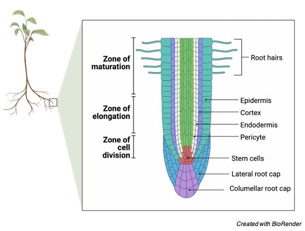
Roots, stems, and leaves are the three primary organ groupings found in plants. These organs, as we know from other areas of biology, are made up of tissues that collaborate to achieve a shared objective (function). Tissues, in turn, are made up of a variety of cells, which are made up of elements and atoms at their most basic level. The many forms of plant tissue, as well as their location and function inside a plant, will be discussed in this section. It’s vital to keep in mind that unique plants may have small changes and alterations to the fundamental tissue types.
The structure and function of plant tissues are used to characterise and classify them. Within a plant, the organs they produce will be arranged into patterns, which will assist in further categorising the plant. The three fundamental tissue patterns seen in roots and stems that serve to distinguish between woody dicot, herbaceous dicot, and monocot plants are an excellent illustration of this.
(1) pith, (2) protoxylem, (3) xylem, (4) phloem, (5) sclerenchyma, (6) cortex, and (7) epidermis are the different plant tissues.
Meristematic Tissues
Meristems or meristematic tissues are tissues in which cells are continually dividing. These areas generate new cells. By comparison, these new cells are compact, six-sided boxlike structures with a lot of tiny vacuoles and a big nucleus. There are no vacuoles in some cases. Vacuoles will develop into a variety of shapes and sizes as the cells age, depending on the cell’s demands. The vacuole has the potential to occupy 95 percent or more of the entire volume of the cell. Apical meristems, lateral meristems, and intercalary meristems are the three types of meristems.
Apical Meristems
At or around the tips of roots and shoots, apical meristems are found. The roots and shoots will grow longer as new cells develop in the meristems. This type of vertical development is referred to as primary growth.
An excellent illustration is the increase in height of a tree. Procambium, protoderm, ground meristems, and procambium, three types of primary meristems, are all developed by each apical meristem. The cells that will become the primary tissues will be produced by these primary meristems.
Lateral Meristems
Plants’ secondary growth is controlled by lateral meristems. Horizontal growth is the most common type of secondary growth. An excellent illustration is the increase in girth of a tree trunk. In the study of plants, there are two types of lateral meristems to be aware of.
The first form of lateral meristem, the vascular cambium, is also known as the cambium. Except at the tips, where the apical meristems are located, the cambium is a thin, branching cylinder that covers the length of the roots and stems of most perennial plants and many herbaceous annuals. The cambium is in charge of producing cells and tissues that increase the plant’s thickness, or girth.
The cork cambium, the second kind of lateral meristem, is a narrow cylinder that runs the length of roots and stems, similar to the vascular cambium. The distinction is that it can only be found in woody plants since it is responsible for producing the outer bark.
Only until the main tissues produced by the apical meristems have begun to mature, will the vascular and cork cambiums, if present, begin to create cells and tissues.
Intercalary Meristems
Because they do not expand in girth, intercalary meristems are seen in grasses and similar plants that do not have a vascular or cork cambium. These plants have apical meristems and the third kind of meristematic tissue at regions of leaf attachment, termed nodes. This meristem is also important for lengthening since it actively produces new cells. The regeneration of cut grass is controlled by the intercalary meristem.
Plant tissues that do not actively generate new cells are known as non-proliferative tissues. Nonmeristematic tissues are the name for these types of tissues. Nonmeristematic tissues are made up of cells produced by meristems and come in a variety of shapes and sizes depending on their function in the plant. Sometimes the tissues are made up entirely of the same cell type, while other times they are mixed. There are simple and complicated tissues to examine, but for the sake of discussion, we’ll start with the basic tissues.
Simple Tissues
(1) parenchyma tissue, (2) collenchyma tissue, and (3) sclerenchyma tissue are the three main kinds of tissue called after the type of cell that makes up its composition.
i. Parenchyma Tissue
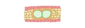
Parenchyma tissue is made up of parenchyma cells. Parenchyma cells are the most common cell type in higher plants, and they may be found in nearly every major organ. When these cells are originally created, they are essentially sphere-shaped. These cells, on the other hand, have thin walls that flatten at the points of contact when a large number of them are packed together. They usually have a lot of sides, with the majority having 14 sides. These cells may store a variety of secretions such as starch, lipids, tannins, and crystals. The tissues present in leaves are formed by parenchyma cells with numerous chloroplasts. Chlorenchyma is the name of this kind of tissue.
Photosynthesis is the primary function of this type of tissue, whereas parenchyma tissues lacking chloroplasts are often utilised for food or water storage. Also known as aerenchyma tissue, certain clusters of cells are loosely packed together with connecting air gaps, such as in water lilies. These cells can also grow irregular inner wall expansions, which enhances the plasma membrane’s overall surface area and promotes the transfer of dissolved chemicals between neighbouring cells. If parenchyma cells are mature, they may divide, which is important for healing damage to plant tissues. The majority of the edible parts of fruit are made up of parenchyma cells and tissues.
ii. Collenchyma Tissue
Collenchyma tissue is made up of collenchyma cells. These cells, like parenchyma cells, have living protoplasm and may exist for lengthy periods of time. Their walls are thicker than those of parenchyma cells, which is their major differentiating feature. The walls appear irregular in cross-section. Collenchyma cells are located immediately beneath the epidermis, and they are typically elongated with flexible as well as robust walls. These cells, together with the tissues they produce, offer flexible support for organs like leaves and flower parts as a plant grows. Celery‘strings’ that get trapped in our teeth are a good example of collenchyma plant cells.
iii. Sclerenchyma Tissue
Sclerenchyma tissue is made up of sclerenchyma cells. These cells contain lignin-embedded secondary walls that are thick and strong. Most sclerenchyma cells are dead by the time they reach maturity, but they nevertheless provide structure and support. Sclerenchyma cells come in two varieties:
a) Sclereids are sclerenchyma cells that are dispersed throughout the body at random. They are sometimes clustered together in distinct zones or areas inside other tissues. They’re usually twice as long as they’re wide. The rough feel of some varieties of pears is one example. The grittiness is caused by sclereid cell clusters. Stone cells are another name for scleereids.
b) Fibers can be found in roots, stems, leaves, and fruits in connection with a wide range of tissues. Fiber cells are often longer than they are wide, with a very small hollow in the centre. Textiles, ropes, strings, and canvas items, to mention a few, are now made from fibres from over 40 different plant families.
Secretory Cells and Tissues
Substances that collect within the cell as a result of biological activities can occasionally harm the protoplasm. As a result, it is critical that these components be separated from the protoplasm from whence they arise, or that they be transported outside of the plant body. Although the majority of these chemicals are waste products, some are necessary for plant function. Citrus oils, pine resin, latex, opium, nectar, fragrances, and plant hormones are among examples. Secretory cells are formed from parenchyma cells and can operate as a tissue or on their own. They might have a lot of commercial value.
Complex Tissue
The term “complex tissue” refers to tissues that have more than one cell type. The fundamental tasks of xylem and phloem, the two most essential complex tissues in a plant, are to transport water, ions, and soluble food items throughout the plant. While apical meristems create some complex tissues, the vascular cambium produces the majority of them in woody plants, and is frequently referred to as vascular tissue.
The epidermis and periderm are two more complicated tissues. All plant organs are protected by the epidermis, which is made up largely of parenchyma-like cells. Specialized cells that enable water and gases to flow in and out of the plant, secretory glands, different hairs, cells in which crystals are collected and separated, and other cells that improve absorption in the roots are all found in the epidermis. The outer bark of woody plants is formed by the periderm, which is mainly cork cells. Because of the pockets of parenchyma cells distributed throughout, it is considered a complex tissue.
Xylem
The xylem is an important plant tissue since it is part of the plant’s “plumbing.” Consider the main axis of stems and roots as a network of pipes. It is made up of parenchyma cells, fibres, vessels, tracheids, and ray cells and transports water and dissolved chemicals throughout the body.
The vessels are long tubes made up of individual cells, with open ends on both ends. There may be bars of wall material stretching across the open space on the inside. From end to end, these cells are connected to form lengthy tubes. Vessel members and tracheids die when they reach adulthood. Tracheids have thick secondary cell walls that taper towards the ends. They don’t have the same end holes as the vessels.
The tracheids’ ends overlap, and there are two pits on each side. Water can flow from cell to cell through the pit pairs. While most xylem conduction is up and down, there is some lateral or side-to-side transmission through rays. Rays are long-lived parenchyma cells that emerge from the vascular cambium in horizontal rows. Rays extend out from the centre of the stems and roots of trees and other woody plants, resembling the spokes of a wheel in cross-section.
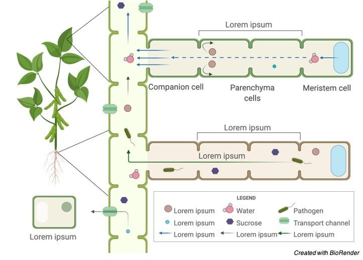
Phloem
Phloem is an equally essential plant tissue since it is a part of the plant’s “plumbing.” Phloem is primarily responsible for transporting dissolved food molecules throughout the plant. The sieve-tube component and companion cells in this conduction system do not have secondary walls. Both xylem and phloem are produced by the vascular cambium’s parent cells. Fibers, parenchyma, and ray cells are generally included.
Sieve tubes are made up of end-to-end sieve tube components. Unlike vessel members in xylem, end walls do not have apertures. The end walls, on the other hand, are lined with tiny holes through which cytoplasm flows from cell to cell. Sieve plates are the name of these porous connectors.
Sieve-tube members do not have nuclei at maturity, despite the fact that their cytoplasm is actively involved in the conduction of food items. It is the companion cells that are nestled between the sieve-tube members that are responsible for food conduction in some ways. A polymer called callose is found in living sieve-tube members. As long as the cell contents are under pressure, the callose remains in solution.
If an insect injures a cell and the pressure decreases, the callose will form as a healing mechanism. The callose and phloem protein, on the other hand, will be transported to the closest sieve plate, where they will form a clog. This stops additional sieve tube contents from leaking, and the injury isn’t always deadly to the plant’s total turgor pressure.
Epidermis
The epidermis is a fascinating plant tissue that is both complicated and intriguing. The epidermis is the outermost layer of cells on all plant organs, according to official definitions (roots, stems, leaves). Because the epidermis is in close touch with the environment, it is subject to its circumstances and limitations. The epidermis is usually one cell layer thick, although there are certain outliers, such as tropical plants, where the layer can be many cells thick and serve as a sponge.
The cuticle is a waxy protective coating formed by cutin, a fatty substance produced by most epidermal cells. One of the primary determinants of how much water is lost by evaporation is the thickness of the cuticle. The cuticle also provides some resistance to bacteria and other disease organisms at no additional cost. The wax palm, for example, produces enough cuticle to be commercially valuable: carnauba wax.
Polishes, candles, and even phonographic recordings are made from other wax materials. The absorptive surface area of root hairs is increased by epidermal cells. Root hairs are tubular extensions of the main root body that are completely made up of epidermal cells. Leaves are not ignored. They feature a lot of tiny pores called stomata that are bordered by guard cells, which are specialised epidermal cells. Because they have a distinctive form and contain chloroplasts, guard cells are distinct epidermal cells. Other modified epidermal cells, such as glands or hairs, may be used to repel insects or minimise water loss.
Periderm
The epidermis is sloughed off and replaced by a periderm in woody plants as the cork cambium begins to develop new tissues to increase the girth of the stem or root. The periderm is made up of cork cells that are semi-rectangular and boxlike. The outermost layer of bark will be this. When these cells reach maturity, they are no longer alive. The protoplasm, however, secretes a fatty material called suberin into the cell walls before the cells die. Suberin protects the tissues beneath the bark by making the cork cells waterproof. The cork cambium produces pockets of loosely packed cork cells in some areas.
Suberin is not incorporated into the cell walls of these cork cells. Lenticels are loose patches that extend through the periderm surface. Lenticels are responsible for gas exchange between the air and the inside of the stem. Lenticels are found at the bottom of deep cracks in tree bark.
Plant Tissue Citations
- Ice-cap. A high-throughput method for capturing plant tissue samples for genotype analysis. Plant Physiol . 2004 Jul;135(3):1162-9.
- Ups and downs of tissue and planar polarity in plants. Bioessays . 2004 Jul;26(7):719-29.
- The lipids of plant tissue cultures . Adv Lipid Res . 1976;14:171-211.
- Rice virus release from the planthopper salivary gland is independent of plant tissue recognition by the stylet. Pest Manag Sci . 2020 Sep;76(9):3208-3216.
Share
Similar Post:
-

Substrate: Definition, Characteristics, and Examples
Continue ReadingSubstrate Definition
The term “substrate” is frequently used in the material sciences to define the basis of a material on which different processing is carried out under specific reaction conditions to produce additional layers and films, such as coatings. As a result, depending on the substrate’s uses and field of research, there are a variety of definitions accessible in the literature. Let us, however, concentrate on the biological side of the substrate.
What is Substrate?
Substrate refers to the underlying substances or layers. The term “substrate” is defined differently in different disciplines of study. It is the main chemical that interacts with the reagent under a set of reaction conditions in key science areas such as chemistry.
To put it another way, the substrate definition in chemistry refers to the chemical reactant that is involved in the chemical reaction and on which an enzyme will operate. The substrate is described as the fundamental surface to which the paint adheres in other connected science disciplines, such as basic engineering.
The compounds that enzymes react with are referred to as substrates in biochemistry. The substrate is the basis on which an immobile material is bonded in ecology. The substrate, in basic terms, is the surface or substance from which an organism develops or receives sustenance. The term “substrate” is sometimes known as “substratum” or “underlayer.”
(1) In ecology, the earthy material where an organism dwells or the surface or medium where an organism develops or is connected is referred to as the substrate. It is the material at the bottom of marine environments (e.g. soil, boulders, sand, gravel) in marine ecosystems. It can be used as a location to live, a stream bed (or an aquarium), or a food source for microorganisms. (2) In biochemistry, a substrate is any chemical that an enzyme reacts with. Substratum and underlayer are synonyms.
Substrate Examples
Below are some examples of substrates in biology areas, including biochemistry, plant ecology, reptile ecosystems, and microbial ecology.
i. In Biochemistry
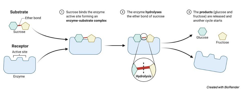
The substrate is defined in biochemistry as any chemical that interacts with an enzyme’s active site. Between the active site and the substrate, a chemical connection is established. Hydrogen bonds, hydrophobic contacts, and weak Van der Waals forces are used to bind the substrate to the active sites in such processes, which are also known as enzyme-catalyzed reactions. After the enzyme-substrate complex is formed, the enzyme exerts a force on the substances, which causes them to be transformed into products.
In such a complex, an enzyme requires a well-defined substrate to carry out its catalytic activities, whereas numerous active spots in the enzymes’ bodies draw substrates to them. Substrate specificity refers to the process through which enzymes activate particular substrates. As a result, the substrates are loaded onto those active sites, allowing for the formation of comparatively weaker connections between them.
ii. Enzyme-Substrate Complex
Enzyme (E) + Substrate (S) → Enzyme-Substrate or ES Complex
The interaction between the active sites and the substrate takes place under certain conditions, resulting in an enzyme-substrate complex, which causes the substrate to become part of the reaction product.
Although the substrate becomes part of the product, the enzyme undergoes multiple conformational modifications, changes in size and shape, and changes in physicochemical characteristics. Refer to the enzyme and substrate schematic diagram below.
The majority of molecules found in the human body are substrates in their early stages. There are numerous reactions that occur in daily life, and the majority of them require a large amount of energy or extended response periods to meet the completion conditions. As a result, enzymes aid the reaction by decreasing the activation energy and increasing their reaction rates, allowing biological and chemical reactions to proceed smoothly.
The physiochemical characteristics of the substrate change once the reaction is complete, depending on the reaction conditions and the type of the product produced. It’s worth noting that many reactions need many steps to complete, which are referred to as intermediate reactions.
Until the last step in such processes, the product of one reaction becomes the reactant of another. As a result, it may be inferred that substrates, in conjunction with enzymes, play a critical part in many of the processes that occur in our environment.
Enzyme and Substrate Concentration
Many researchers have explained that the availability of the appropriate amount of substrate is essential for an enzyme’s successful functioning, as a rise in the amount of substrate increases the rate of concentration of enzyme activity. Even if there are plenty of substrates available, the rate of reaction will grow after some time until it reaches a certain value, but the rate of enzyme activity will not change. The buildup of substrates in the enzyme’s active sites is the cause of this. As a result, once you reach that level, the enzyme activity will remain constant.
Substrates play a key role in the manufacturing of a variety of sweetening agents, as well as the modification of antibiotics and the production of various washing agents. They also have a variety of uses in the therapeutic, forensic, and environmental fields.
ii. In Plant Ecology
The substrates on which diverse plants, bacteria, and reptiles grow are the most palpable components of our ecosystem, and hence, the substrate’s influence on ecology is immense. Because it is the mechanism through which plants and other creatures receive water and nutrients from the soil, the substrate is critical to their development and nutrition.
It’s worth noting that substrates differ from natural soil in terms of the percentage of organic matter present, as natural soil has a concentration of organic matter of 1-3 percent, whereas substrates have a concentration of organic matter of at least 70%.
The substrates are divided into two kinds on a global scale. There are two types of substrates: universal and specialised. They’re frequently found in peat, perlite, and organic fertiliser mixtures.
The substrates can also be distinguished from one another based on the plants to which they are intended to be applied. Acidic plants, green plants, blooming plants, cactus, succulents, gardens, orchids, carnivorous plants, rose bushes, and seed buds all thrive on them.
iii. In Reptile Ecosystem
The reptile enclosure is primarily made up of surfaces for reptiles. According to many sources, the relationship between the reptile and the substrate must be carefully examined because not all substrates are suitable for all reptiles. The substrate composed of synthetic biodegradable materials, for example, is not ideal for lizards since they sniff with their tongue and the substrate may adhere to it, causing significant gastrointestinal disease.
As a result, regularly monitoring the animal and its natural environment is a smart place to start when choosing a substrate for the reptile. Some of the most frequent substrates for reptiles are silica desert sands, calcium sands, wood fibre substrates, beech chips, bark substrates, and coco fibre.
iv. In Microbial Ecology
Microbes generate enzymes in order to get nutrition and energy by breaking down complicated organic substrates. As a result, it’s been assumed that the extracellular enzymes’ activity is heavily influenced by the substrate composition. The researchers discovered that there are a few key processes that may be used to investigate how differences in substrate composition impact enzymic activity.
The resource limitation concept and the substrate simulation model are the mechanisms. Scientists think that the variety and amount of substrates accessible to microorganisms have a significant impact on their microbial activity, and that both of these processes are at play.
For example, increasing the variety of the substrate increases enzyme activity since the substrate is available to numerous enzymes, as the substrate simulation model has successfully demonstrated. The substrate stimulation concept is possible because the substrate’s variety increases the number of niches available to microorganisms, opening the way for the creation of a more diverse community of bacteria.
Similarly, the resource constraint model accurately anticipated the kind and quantity of enzyme activity, as well as its dependency on the composition of the substrates supplied.
Synergistic effects can emerge from the addition of complementary resources, such as the addition of carbon and nitrogen molecules combined, can have far more beneficial impacts on enzymic acidity than feeding both carbon and nitrogen alone, as evidenced by the resource restriction model. As a result, it is clear that the substrate composition and concentration have a significant impact on the enzymes’ catalytic activity. Furthermore, maintaining the balance between their activities necessitates the use of an appropriate enzyme and a substrate.
Biological Importance of Substrate
A substrate is the earthy substance or surface on which different microorganisms, such as plants, fungi, and algae, live, grow, and adhere. For example, the algae on the rock may serve as a substrate for another living creature that lives on top of the algal underlayer, while the rock itself could be considered a substrate for the algae.
The substrate in an aquatic environment is the material that forms the bed of a stream or the bottom of the ocean. As a result, it encompasses rocks, sand, gravel, and soil. As a result, they are critical as a source of minerals and nutrients, particularly for species that live and grow in the area. Bottom dwellers are additionally protected from predators because they may camouflage and are difficult to spot near the bottom.
Although there are several instances of substrates in various disciplines, there are many typical substrates that we encounter in our daily lives. Enzymes like maltase and salivary amylase use carbohydrates as substrates, such as glucose, starch, and sucrose. Proteins and peptides are the substrates of the enzymes trypsin and chymotrypsin, which are found in a variety of foods such as cereals and meats that humans ingest on a regular basis. The Lipase enzyme’s substrates are lipids.
Substarte in Summary
As a result of the preceding explanation, it can be inferred that the underlying substances or layers are referred to as substrates, albeit the terminology differs depending on the field. Substratum is a term that has frequently been used as a synonym for substrate. A substrate is the molecule that an enzyme interacts with. Furthermore, each enzyme requires a well-defined substrate to carry out its catalytic activities, despite the fact that the enzymes’ bodies include several active regions that draw substrates to them. Substrate specificity refers to the process through which enzymes activate particular substrates.
through which enzymes activate particular substrates. The majority of molecules found in our bodies start off as substrates. The interaction between the active sites and the substrate takes place under certain conditions, resulting in an enzyme-substrate complex, which causes the substrate to become part of the reaction product. Many researchers have explained that the availability of the appropriate amount of substrate is essential for an enzyme’s successful functioning, as a rise in the amount of substrate increases the rate of concentration of enzyme activity.
Substrates have a wide range of uses that differ from one field to the next. Substances that operate on the active sites of enzymes are referred to as substrates in biochemistry. As a result, new bonds between the active sites and the substrates are generated. Enzyme-catalyzed reactions are another name for these types of reactions.
Substrates are the surfaces on which different microorganisms, such as plants, fungi, and algae, dwell in ecology. As a result, any other organism that lives on top of algae will use the algae on the rock as a substrate, while the algae itself will use the rock as a substrate.
Microbes generate enzymes in order to get nutrition and energy by breaking down highly complicated organic substrates. The compositions of the substrates have a big impact on the extracellular enzymes’ activity.
Substrate Citations
- Substrate-Enzyme Interactions in Intramembrane Proteolysis: γ-Secretase as the Prototype. Front Mol Neurosci . 2020 May 19;13:65.
- Alternative Substrate Metabolism in Yarrowia lipolytica. Front Microbiol . 2018 May 25;9:1077.
- The substrate repertoire of γ-secretase/presenilin. Semin Cell Dev Biol . 2020 Sep;105:27-42.
Share
Similar Post:
-

Ploidy: Definition, Characteristics, and Examples
Continue ReadingPloidy Definition
The number of homologous chromosomal sets that make up a cell’s or organism’s genome.
The number of sets of homologous chromosomes in a cell’s or organism’s genome is referred to as ploidy.
The number n is assigned to each set. As a result, one chromosomal set, 1n, is classified as monoploid.
The term haploid, on the other hand, refers to gametes that have only half of the normal sets of chromosomes seen in an organism’s somatic cells.
The combination of two haploid gametes, such as female and male gametes, produces a zygote with two sets of chromosomes, keeping the parents’ original chromosomal number.
The two sets of chromosomes would be homologous, meaning that the chromosomes from the female gamete would correspond to the corresponding chromosomes from the male gamete based on morphology and gene loci linear sequence.
Diploid refers to a cell or organism with two sets of homologous chromosomes, 2n.
Polyploid refers to an organism’s genome having numerous sets of paired chromosomes. Triploid means three sets of chromosomes, whereas tetraploid means four sets of chromosomes.
A huge number of sets can be labelled with a number (for example 15-ploid for fifteen sets).
Ploidy Citations
Share
Similar Post:
-

Realized Niche: Definition, Characteristics, and Examples
Continue ReadingRealized Niche Definition
The environmental location that a species occupies and lives in might be characterized as a realized niche. The term “post-competitive” refers to a niche that has been identified. The existence of limiting variables (such as food, light, water, the presence of other species, and so on) forces species or creatures to migrate to certain habitats where they can flourish. As a result, a realized niche is defined as the area in the environment where a species is most suited to perform its function and reproduce.
When seen from the perspective of the basic niche, the idea of a realized niche becomes clearer. The environmental location of a species in the ecosystem is represented by both realized and basic niches. The basic niche, on the other hand, encompasses all of the environmental circumstances under which a species may survive and reproduce. This comprises both biotic (such as the existence of food, such as grass, or abiotic (such as temperature, humidity, and sunshine in the case of carnivores) and abiotic (such as temperature, humidity, and sunlight) factors.
The basic niche is the perfect environment for a species to thrive in the absence of competition. However, we know that several species may coexist in a single environment. As a result, there would be rivalry for food, mates, and other limited resources, affecting the population numbers of the many species living in the same area. This competition might lead to the creation of a niche. The size of a realized niche is generally less than the size of a basic niche.
Furthermore, due to the number of rivals and predators present in that specific habitat, the realised niche of the same species residing in various places might change. The portion of a basic niche that an organism occupies as a result of limiting constraints in its environment (biology definition). A limiting factor that restricts or narrows an organism’s ecological niche is the existence of competing species in the environment. The organism tends to occupy and perform an ecological function where it is most highly adapted to a realised niche.
What is Realized Niche?
A niche is the method through which a species or an individual interacts with its surroundings. To put it another way, a niche describes how a species lives in its habitat and what function it plays in the community. A niche has various behavioural characteristics, such as food-finding strategies, movement, and survival. An organism’s niche is also defined by the temperature limitations of its environment.
Regardless of whether the temperature is above or below a particular threshold, the species will either be unable to live because some biological functions will be disrupted, or worse, will come to a halt. In ecology, a niche is described as a biological population’s reaction to limiting constraints such as the existence of rivals and resource distribution. Another example is when the population of a species grows due to the absence of predators, diseases, and parasites.
As a result, the species will thrive in its niche. An increase in predators or the danger of sickness caused by parasites and pathogens, on the other hand, will result in a decline in the population of that species. As a result, the species would have to adapt, such as by developing modifications that increase disease immunity. Alternatively, they would have to relocate to a more suitable area to ensure the survival of their species. The niche of that species will then be a favourable environment.
Scientists have coined terms like Grinnellian niche (1917), Eltonian niche (1927), and Hutchinsonian niche to describe niches (1957). The Grinnellian niche is established by the species’ behavioural adaptations and the ecosystem in which it dwells. The relationship between a species with food and its adversaries in the biotic environment determines its Eltonian niche.
The Hutchinsonian niche is a more general concept that tries to explain why so many species live in the same habitat. He also created the word “hyper-volume,” which refers to the multidimensional space in which resources like nutrients, water, and light are stored. Hutchinson referred to the hyper-volume as an n-dimensional hyper-volume since it is multidimensional with no limits on the dimensions. The environmental circumstances and resources in which species can coexist and thrive are the subject of this book (Chase & Leibold, 2003).
Fundamental Niche vs Realized Niche
The basic niche encompasses all of the environmental circumstances in which a species may readily live and reproduce in order to perpetuate itself. The realised niche, on the other hand, is the actual habitat of the species. On Earth, species must exist in a habitat that includes other species or rivals. Other variables, such as temperature, topography, and resources, shape a species’ place in its habitat in addition to competition.
The competitive exclusion principle may apply if many species occupy the same niche within their environment. One species will outcompete the others if this concept is followed. The outcompeted species will have two options: either die out or adapt. Different species may be able to cohabit and avoid competition by utilising resources in a flexible manner. To put it another way, species must change their realized niches in order to attain this level of cohabitation.
The fundamental niche differs from the realized niche in that the realized niche is the actual habitat in which the species lives, but the fundamental niche is any sort of environmental circumstance in which a species can survive. A pre-competitive niche, also known as a basic niche, is characterized by a collection of circumstances and sources that allow a species to exist, thrive, and reproduce. Because there is no rivalry for resources or predators, the species can benefit from both biotic and abiotic needs for long-term survival in the environment.
The red-winged blackbird is an example of a basic niche. During the early spring, the marches are dominated by bird species. The red-winged blackbirds established a permanent home in that region. The tri-colored blackbirds, on the other hand, arrive in the wetlands later in the summer. Because they are more aggressive, they have the best territory.
Every population on the planet is affected by a variety of environmental variables. A realised niche is a post-competitive niche that falls within the basic niche umbrella. When a species in a basic niche faces the pressure of co-existing with other species in the environment, a realized niche is established. The species has to adapt to a reduced niche.
The cohabitation of wolves and coyotes in North America is an example of how a realised niche gets established. Coyotes would compete for food and territory because both animals live in the same region. Because of wolves’ more aggressive disposition, the realized niche for coyotes was tiny. When European immigrants arrived on the continent, wolves were hunted to extinction. This benefited coyotes, and their realized niche grew as a result.
The basic and realised niches are comparable in that they are both sorts of ecological roles inhabited by similar species. Here’s a comparison chart to help you distinguish between a realized niche and a basic niche.
Realized Niche Examples
Scientists have looked at the realized niches of various species to see how they are distributed in different settings. The rat’s gut, for example, is a basic realised niche that limits the number of species that may dwell there. Potential rivals in the mouse gut include acanthocephalans (a worm family) and tapeworms.
The parasitic worms in each of these categories get their nutrition from the blood in the intestinal walls. Carbohydrate availability in the intestine varies by location; more carbs are accessible in the front than in the back. When both acanthocephalans and tapeworms are present in a mixed infection, the niches of both groups are constricted. The former species’ realized niche is limited to the anterior region of the intestine, whereas the latter species’ realized niche is narrowed towards the posterior portion.
Based on Werener’s research, another example of a realised niche in bluegill fish (Montana State University, n.d.). By separating a pond with a net and introducing a predator (largemouth bass) to one side exclusively, he established two separate ecosystems. Bluegills of three sizes (small, medium, and big) were investigated to see how their interactions with food changed in the presence and absence of a predator. Plankton, plants, and benthos were all introduced as food sources. Because there were no predators, a basic niche developed, and all sizes of fish ate benthos.
The presence of a predator had no effect on big bluegills because they were not sensitive to predation. The diet remained unchanged. The medium bluegills were particularly susceptible, so they began to feed on plankton. Small bluegills were the most vulnerable, and they preferred plants as their diet because they allowed them to hide. The realised niche for smaller bluegills was reduced to the least energy-dense food.
Realized Niche Width
The phrase “realized niche width” refers to the actual space used by an organism in the environment in which it dwells. It may also be characterised by the resources that a species can use as a consequence of restricting pressure from other species. The niche width of specialised species is limited, whereas the niche width of generalist species is wide. The measurement of realized niche width might reveal overlapping species niches.
Realized Niche Citations
- Niche Contractions in Declining Species: Mechanisms and Consequences. Trends Ecol Evol . 2017 May;32(5):346-355.
- The realized niche of adult neural stem cells. Stem Cell Rev . 2006;2(3):233-40.
- Insect communities in saline waters consist of realized but not fundamental niche specialists. Philos Trans R Soc Lond B Biol Sci . 2018 Dec 3;374(1764):20180008.
Share
Similar Post:
-

Semilunar Valve: Definition, Characteristics, and Examples
Continue ReadingSemilunar Valves Definition
The human heart is made up of four chambers: two atria and two ventricles, each with its own set of functions. Each of the heart’s four chambers has a different role in blood circulation. Through the superior vena cava and inferior vena cava, the atria collect blood from various areas of the body, which is subsequently pushed into the lungs by the ventricles. The direction of blood flow inside the heart is controlled by valves located in various regions of the heart. Within the heart, cardiac valves allow blood to flow in one direction while preventing it from returning.
What is Semilunar Valve?
A mammal’s heart includes four valves: The atria and ventricles are separated by the two atrioventricular valves (AV valves). They restrict blood from returning from the ventricles to the atria. The bicuspid (mitral) and tricuspid valves are the two kinds.
During systole, the two semilunar valves (SL valves) are positioned in the arteries, enabling blood to flow from the heart but preventing it from returning to the ventricles. The aortic semilunar valve, which protects the point of connection between the aorta and the left ventricle of the heart, and the pulmonary semilunar valve, which protects the point of attachment between the pulmonary artery and the right ventricle of the heart, are the two semilunar valves. Only when the semilunar valves are closed do the ventricles fill with blood.
Heartbeats are the noises produced when valves shut. During a heartbeat, two different noises are heard: the first happens when the tricuspid and bicuspid AV valves shut, and the second occurs when the aortic and pulmonary SL valves close.
The action of the heart muscle and the numerous blood arteries around it causes cardiac sounds. The first cardiac sound is produced when the ventricles contract and the valves close owing to blood backflow.
When the aortic and pulmonary arteries close quickly, the second sound is produced by the movement of blood inside elastic large vessels (such as the aorta and pulmonary artery) as well as the two ventricles. Valves do not make a sound when they open. As a result, the noises are only created when the valves close, not when they open.
Cusps or flaps in the heart valve near the base of the aorta and the pulmonary artery prevent blood backflow during systole (biology, anatomy definition). Semilunar valves are paired cardiac valves located at the aorta and pulmonary artery bases. They make up the heart valves in humans, together with atrioventricular valves.
Semilunar Valves Structure
Flaps or leaflets make up the semilunar valves in the heart. Leaflets are termed “semilunar” because their edges are linked to the artery wall in a half-moon shape. Each aortic and semilunar valve leaflet contains a fibrous tissue core and is lined by an endothelial tissue with an elastin coating. As a result, it’s known as the backbone, and it’s made up of a collagen layer that makes it looser.

Connective tissues make up the semilunar valves. They are found at the junction of the pulmonary artery and the aorta with the left and right ventricles. They act as a barrier between the pulmonary artery and the aorta and the ventricles, preventing blood from flowing in the incorrect direction. A tiny portion of the artery wall is considered a ventricular structure as a result of their merger.
The aortic and pulmonary valves are the two types of semilunar valves. The aortic valves, which are located between the left ventricle and the aorta, contain three cusps. The aortic valves allow blood to flow from the ventricle into the arteries when they are open. It stops blood from returning to the ventricle when it is closed. The cardiac valve that connects the right ventricle to the pulmonary artery is known as the pulmonary valve. The pulmonary valve, like the aortic valve, contains three cusps. It also opens at the start of ventricular systole and shuts at the conclusion. The second heart sound is caused by the closing of these two valves.
Semilunar Valves in Humans
Both the aortic and pulmonary valves are made up of three cusps. Endocardium foldings cover the cusps of the valves of the heart. Semilunar valve cusps are thinner than atrioventricular valve leaflets, but their structure is identical, with the exception that they lack chordae tendineae. The aortic valve allows blood to flow from the left ventricle into the arteries in one direction.
Blood can not flow back into the ventricle when the left semilunar valve is closed. Between the right ventricle and the pulmonary artery is the pulmonary vessel. As a result, it’s also known as the right semilunar valve. The pulmonary valve, like the aortic valve, stops blood from flowing back into the ventricle. The second heart sound is produced by the closing of the two semilunar valves.
Semilunar Valves of Other Animals
Humans and other animals have small variations in the form of cusps and the structure of semilunar valves. In swine, the noncoronary cusp is smaller than the other cusps, but in humans, the three cusps are all the same size. The aortic valve cusps of sheep are narrower than those of humans. In addition, when compared to other animals, humans have far fewer muscles that support the valves. In humans, the aortic valve is larger in diameter than in other species. The length of the membrane septum between the aortic and mitral valves varies by species, and it is nonexistent in sheep.
Semilunar Valves vs Atrioventricular Valve
The two atria and ventricles are separated by the atrioventricular valves (mitral and tricuspid valves). They stop blood from flowing backwards from the ventricles to the atria. Semilunar valves (aortic and pulmonary valves, respectively) divide ventricles from major arteries like the aorta and pulmonary arteries, limiting blood backflow from arteries to the ventricles.
Leaflets make up atrioventricular valves, whereas cusps make up semilunar valves. Because of their unique structure and placement inside arteries, atrioventricular valves are physically supported by a specific system, whereas semilunar valves are self-supporting.
The atrioventricular valves are connected to the ventricle walls via the chordae tendineae. This attachment keeps valves from inverting. When the heart muscles contract, the pressure created by the flow of blood affects the valve’s opening and closing completely. The mitral valve, which is made up of two leaflets, the aortic and posterior leaflets, is positioned on the left side and allows blood to flow from the left atrium to the left ventricle.
The tricuspid valve is made up of three leaflets: the anterior leaflet, the posterior leaflet, and the septal leaflet. The tricuspid valve is made up of three leaflets: the anterior leaflet, the posterior leaflet, and the septal leaflet. On the right side of the heart, the tricuspid valve allows blood to pass from the right atrium to the right ventricle.
Chordae tendineae do not connect semilunar valves to the walls of arteries. Instead, they’re found at the aorta’s and pulmonary artery’s ends. The aortic valve is a valve that unites the left ventricle and aorta. The right coronary cusp, the left coronary cusp, and the non-coronary cusp are the three cusps that make up the heart.
When the pressure in the left ventricle exceeds the pressure in the aortic blood, the aortic valve opens. When the pressure in the left ventricle drops, the aortic valve shuts as a result of ventricular relaxation, preventing blood from flowing back into the ventricle. The pulmonary valve is a canal that connects the right ventricle with the pulmonary artery on the right side of the heart.
Semilunar Valves Function
The myocardium contracts during systole, pushing the semilunar valve’s cusps to the exterior. The pressure on the cusps lessens when the heart muscles relax, allowing them to shut again. Semilunar valves allow blood to flow in one direction to big arteries, delivering blood to distant parts of the body while also preventing blood from large vessels such as the aorta and pulmonary artery from flowing back into the heart’s ventricles.
As a result of ventricular contraction, the pulmonary semilunar valve opens, allowing blood to flow in one direction to the aorta and pulmonary arteries. When the ventricles relax, the semilunar valves close because the pressure drops, resulting in the second heart sound (dub) P2.
The aortic valve is located at the point where the aorta and the ventricle connect. When the ventricle contracts, the aortic semilunar valve opens, enabling blood to flow in one direction from the ventricle to the aorta. When the ventricle muscles relax, the pressure drops, and the aortic valve closes, resulting in the second sound (dub) A2. The aortic valve is positioned between the tricuspid and mitral valves in the middle of the heart. As a result, it is known as the heart’s centrepiece and is thus regarded as the most significant valve in the heart when evaluating the heart’s physiological function.
Semilunar Valves Dysfunction
Many people die each year as a result of valve disorders. Valve disorders are often defined by a lack of normal valve opening or closure, which can be caused by a blockage of blood flow, stenosis (valve narrowing), a fault in valve closure, or regurgitation (valve allowing the backflow of the blood).
Valve dysfunction worsens over time and can eventually result in ventricular compression. Valve stenosis is the most prevalent type of valve illness, in which the valve narrows as a result of thickening, and symptoms include angina, heart failure, and syncope. When the valve can not open correctly during systole, the ventricle contracts harder in order to transport blood to the lungs and other areas of the body.
Valve dysfunction can be detected clinically and by echocardiography, which is used to determine the disease’s development and severity. The majority of valve dysfunction illnesses are caused by hereditary abnormalities. Valvular illnesses are generally treated by surgical valve repair or replacement, but in rare cases, such as infective endocarditis, antibiotics may be required.
The term “regurgitation” refers to a valve malfunction that permits blood to flow in the incorrect direction. Any valve, including the pulmonary, aortic, mitral, and tricuspid insufficiencies, might be affected by this condition. Stenosis, like regurgitation, may damage any valve.
Infectious endocarditis can cause inflammation of one of the valves, which can lead to bacteria infecting the injured valves. Nonbacterial thrombolytic endocarditis is another kind of endocarditis that affects healthy valves.
Valve dysfunction can be inherited (e.g., pulmonary regurgitation) or acquired as a result of other diseases, including hypertension, infective endocarditis, and connective tissue disorders. The kind and degree of the illness, as well as the afflicted valve, determine the clinical symptoms of valve dysfunction. Fever can be caused by valve illness caused by bacterial infective endocarditis, whereas dyspnea might be caused by aortic regurgitation or stenosis.
Valve malfunction can cause embolism as a result of uncontrolled blood flow, which can lead to heart failure.
The most frequent semilunar valve failure is bicuspid aortic valve dysfunction, which is a congenital cardiac abnormality. It occurs when the aorta develops with two cusps instead of three cusps owing to the fusion of two cusps during the embryonic phases of the heart’s development. This disease is generally undetectable until calcific aortic stenosis develops after ten or more years.
Pulmonary atresia, in which the pulmonary valve is entirely blocked, is another semilunar malfunction.
The semilunar valve that has failed might be repaired or replaced. The restoration of the form and function of a semilunar valve is called repair. Valve repair is commonly used to address regurgitation and, to a lesser extent, stenosis. The defective valve is replaced with a prosthetic or a native valve during valve replacement. Mechanical or tissue valves are the two types of artificial heart valves.
Young people choose mechanical valves because they last longer than tissue valves. Furthermore, they raise the danger of blood clot development, which might result in an embolism. As a result, chronic anticoagulant usage is linked to this type of artificial valve. Mechanical valves are metal valves that are not the same as conventional valves.
Animal pericardial or valve tissues make up tissue valves. Because animal tissue artificial valves are made out of leaflets that form cusps, they mimic natural heart valves.
Semilunar Valves Citations
- Echocardiographic assessment of pediatric semilunar valve disease. Echocardiography . 2017 Sep;34(9):1360-1370.
- Ablation above the semilunar valves: when, why, and how? Part II. Heart Rhythm . 2008 Nov;5(11):1625-30.
- Quadricuspid semilunar valves: report of two cases. Can J Cardiol . 2003 Jul;19(8):938-42.
- The anatomy and development of the cardiac valves. Cardiol Young . 2014 Dec;24(6):1008-22.
Share
Similar Post:
-

Trait: Definition, Characteristics, Types, and Examples
Continue ReadingTraits Definition
The term “trait” is a well-known one in the English language. A noticeable attribute of someone or something is characterised as a characteristic. In general, the English term corresponds to the biological definition. In biology, a trait is a distinctive property that an organism might possess or represent.
What are Traits?
Traits are traits or attributes of an organism that are influenced by the environment and/or expressed by genes. Physical features of an organism, such as hair colour, leaf form, size, and so on, as well as behavioural characteristics, such as bird nesting, are examples of traits. The word “traits” comes from the Latin word “tractus.”
Traits Type
An organism can exhibit a wide range of characteristics. All characteristics are either genetic traits (traits determined by an organism’s genes) or behavioural traits (traits determined by the interaction of genes with the environment).
i. Genetic Traits
The genes that one gets from both parents influence one’s genetic characteristics. Individual and independent traits are defined by genes, which are encoded by unique portions of our DNA.
These genes code for our genotype, which is an organism’s genetic make-up. Because the genotype influences the phenotype, phenotypic traits are the physical characteristics that an organism exhibits as a result of the genetic features it acquired.
ii. Behavioral Traits
Behavioral characteristics are behaviours that are seen in creatures throughout their species. This is a learned or instinctive feature that is inherited through observation or the environment. Behavioral characteristics, on the other hand, are a mix of inherited behaviours based on genetics and acquired behaviours based on social cues and the immediate environment.
The environment has a significant impact on how characteristics are manifested in the end. Certain characteristics, whether behavioural or genetic, might look one way under normal circumstances, but change depending on the organism’s surroundings.
These environmental variables can be regulated by some motile creatures, such as animals and people, if they are able to move away from them. Others, on the other hand, do not have this option and must comply and adapt to the circumstances. This has the potential to alter how physical characteristics are portrayed in future generations.
Environment-based characteristics may be extremely beneficial to wild and domestic animals, as well as people. Predators are less likely to target baby snakes with scale patterns that fit in with their surroundings. Camouflage is an excellent environmental characteristic that can aid creatures in surviving. Other physical characteristics, such as the beauty and patterns of a peacock’s feathers, might aid in selecting a partner.
Traits Examples
Physical characteristics are readily apparent in ordinary creatures. Many living creatures exhibit features that they pass on to future generations. Plants, for example, have a variety of characteristics that the typical person may observe. Leaf forms, flower hues, and even the branching patterns of some trees are all examples of physical features that have been passed down through the generations.
i. Mendel’s Pea Plant Traits: Mendel’s Pea Plant Traits depict the many phenotypes or physical characteristics that a pea plant might have depending on whether its genotype is dominant or recessive. Based on the genes they acquired, the plants can be tall or short, yellow or green, and even axial or terminal.
ii. Dog Coat Traits: Many animals exhibit physical characteristics as well. These characteristics are handed down through the generations from the parents to their kids and future generations. Parent organisms can generate children with physical characteristics that are vastly different from those of other offspring from the same parent.
iii. Monogenic Traits: well-known physical genetic characteristics that people pass on to their kids. They’re instances of monogenic traits, which are traits that are determined by a single gene.
iv. Polygenic Traits: Polygenic characteristics are those that are influenced by several genes (multiple non-allelic genes). Humans’ height, skin colour, hair colour, and eye colour are all examples. Multiple genes, rather than a single gene, determine the characteristics.
v. Behavioral Traits: Behavioral characteristics in animals can include everything from how they move to how they defend themselves from predators and stimuli. Opossums are known for pretending to be dead in order to avoid being eaten by predators. Their kids watch and imitate this behavior, which has been passed down through the generations.
Other creatures, such as hissing cockroaches, utilise sound to deceive predators into believing they are more deadly than they look. These noises are learnt by their children and passed down through the generations.
Some animals learn and develop characteristics as a result of their environments. Internal changes such as hormones and body temperature, as well as exterior changes such as physical characteristics, are examples of environmental features. Animals who live in locations where they experience all four seasons, for example, will undergo bodily changes as winter approaches.
Camouflage is used by animals like the snowshoe hare to evade predators. As a result, as the seasons change, they alter the colour of their fur coats, which are white in the winter and brown in the fall and spring, to better blend in with their environment.
Traits Citations
- Do traits separated by metamorphosis evolve independently? Concepts and methods. Proc Biol Sci . 2019 Apr 10;286(1900):20190445.
- Genome-wide association studies for complex traits: consensus, uncertainty and challenges. Nat Rev Genet . 2008 May;9(5):356-69.
- Plant traits inform predictions of tundra responses to global change. New Phytol . 2019 Mar;221(4):1742-1748.
Share
Similar Post:
-

Turgor Pressure: Definition, Characteristics, and Examples
Continue ReadingTurgor Pressure Definition
Turgor pressure is the pressure that water exerts on the cell wall in a biological environment. As an illustration of turgor pressure, consider a balloon being inflated with water. As more water is drawn in, the balloon expands. The turgor pressure exerted against the wall is analogous to the pressure produced by the water against the balloon’s walls.
What is Turgor Pressure?
The turgor pressure definition in biology refers to the pressure that a fluid (such as water) exerts against the cell wall. Hydrostatic pressure is another name for it. The density of the liquid, the acceleration of gravity, and the depth of the fluid column may all be used to calculate the pressure in a liquid at rest.
Osmotic pressure refers to the pressure exerted by a fluid containing water as a result of inflow over a semi-permeable barrier. Water diffuses to where there are more solutes due to variations in solute concentrations across solutions. The ability of water to flow from one location to another is referred to as “water potential.” The cell becomes turgid when water is introduced. Turgidity is a state in which a cell is turgid or enlarged.
The importance of turgor pressure to the plant’s essential activities cannot be overstated. It stiffens and rigidifies the plant cell. The plant cell becomes floppy without it. Plants may wilt as a result of prolonged flaccidity. Stomate formation is also influenced by turgor pressure. The guard cells’ sturdiness provides a space for gas exchange. Photosynthesis might be aided by the entry of carbon dioxide. Apical development, nastic movement, and seed dissemination are the other roles.
Turgor Pressure Etymology
Turgor is derived from the Latin word turgre, which means “to swell.” The word pressure originates from the Latin words pressūra, pressus, and premere, which imply “to press.” Hydrostatic pressure, pressure potential, and wall pressure are all terms used to describe turgor pressure.
Turgor Pressure and Osmosis
Water mobility is regulated by the cell in nature. Water may diffuse through a biological membrane since it is a tiny molecule. Osmosis is the passive movement of molecules of water from a low-solute-concentration area to a high-solute-concentration area across a membrane. Osmosis is the net flow of water molecules through a membrane from one area of higher water potential to another area with lower water potential, according to another definition. Turgidity is caused by a net positive flow of water into the cell. Excessive osmosis in animal cells might result in the cell bursting. The cells of plants do not rupture. Turgor is required for structural integrity and stiffness in plant cells.
Turgor Pressure in Plants
In contrast to the wilted plant on the left, which contains turgid cells, the plant on the right has lost its turgor. Turgidity aids the plant’s upright position. When a cell’s turgor pressure is lost, the cell becomes floppy, causing the plant to wilt.
On a cellular level, the plant has characteristics that allow it to control internal turgor pressure. They have cell walls that protect their cells from lysis (bursting) when there is a lot of water in the environment. Their cell’s vacuole is bigger than any of the subcellular components. Osmoregulation causes water inflow in the vacuole.
Turgor Pressure and Cell Wall
The plant cell wall is responsible for the turgidity and stiffness of the plant. The plant cell’s cell wall prevents it from bursting owing to the entry of water. The cell is able to endure the osmotic pressure imposed by the water molecules rather than exploding. As a result, the cell remains turgid. A single layer of cell wall exists in some plant cells. Primary and secondary cell walls are seen in other plant cells. The second cell layer contains a lot of lignin, which serves to keep the cell watertight.
Turgor pressure occurs when a plant’s cells become turgid due to the pressure exerted by water molecules on the cell wall. In contrast to a mammal cell, its cell membrane prevents it from bursting despite considerable water inflow. Plant cells have both a cell membrane and a cell membrane, but animal cells only have one. The cell wall protects the cell membrane by acting as a barrier. It aids in the resistance to osmotic pressure, which is caused by the osmotic flow of water caused by differences in solute concentrations between extracellular and intracellular fluid.
The cell wall of a plant is a strong, inflexible structure made primarily of cellulose. A single or double layer of cellulosic material might be used. The main cell wall (also known as the primary cell wall) secretes a secondary cell wall underneath and on top of the cell membrane as the plant cell grows. The high lignin concentration of the “secondary cell wall” contributes to cell waterproofing.
Osmoregulation by Vacuoles
Osmoregulation is the process of adjusting water potential in order to maintain an optimum osmotic pressure inside the cell. It is the mechanism by which a cell maintains a proper concentration of solutes and water in proportion to the surrounding fluid. Water potential, as previously stated, is the ‘’tendency” of water to migrate from one location to another. Plants may regulate their water potential through osmoregulation, and the vacuole is a crucial cytoplasmic component in this biological process.
A plant vacuole is a large cytoplasmic membrane-bound vesicle. Water, inorganic compounds, and organic molecules are all found in the vacuole. It regulates the osmotic flow of water to maintain turgor pressure. It has the ability to absorb and store ions, carbohydrates, and other solutes. As a result, the intracellular fluid becomes hypertonic in comparison to the external fluid (which, in this case, is hypotonic relative to the cell). Water pulls in because there are more solutes inside the cell than in the extracellular fluid. Osmotic pressure, also known as turgor pressure, is the outcome of a positive net inflow of water.
Tension
Plant cells have a cell wall that protects them from large water influxes that animal cells are vulnerable to, but it cannot protect them from drought or water scarcity. Water molecules will tend to migrate out of the cell if there isn’t enough water in the extracellular fluid, resulting in a neutral or negative net water movement and a low turgor pressure. In an isotonic solution, a plant cell’s turgor pressure might drop, causing it to become flaccid. If this was done for a long time, the plant would finally get sick and withered. The situation may be improved if there was enough water available.
Turgor Pressure and Stomata
Guard cells utilise turgor pressure to produce a stoma, or opening. An open stoma (above) develops when K+ ions reach the guard cells, permitting water to flow into the cells, as shown in this schematic figure. The guard cells’ turgor pressure rises as a result of this. When K+ ions exit the cell, a closed stoma occurs. Water is ejected as a result of this. As a result, the turgor pressure in the guard cells drops.

Stomates are the small holes that enable gas exchange in plants. They are usually located on the lowest epidermal layer of the leaf’s surface. They can also be seen on the stems of certain plants. Stomates are openings that occur when two guard cells are opened. When the two guard cells are turgid, they produce an opening.
The osmotic pressure sucks water in, causing the guard cells to expand in bulk, or swell. Because the inner wall of the pore is more stiff than the wall on the opposite side of the cell, the swelling forces the guard cells to bend apart. Stomates rely on the opening formed by the turgid guard cells to operate properly. It provides a pathway for carbon dioxide to enter through these holes. One of the reactants in photosynthesis is carbon dioxide. Plants, in turn, waste oxygen as one of the byproducts of photosynthesis through the stomates.
Negative Turgor Pressure
While most cells have a positive turgor pressure, the xylem of a transpiring plant has a negative turgor pressure. The xylem is a vascular tissue that transports water and nutrients from the roots to the shoots and leaves, so this is not surprising. Evaporation is how the plant loses water as it transpires. The stomates allow the water vapour to escape. The xylem experiences high surface tension and negative turgor pressure because to water loss through transpiration. This allows water to flow from the roots to the plant’s apical portions.
Function of Turgor Pressure in Plants
i. Rigidity: Turgor pressure is essential for plants, particularly those that dwell on land. This pressure gives them the turgidity and stiffness they require to stand upright against gravity while positioning themselves towards the source of light.
ii. Stomates Formation: Turgor pressure is important for transpiration, water transport, and photosynthesis because it is used by guard cells to generate stomates.
iii. Nastic Movements: Some plants utilise a process similar to that used by stomates to get into a sleeping posture at night. These plants are erect throughout the day to collect light for photosynthesis. Their leaves and petals close and droop at night, indicating that they are sleeping. The pulvinar cells near the base of a plant leaf (or leaflet) or at the apex of the petiole appear to be involved in this movement (called nyctinasty, a type of nastic movement). Plant drooping is also evident in Mimosa pudica, where the leaves lose turgor pressure while the pulvinar cells increase it in reaction to contact.
iv. Apical Growth: Turgor pressure is also linked to plant growth. The pressure causes the cell wall to expand. As a result, this pressure is responsible for root tip apical development.
v. Seed Dispersal: Turgor pressure is employed to disperse seeds in Ecballium elaterium (squirting cucumber). The fruit detaches from the stem due to a build-up of pressure inside the fruit. Once a result, as the fruit falls to the ground, the seeds and water inside are expelled. As a result, the pressure inside the fruit ranges between 0.003 to 1.0 MPs.
Turgor Pressure Citations
- Interplay between turgor pressure and plasmodesmata during plant development. J Exp Bot . 2020 Jan 23;71(3):768-777.
- Turgor Pressure and Possible Constriction Mechanisms in Bacterial Division. Front Microbiol . 2018 Jan 31;9:111.
- Regulation of microbial growth by turgor pressure. Curr Opin Microbiol . 2018 Apr;42:62-70.
Share
Similar Post:
-

Variation: Definition, Meaning, and Examples
Continue ReadingVariation Definition
A divergence or deviation from a recognised norm or standard (e.g., in structure, form, or function).
In genetics: Differences in genetics within and between species or populations. A mutant or a variation in genetics.
Differences or variations from a known norm or standard are referred to as variations. It might be a difference in the structure, shape, or function of an organism that distinguishes it from other organisms of the same species or group.
Variety in genetics refers to an individual who differs in some manner from others of his or her species.
The most prevalent source of genetic variation is a mutation in a gene that codes for a protein or RNA.
Variations can be represented in an individual’s phenotype, such as a change in size, colour, or pattern, or they can be detected exclusively by DNA or protein sequencing.
A particular variant can result from mutations in a single gene or through interactions between numerous genes.
Variations among populations of the same species residing in different locations of the world can be distinguished.
Variation can thus be quantified at two levels: at the individual level, i.e. variations between individuals in a family or population, and at the population level, i.e. differences across populations in various locations.
Word origin: Latin variātiōn– (s. of variātiō), equiv. to variāt(us).
Variation Citations
Share
Similar Post: