Category: Biology

Category: Biology

  • Polyploidy: Definition, Types, Causes, and Examples

    What is Polyploidy?

    As we all know chromosomes make up the important part of our cells by giving instructions for the production of certain enzymes and also in carrying the hereditary characters for future generation.

    Any change or mutation in the structure or number of a chromosome results in chromosomal disorders.

    Where any change in number of chromosomes is referred to as ploidy.

    If there is addition of one or more numbers in a chromosome it is referred to as polyploidy.

    Characteristics of Polyploidy

    Polyploidy is a condition where the diploid cell consists of more than a 2n number of chromosomes than normal.

    It results in many disorders and abnormalities in functioning of a cell. Where the cell with this polyploid condition has three or more extra chromosomes.

    This condition often occurs in plants and other lower vertebrates like fishes and in amphibians. In plants it also especially occurs in angiosperms.

    The most important type is allopolyploid which results in doubling or duplication of all chromosomes in a hybrid plant.

    The polyploidy in plant is very beneficial as it induces the vigorous fertility in plants and gives good yield especially in angiosperms. so the farmers often feed the plant with some chemical polypoid triggers like colchicine.

    Polyploid in animals is also not very common because it causes affects in most of the species.

    Cause of Polyploidy

    One of the major causes of ploidy is due to the non-disjunction of sister chromatids. This occurs mostly during the meiotic cell divisions.

    Before undergoing meiosis, the chromosome number is doubled during gamete formation. These mutations in apical meristems results in doubling of growth in plants.

    Polyploidy - research tweet 1

    Allopolyploidy, which is a type of polyploidy results as a hybridization between two genome sets.

    The combination of initial sterile hybrids with the other set of chromosomes results in sterile and genetically unstable condition of the plants; This on undergoing meiosis further doubles the chromosomes and creates a stable plant.

    However, artificial polyploidy has been used in researches now a days for good commercial and economic purposes. Mostly the farmer uses the colchicine, the chemical inducer for the crops which interferes with the meiotic spindle formation.

    Even the crosses are made between polyploids to increase the value of the vegetative parts of the plant. Usually banana, Bermuda is crossed between diploid and tetraploid species by the cultivars to increase the yield.

    Types of Polyploidy

    Generally, there are two types of ploidy they are autopolyploidy and allopolyploidy which is also known as amphipolyploidy. These are also subdivided into many categories.

    1. Autopolyploidy

    This condition results when there are multiple set of chromosomes and these chromosomes which form a multiple set are derived from a single genotype, which results in somatic doubling of cells and end up in formation of zygotes at the apical meristems.

    Here the doubling occurs evenly and the existing chromosomes are doubled up, which results in multivalent pairing at the stage of meiosis.

    There are two types of allopolyploidy as

    a. Strict Autopolyploidy: This condition results in the doubling of chromosome evenly and it also occurs within the same individual.

    b. Interracial Autopolyploidy: This condition occurs as a result of cross between different individual but these individuals have similar genotype.

    2. Allopolyploidy

    This condition occurs as a result of meiotic recombination’s between different genomes, so that the resultant obtained will be either odd or even in number.

    This condition often results in bivalents of the species which increases the rate of fertility in offspring. Mostly allopolyploidy causes only reduced vigour in plants.

    The economically important plants like strawberry, wheat, cotton, mustard, blueberry is grown with the help of allopolyploid inducing fertilizers which gives better yield.

    Importance of Polyploidy

    Polyploidy is one of the important and major cause the evolution the species. It is very important for the plants in increasing their vigour and size.

    This phenomenon is similar to that of heterosis which helps in improving the economically important crops and flowering plants.

    Polyploidy in Humans and Animals

    As said earlier polyploid is a condition where an organism have a greater number of chromosomes than normal.

    Most species of eukaryotes have their cell nuclei as a diploid condition which means that they have two set of chromosomes.

    Each of them inherit their genes from each of the parents. As polyploid is most common in plants, it also occurs in humans and animals at some cases.

    Polyploid generally occurs due to an abnormal cell division during the phases of mitosis or meiosis.

    Polyploidy commonly occurs in highly differentiated tissues of our body such as in liver, heart muscles and in bone marrow and in the placenta regions. In animals such as lower vertebrates like fishes such as gold and salmon fish and also in amphibians such as salamanders.

    It is rare occurring in human because it leads to death of the organism by duplicating the number of chromosomes.

    Whereas in plants it results in diploid, triploid, tetraploid and hexaploidy which is often followed in food crops

    Polyploidy vs Aneuploidy

    Aneuploidy is a condition where the individual is having an extra number of chromosomes or loss or deletion one chromosome which leads to monosomy or deletion or addition of a chromosome to a normal pair results in trisomy.

    These conditions give rise to various disorders such as Downs syndrome, Klinefelter’s syndrome and turners syndrome.

    Where as Polyploidy is a condition where there will be addition of two or more set of the chromosomes and this condition mostly occurs only in plants and it is also beneficial among them by providing excess yield than normal and this condition is mostly possible with hybrid plants.

    Polyploidy Citations

    Share

    Similar Post:

    Continue Reading
  • Down Syndrome: Definition, Causes, Symptoms, and Diagnosis

    What is Down Syndrome?

    We know that abnormality in a structure or function of a chromosome leads to various chromosomal abnormalities or disorders. These disorders sometimes make a human an unfit to live a comfortable life with a good healthy lifestyle.

    However, these disorders sometimes lead to mortality of an individual even at birth or it also results in lower lifespan of a person.

    Down syndrome is also a one such genetic disorder which results in various physical and mental retardation.

    This condition is as a result of addition of an extra chromosome in the chromosome number 21 which is also known as trisomy of the chromosome 21.

    This is considered as one of the leading genetic disorder in the world.

    This condition was first observed by the physician named Langdon Down.

    The important physical condition that affects the individual due to this disorder is mental retardation.

    The individuals affected to this disease are mostly suspectable to other diseases such as leukaemia and Alzheimer’s disease.

    This condition has no treatment or medication and it’s a life long syndrome. But people with this syndrome are practised in speech and language therapies to improve their intellectual activities.

    Down syndrome can be diagnosed at the time of pregnancy itself, many such population can be reduced by the decision of the parents.

    Causes of Down Syndrome

    Down syndrome is due to the addition of one extra chromosome to an 21st chromosome, which is resulted due to the aneuploidy of the chromosome.

    This extra chromosome is present in all cells of the body in the chromosome number 21.

    This condition occurs 1 in 800 lives. The important risk factor to be consider here is the age of the mother, because the mothers who are above the age of 35 can pass this condition to their children.

    Down Syndrome - research tweet 1

    Downs syndrome is due to abnormal cell division in the mitotic and meiotic phases, where the chromosome pair are separated and each chromosome are allowed a get another copy of the chromosome. Which results in giving rise to unequal set of chromosomes. This process is generally known as non-disjunction of chromosomes.

    Non-disjunction of chromosomes results when the chromosomes are segregated before all of the segregated chromosomes.

    The daughter cells contain less chromosome with one extra chromosome, where the kinetochores are attached to the microtubules during metaphase at the opposite poles.

    Down syndrome also occurs if translation occurs between two parts of the chromosome. In this case the number of chromosomes is normal but they have an additional part along with them.

    But there will be no change or missing of any information but there will be extra segment which results in mental retardation. Of the all-inherited syndromes, Down syndrome does not occur mostly through inheritance. Where as the translocated downs syndrome can be passed from parents to offspring and it is inherited, but here the inherited parent will not show any symptoms for this condition, as the gene is found hidden in them and they are considered as carrier of this disease.

    The individuals having this syndrome has a life span of about 60 years approximately.

    Types of Down Syndrome

    Down syndrome is generally classifying as three types as Trisomy of chromosome 21, Mosaicism, translocation.

    I. Trisomy of Chromosome 21

    Trisomy of chromosome 21 is the most happening and common type of downs syndrome. Here the extra number of chromosomes is present in the chromosome number 21 which leads to 47 number instead of 46 number of chromosomes.

    The main reason for this condition is due to the non-disjunction of chromosomes during meiosis at the time of gamete formation.

    Mosaicism

    This condition occurs when non-disjunction of chromosomes occur in the mitotic division during the formation of zygote, at the time of fertilization.

    This is the rarest type of syndrome occurring in humans. It is considered as mosaicism because some cells have normal 46 number of chromosomes where as the other cells contain extra chromosome which means 47 chromosomes.

    Symptoms are not much severe in mosaicism. This condition is very rare among individuals.

    Translocation

    This condition occurs when the translocation occurs in the long arm of the chromosome. Where the q arm of the chromosome 21 gets translocated to the other chromosome. Where this portion of chromosome get located often to the chromosome 14.

    This condition is not due to the mothers age or any other factors it is because of the inherited conditions.

    Symptoms of Down Syndrome

    People who are suffering with downs syndrome has many abnormalities in both physical and mental condition.

    They have very poor immunity, so that they get easily affected with many diseases, such as sleep apnea, gastrointestinal defects, heart defects, thyroid infections.

    They are also easily affected with Alzheimer’s and leukaemia.

    The people with these conditions can be identified easily through their physical appearance and their activities. After every stage of life, they come across different symptoms.

    The infants with this syndrome start growing slowly and they remain short as compared to others of same age.

    The main symptoms of this syndrome are stunted growth with short stature, and fold of a flap of skin above the eyes with slanted eyes, protrusion of tongue and flattened nose, mental retardation, cardiac deformities, poor muscle tone but they have good flexibility, and as they have poor language development, they need language therapy and cognitive impairment.

    Diagnosis of Down Syndrome

    Children with this syndrome are often diagnosed after birth. But due to their different physical structures they are easily identifiable.

    If these disorders are identified after birth they cannot be treated or cured. But the activities of the individual can be corrected through undergoing therapies.

    It can also be diagnosed before giving birth by amniocentesis, where the amniotic fluid is taken from the mother’s womb with the help of the needle and they are tested by karyotyping as the genetic content of the amniotic fluid the foetus is same.

    If the foetus is found to be abnormal, then it is aborted with the parents decision. However, this condition is becoming more common in every region in the world.

    Down Syndrome Citations

    Share

    Similar Post:

    Continue Reading
  • Mitochondrial Disease: Causes, Symptoms, and Diagnosis

    What are Mitochondrial Diseases?

    Mitochondrial Diseases are varied and are specific organelles in each cell of the body (except red platelets which do not have mitochondria).

    Mitochondria produce over 90% of the energy needed by the body. Mitochondrial brokenness exhausts cells of energy causing cell harm and even cell passing.

    Because of the great energy necessities of the body, the mitochondrial disease regularly influences these pieces of the body causing Brain and Muscle disease.

    Different organs are much of the time influenced including eye, ears, heart, liver, gastrointestinal plot, liver, kidney, endocrine framework, and blood.

    Symptoms of Mitochondrial Disease

    A few indications of mitochondrial diseases are inconspicuous while others are long lasting and effectively noticeable.

    Mitochondrial disease can either be seen in a grown-up or kid; typically, adolescence mitochondrial disease is more astringent and incorporates neurological, cardiovascular, and liver disorders.

    A broad cluster of manifestations might be evident in youth mitochondrial disease, counting laziness, hypotonia, inability to grow, seizures, cardiomyopathy, deafness, visual deficiency, development issue, and lactic acidosis.

    On the other hand, most patients with mitochondrial disease don’t show entirely perceptible indications.

    Most conspicuous side effects can generally be credited to different causes; nonetheless, further examination may prompt mitochondrial changes being the authoritative reason, particularly these indications inside the accompanying frameworks.

    Mitochondrial diseases of the focal sensory system or the fringe sensory system normally show the cliché youth indications, for example, formative deferral or relapse, seizures, and development issues.

    In grown-up beginning mitochondrial disease, manifestations would rather include stroke or stroke-like scenes.

    Indicative or non-symptomatic fringe neuropathy might be uncovered upon physical assessment or through nerve conduction examines.

    Due to mitochondrial illnesses inside the hear-able framework, sensorineural deafness because of cochlear brokenness and brokenness of mitochondria inside cranial nerve VIII may happen.

    Since ocular muscles contain the most elevated thickness of mitochondria per cell of a muscle and subsequently utilize a lot of adenosine triphosphate, mitochondrial disease inside the ocular framework are fairly bountiful.

    Mitochondrial Disease Diagnosis

    Actual assessment and research center tests are important to portray association of different organs and to arrive at the right determination.

    Lab concentrates commonly include: blood tests, MRI or CT exam, heart tests, ophthalmological and neurological assessments.

    At long last, hereditary testing of blood, pee, or muscle is performed to pinpoint the specific change answerable for a particular disease.

    Treatment of mitochondrial diseases is restricted. Treatments to treat explicit manifestations and indications of mitochondrial diseases are vital.

    For instance, in mitochondrial patients, epilepsy commonly reacts to anti-convulsant drugs while insulin and other standard medicines are successful for diabetes mellitus.

    Retinitis pigmentosa is an acquired mitochondrial condition that includes the two eyes. In the event that it begins in one eye, it generally moves to the following eye.

    There are around 75,000 individuals in the United States with retinitis pigmentosa (RP). Since retinitis pigmentosa starts as pole degeneration, the patient first notification is expanding trouble in night vision, trailed by trouble finding in the fringe.

    Gradual constriction causes disruption in the visual field leading to tunnel sight.

    A little space of focal vision in the two eyes generally endures for quite a long time. By and large night visual impairment goes before limited focus by years or even many years.

    All out visual deficiency, in the long run, follows much of the time. For quite a long time, Vit. A treatment has been suggested for some RP patients, in view of examination tracing all the way back to the mid 1990s. , it has been the solitary treatment found that eases back the RP cycle.

    Causes of Mitochondrial Disease

    Mitochondrial issues might be brought about by transformations (gained or acquired), in mitochondrial DNA (mtDNA), or genes in the nucleus that code for mitochondrial segments.

    They may likewise be the consequence of procured mitochondrial brokenness because of unfavourable impacts of medications, contaminations, or other ecological causes.

    Oxalate may enter cells where it is known to cause mitochondrial brokenness.

    Illustration of a family for a hereditary characteristic acquired by mitochondrial DNA in creatures and people.

    The posterity of the male with the characteristic doesn’t acquires the attribute.

    The posterity of the females with the characteristic consistently acquires the attribute (freely from their own sexual orientation).

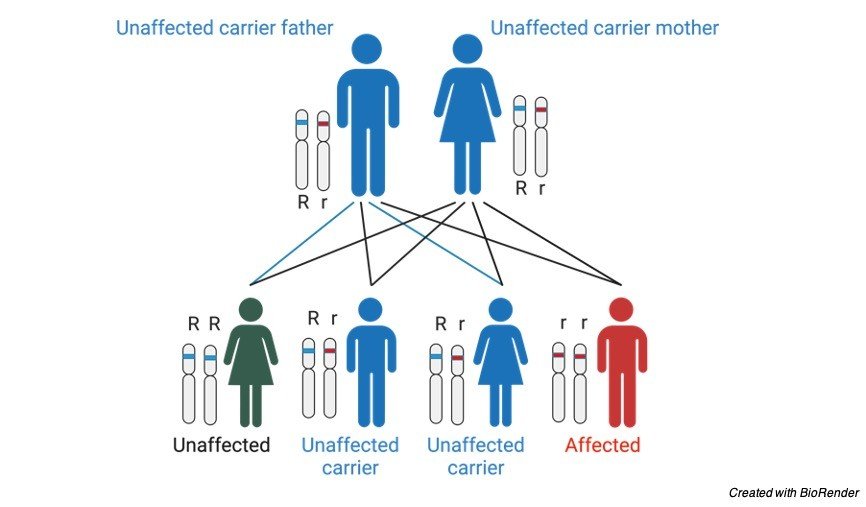
    Nuclear DNA has two duplicates for each cell (with the exception of sperm and egg cells), one duplicate is acquired from the dad and the other from the mother.

    Mitochondrial DNA, notwithstanding, is acquired from the mother just (for certain special cases) and every mitochondrion normally contains somewhere in the range of 2 and 10 mtDNA duplicates.

    During cell division the mitochondria isolate arbitrarily between the two new cells.

    Those mitochondria make more duplicates, typically arriving at 500 mitochondria per cell. As mtDNA is replicated when mitochondria multiply, they can collect irregular changes, a marvel called heteroplasmy.

    If by some stroke of good luck, a couple of the mtDNA duplicates acquired from the mother are deficient, the mitochondrial division may cause a large portion of the faulty duplicates to wind up in only one of the new mitochondria (for more nitty gritty legacy designs, see human mitochondrial hereditary qualities).

    The mitochondrial disease may turn out to be clinically evident once the quantity of influenced mitochondria arrives at a specific level; this wonder is designated “limit articulation”.

    Mitochondria have large numbers of a similar DNA fix pathways as cores do—however not every one of them; subsequently, transformations happen more often in mitochondrial DNA than in atomic DNA (see Mutation rate).

    This implies that mitochondrial DNA problems may happen suddenly and moderately frequently.

    Imperfections in proteins that control mitochondrial DNA replication (which are all encoded for by qualities in the atomic DNA) may likewise cause mitochondrial DNA transformations.

    Most mitochondrial capacity and biogenesis are constrained by nuclear DNA.

    Human mitochondrial DNA encodes 13 proteins of the respiratory chain, while a large portion of the assessed 1,500 proteins and parts designated to mitochondria are encoded.

    Deformities in atomic encoded mitochondrial qualities are related to many clinical disease aggregates including pallor, dementia, hypertension, lymphoma, retinopathy, seizures, and neurodevelopmental diseases.

    Mitochondrial Disease Citations

    Share

    Similar Post:

    Continue Reading
  • Trophic Level: Definition, Pyramid, and Examples

    What is Trophic Level?

    Trophic level is a taking care of positions in a web or evolved way of life of the environment or step in a nutritive series.

    Gathering of living beings characterized into these levels based on their taking care of conduct and for the most part sea-going environment have not in excess of seven trophic levels inside a natural way of life.

    These trophic levels can be shown by trophic pyramid and life forms are assembled concurring to the job they play in the food web.

    Types of Trophic Level

    The main level which is the essential makers frames the foundation of the trophic pyramid.

    Essential makers can make their own food or portrayed as autotrophic, and they convert energy from the sun into food energy through the interaction of photosynthesis.

    Instances of oceanic essential makers are phytoplankton and ocean growth.

    The second trophic levels of sea-going biological systems are the herbivorous buyers, for example, zooplankton and cockles that devour the essential makers.

    First level meat eating purchasers structure the third trophic level which incorporates adolescent phases of bigger creature, for example, fish and jellyfish just as little fish, scavangers and ocean stars.

    This third trophic level burn-through the zooplankton and cockles.

    Next is the fourth trophic level which is the second level flesh eating purchasers incorporate the bigger fish that burn-through the little fish, jellyfish, shellfish and ocean stars.

    Bigger fish eaten by the fifth trophic level, the third level of flesh eating buyers and the instances of this level is squid and octopus.

    Ultimately the 6th trophic level on the highest point of the trophic pyramid are shark, dolphin and gooney bird that is the top rapacious or summit hunters.

    In the oceanic biological system trophic levels, not all the top hunters live in the water.

    Decomposers, mostly microbes that separate dead living beings exist on each trophic level.

    They assume significant part during the time spent delivering supplements to support makers and shoppers that feed through retaining natural material in the water segment as they separate waste or dead tissue.

    Trophic Level: Where it Gets Food?

    First trophic level: primary makers. Makes its own food (autotrophic) phytoplankton, Ocean growth.

    Second trophic level: herbivorous consumers (primary) consumes makers zooplankton, cockles.

    trophic level - research tweet 1

    Third trophic level: first level predatory customers (auxiliary) consumes primary. Adolescent phases of fish and jellyfish, little fish, scavengers and ocean stars.

    Fourth trophic level: second level meat eating customers (tertiary buyers) consumes auxiliary. Bigger fish.

    Fifth trophic level: third level flesh eating purchasers (quaternary customers) consumes tertiary. Squid, octopus, bigger fish.

    Sixth trophic level: top meat eating (quinary customers) consumes quaternary. Shark, dolphin, gooney bird.

    How Ecological Productivity of Aquatic Environments Maintained?

    Ecological productivity characterized as the exchange of energy between trophic levels or at the end of the day is the productivity with which energy moved starting with one trophic level then onto the next trophic levels.

    There are two significant physical science laws in the investigation of energy course through the environment.

    First thermodynamics law expresses that energy can’t be made or annihilated and it can just changed starting with one structure then onto the next.

    Sun is the wellspring of energy that consumed by the essential makers where in it is changed over to put away substance energy for the working of an environment.

    The second law of thermodynamics expresses that at whatever point energy is changed, there is a misfortune energy through the arrival of warmth and loss of energy additionally happens during breath and development.

    Energy misfortune happens when energy is moved between trophic levels where more what’s more, more energy lost as one maneuver up through trophic levels when one creature takes care of off another creature.

    In oceanic environments, phytoplankton complete the majority of the photosynthesis that happens.

    The majority of phytoplankton essential creation is burn-through and utilized for energy on the grounds that phytoplankton are little creatures with straightforward designs.

    Even though phytoplankton are little yet, they become quickly and they support huge populaces of herbivores.

    This is the explanation why oceanic biological systems can uphold more trophic levels than the land environments.

    Oceanic environments normally have higher productivity in contrast with land biological systems in light of the fact that higher extent of ectotherms and makers like the green growth needs ligin.

    The primary makers with 1000 units energy moved 10% to 100 units energy for herbivorous. Constantly diminished of 10% energy to 10 units and 1 unit energy for first request carnivores and up to second request carnivores.

    The entirety of the purchasers as it climbs from one trophic level to another trophic level varies in its healthful relationship with the maker.

    Energy is left behind a food chain or web from lower to higher trophic levels changes from 5 to 20 percent, normal 10% of the energy at one level accessible to next trophic level.

    The other 90% of energy utilized for metabolic cycles or emitted as warmth to the climate.

    Trophic Level and Energy Balance

    Each trophic level loses energy so it frequently showed as a triangle with essential makers framing the base.

    The more trophic levels present, the less energy preserved at higher trophic levels.

    Each trophic level backings fewer living beings as the consequence of just normal 10% of the energy from a life forms moved to its shopper.

    For instance in the amphibian trophic level, a high level shopper like shark or fish is upheld by a huge number of essential makers, for example, phytoplankton from the foundation of trophic pyramid or the food web.

    Estimation of energy move effectiveness between two progressive trophic levels is named as the trophic level exchange effectiveness (TLTE) and is characterized by the equation.

    Trophic level Exchange Effectiveness

    Trophic level exchange effectiveness is a proportion of how much energy happens at one level partitioned by the energy at the level underneath it times hundred.

    Ecological effectiveness portrays proficiency with which energy moved starting with one trophic level then onto the next trophic levels and controlled by a blend of efficiencies identifying with organismic asset obtaining and osmosis in an environment.

    Trophic productivity fuses with three sorts of effectiveness.

    First is the extent of accessible energy that is burned-through known as utilization effectiveness.

    Second, the extent of ingested food that is absorbed or absorption effectiveness.

    Thirdly is the extent of acclimatized food that goes into new customer biomass known as creation productivity.

    Ecological productivity is the exchange of energy between trophic levels or the energy streams inside a food web.

    A lot of energy are lost starting with one trophic level then onto the next level as energy streams from essential makers to the different trophic levels of buyers and decomposers.

    Level of energy moved shifts from 5 to 20 percent, normal 10% from one trophic level to higher levels and the rest misfortune as a warmth.

    The main consideration that restricts the length in a food chain or food web is the low effectiveness of energy move between trophic levels.

    There isn’t sufficient energy to help another trophic level after four up to six energy moves.

    Sea-going biological systems can uphold more trophic levels contrast with the land environments due to higher productivity.

    Trophic Level Citations

    Share

    Similar Post:

    Continue Reading
  • Detritivore: Definition, Function, and Examples I Research...

    Detritivore Definition

    A detritivore is a heterotrophic living being, which gets its nourishment by benefiting from waste.

    Debris is the natural matter comprised of dead plant and creature material.

    Detritivores may likewise acquire nourishment by coprophagy, which is a taking care of technique including the utilization of dung.

    Detritivores are regularly invertebrate bugs like parasites, creepy crawlies, butterflies, and flies; mollusks like slugs and snails; or soil-abiding worms, millipedes and woodlice.

    Detritivore Examples

    Instances of detritivores in marine conditions are scavangers like crabs and lobsters, echinoderms, for example, ocean stars or ocean cucumbers.

    detritivore - research tweet 1

    A considerable lot of these marine detritivores involve a comparative specialty to earthbound soil-tenants, living on or inside the seabed known as the benthos. These creatures are frequently called “bottom dwellers”.

    Then again in amphibian biological systems, fixed polychaete worms, barnacles and a few corals determine their energy through channel benefiting from skimming natural rubbish called “marine snow”.

    Detritivore or Decomposer

    The expressions “detritivore” and “decomposer” are particular in their importance; albeit the words are frequently utilized conversely, detritivores are actually a part of decomposers.

    It is valuable to take note of that, not at all like detritivores, genuine decomposers like organisms, microorganisms or protists, use saprotrophic taking care of, in which they assimilate supplements through extracellular processing, as opposed to by oral ingestion.

    Likenesses can notwithstanding be drawn among detritivores and scroungers.

    However while both of these taking care of systems include the utilization of dead plant and creature matter, foragers will in general benefit from a bigger scope than detritivores, practicing on remains and excrement.

    Detritivore Features

    Detritivores and decomposers add to the breakdown of the entirety of the dead and rotting material in any biological system.

    In this manner they assume a significant part in the cycling of supplements and are a fundamental piece of most biogeochemical cycles, for example, the carbon cycle, nitrogen cycle and the phosphorus cycle.

    Detritivores feed on material from essential makers just as herbivores and carnivores, and in this way are available all through all trophic levels in a biological system.

    Just as burning-through energy from different life forms, they are generally eaten by optional buyers, thus they are a fundamental segment of the environment energy cycle.

    Not exclusively is the reusing interaction a significant piece of decay, the evacuation of dead material is critical to stop the spread of illness.

    Furthermore, detritivores that live inside the dirt, for example worms, circulate air through and stir up the dirt with their development, which is significant for the development of plants.

    Importance of Detritivore: Worms

    Worms are quite possibly the main soil-staying detritivores. They devour a lot of natural matter and soil and are available all through all layers of their substrate.

    Epigeic worms live on a superficial level, while endogeic worms live in the upper layer of soil.

    These two sorts of worm will in general benefit from dead grass, fallen leaves and other bioorganic materials like parasites, green growth and the microorganisms that are additionally benefiting from these substances.

    Anecic worms live profound inside the dirt, fundamentally comprising off crude soil, albeit this contains numerous microorganisms, parasites and green growth.

    Worms take in food through their mouths, and ‘suck’ it into the stomach related framework utilizing a strong activity. As the material goes through their stomach related framework into their ‘gizzard’, it is dependent upon stomach related compounds just as the pounding activity coming about because of the presence of coarseness and sand from the dirt.

    A ‘cast’ is dropped of the backside of the worm. The cast is fundamentally a more handled form of the admission soil, with more modest supplement particles accessible for decay by different creatures.

    The presence of the microorganisms inside the waste devoured by the worms additionally accelerates the disintegration interaction all through absorption. Due to their taking care of system, worms are continually moving.

    This development assists with stirring up and circulate air through the dirt, which additionally further develops water take-up and transportation of supplements, permitting plants to develop all the more effectively.

    Detritivore and Decomposition Cycle

    Garbage is comprised of Particulate Organic Material (POM) that is shaped from tissues kept when plants and creatures kick the bucket or when they shed skin or prongs through shedding, just as fecal matter and microorganisms.

    The provinces of microorganisms inside the garbage add to its healthy benefit.

    In earthbound conditions, debris might be available as humus (the dead materials blended in with soils), or as leaf litter.

    In sea-going conditions, the garbage is suspended as “marine snow”, which at last tumbles to the seabed. The entirety of this material contains energy, just as the supplements that were available in the waste material and the assortments of the perished.

    These substances are important items in all energy and supplement cycles, albeit the minerals and mixtures should be made accessible by actual breakdown and biochemical change of the material through disintegration or remineralisation.

    Detritivores feed on the bigger particulate material in the beginning phases of disintegration, and in doing as such, section the material in to more modest measured pieces.

    The discontinuity cycle expands the surface region accessible, for assault by microscopic organisms and different microorganisms, and subsequently helps and paces up the decay interaction.

    Absorption by the detritivores additionally separates a few sugars, proteins and lipids present in the garbage in to less difficult substances.

    The water-dissolvable supplements delivered from this drain into the dirt and increment the dirt mineral substance.

    Simultaneously, the detritivores are extricating nourishment for their own life cycles, and in doing as such, contribute their biomass to the natural way of life when they are eaten by buyers.

    Constantly all through the interaction, decomposers like organisms and different microorganisms, otherwise called saprophytes, perform genuine disintegration, utilizing synthetic mixtures and stomach related catalysts to change over material discharged by the detritivores into additional less complex substances like inorganic carbon.

    These cyclic substances are vital for all life, for instance, inorganic carbon delivered from breath of decomposers is taken in by plants and is utilized to perform photosynthesis.

    Progressive deterioration of the altered natural matter outcomes in humification – the development of humus – a sort of soil with a high mineral substance and dependability.

    Detritivore Citations

    Share

    Similar Post:

    Continue Reading
  • Stem Cells: Types, Therapy, Uses, and Function

    What Are Stem Cells?

    Stem cells are human cells that can form into a wide range of cell types. This can go from muscle cells to synapses. At times, they can likewise fix harmed tissues.

    Scientists accept that stem cell-based treatment may one day be utilized to treat genuine ailments, for example, loss of motion and Alzheimer’s disease.

    Stem cells are unspecialized cells that form into the specific cells that make up the various sorts of tissue in the human body. They are described by the capacity to recharge themselves through mitotic cell division and separating into an assorted reach of particular cell types.

    They are indispensable to the turn of events, development, support, and fix of our minds, bones, muscles, nerves, blood, skin, and different organs.

    Stem cells are found in every last one of us, from the beginning phases of human improvement to the furthest limit of life.

    Stem cell research holds enormous guarantees for the advancement of novel treatments for some genuine illnesses and wounds.

    While stem cell based medicines have been set up as a clinical norm of care for certain conditions.

    For example; hematopoietic stem cell transfers for leukaemia and epithelial stem cell-based medicines for consumes and corneal issues, the extent of potential stem cell-based treatments has extended as of late because of advances in stem cell research.

    It has been as of late that researchers have perceived stem cells well enough to think about the potential outcomes of developing them outside the body for significant stretches of time.

    With that development, thorough examinations can be directed, and the chance of controlling these cells in such a way that particular tissues can be developed is genuine.

    Type of Stem Cells

    Stem cells are isolated into 2 principal structures. They are undeveloped stem cells and grown-up stem cells.

    Stem Cells - research tweet 1

    I. Embryonic Stem Cells

    The Embryonic stem cells utilized in research today come from unused incipient organisms.

    These outcome from an in vitro treatment system. They are given to science.

    These early-stage stem cells are pluripotent.

    This implies that they can transform into more than one kind of cell.

    II. Adult Stem Cells

    There are two sorts of adult stem cells. One sort comes from completely created tissues like the mind, skin, and bone marrow.

    There are just little quantities of stem cells in these tissues.

    They are bound to produce just particular sorts of cells.

    For instance, a stem cell that comes from the liver will just make more liver cells.

    The subsequent kind is initiated pluripotent stem cells. These are grown-up stem cells that have been changed in a lab to be more similar to early-stage stem cells.

    Researchers originally revealed that human stem cells could be changed in this manner in 2006.

    Instigated pluripotent stem cells don’t appear to be not the same as undeveloped stem cells, yet researchers have not yet discovered one that can foster each sort of cell and tissue.

    Stem Cells in Medication

    The stem cells presently used to treat sickness are hematopoietic stem cells. These are the platelet shaping grown-up stem cells found in bone marrow.

    Each sort of platelet in the bone marrow begins as a stem cell.

    Stem cells are youthful cells that can make other platelets that develop and capacity depending on the situation. These cells are utilized in methods, for example, bone marrow transfers.

    These assistance individuals with disease make fresh blood cells after their own hematopoietic stem cells have been killed by radiation treatment and chemotherapy.

    They may likewise be utilized to treat individuals with conditions like Fanconi sickliness. This is a blood problem that makes the body’s bone marrow fizzle.

    Stem cells may help your wellbeing in the future from multiple points of view and through numerous new medicines. Specialists imagine that stem cells will be utilized to assist with making new tissue.

    For instance, one day medical care suppliers might have the option to treat individuals with persistent coronary illness. They can do this by developing sound heart muscle cells in a lab and relocating them into harmed hearts.

    Different medicines could target sicknesses, for example, type 1 diabetes, spinal rope wounds, Alzheimer infection, and rheumatoid joint inflammation.

    New medications could likewise be tried on cells produced using pluripotent stem cells.

    Potential Stem Cells Therapy

    Various stem cell therapeutics exist, yet most are at trial stages or potentially expensive, with the exceptional case of bone marrow transplantation.

    Clinical analysts expect that grown-up and early-stage stem cells can soon treat cancer, Type 1 diabetes mellitus, Parkinson’s sickness, Huntington’s infection, Celiac Disease, cardiovascular disappointment, muscle harm and neurological problems, and numerous others.

    They have proposed that before stem cell therapeutics can be applied in the clinical setting, more exploration is important to comprehend stem cell conduct upon transplantation just as the systems of stem cell association with the ailing/harmed microenvironment.

    Bone marrow transfers (BMT) are a notable clinical utilization of stem cell transplantation.

    BMT can repopulate the marrow and reestablish all the distinctive cell kinds of the blood after high dosages of chemotherapy and additionally radiotherapy, our primary guard used to kill endogenous cancer cells.

    Stem Cells Therapy in Diabetes

    Diabetes influences a huge number of individuals in the world and is brought about by the unusual digestion of insulin.

    Ordinarily, insulin is created and emitted by the cellular structures called the islets of Langerhans in the pancreas. As of late, insulin communicating cells from mouse stem cells have been created.

    What’s more, the cells self-gather to frame structures, which intently look like ordinary pancreatic islets and produce insulin.

    Future examination should explore how to upgrade conditions for insulin creation determined to give a stem cell-based treatment to get diabetes swap the consistent requirement for insulin infusions.

    Why Stem Cell Research is Difficult?

    Stem cells need substantially more examination before their utilization can be extended.

    Researchers should initially study how to create early-stage stem cells. This will assist them with seeing how to control the sort of cells made from them.

    Another test is that the early-stage stem cells accessible today are probably going to be dismissed by the body.

    What’s more, a few groups discover it ethically disturbing to utilize stem cells that come from undeveloped organisms.

    Researchers additionally face difficulties when utilizing grown-up pluripotent stem cells.

    These cells are difficult to fill in a lab, so scientists are investigating approaches to work on the interaction.

    These cells are likewise found in modest quantities in the body. 

    Stem Cells Citations

    Share

    Similar Post:

    Continue Reading
  • Phenylketonuria: Types, Symptoms, Causes, and Treatment

    What is Phenylketonuria

    Phenylketonuria is an inborn error of phenylalanine metabolism which is due to the decreased metabolism of the amino acid.

    If left untreated the conditions may lead to seizures, intellectual disability, mental problems and behavioral disorders.

    It also leads to lighter and musty smell skin.

    A baby which was born to a mother who is heterozygous carrier of this disease also gets affected with small head and low weight at birth.

    Cause of Phenylketonuria

    Phenylketonuria is an inherited genetic disorder due to a mutation in a PAH gene which causes low levels of the secretion of an enzyme phenylalanine hydrolase.

    This is generally an autosomal recessive condition where both the copies of gene have to be defected to acquire this condition.

    Types of Phenylketonuria

    There are two types of phenylketonuria namely classic PKU and variant PKU depending upon the function of the enzyme.

    However, the individual with only one copy of mutant gene do not have any symptoms. In many countries the screening test is done to the new born to protect them from severe symptoms.

    It affects 1 in 10,000 people every year. However early screening can help to start the treatments quickly and helps in avoiding death of an individual in early ages.

    Symptoms of Phenylketonuria

    Phenylketonuria causes mild to severe ranges. The severe form of phenylalanine is known as classic variant.

    If the baby is suffering from this classic variant it shows only mild symptoms and does show severe symptoms. 

    If it left untreated it leads to severe symptoms such as seizures, tumors, trembling and shaking of the limbs, stunted growth, hyperactivity, and also eczema in the skin and musty like odor in skin, urine and even in breath of an affected individual.

    If Phenylketonuria is not diagnosed at an early stage it can also leads to irreversible brain damage and disabilities of intellectual abilities and also other behavioral problems in the adult children.

    The other type of Phenylketonuria is also known as variant Phenylketonuria which does not cause any severe symptoms or Non Phenylketonuria hyperphenylalaninemia.

    This occurs mostly in babies who has too much phenylalanine but they cause only mild symptoms in infants.

    On following a proper diet from their Early stages Phenylketonuria can be prevented from becoming severe and also can be diminished.

    Pedigree Analysis of Phenylketonuria

    As we know this condition is caused due to an autosomal recessive condition which means that where the parents are in a heterozygous condition of being a carrier, it affects the child.

    The graph for this is listed below.

    Phenylketonuria - research tweet 1

    Frequency of Getting Phenylketonuria

    The frequency of obtaining this disease ranges according to the geographical regions. Phenylketonuria occurs 1 in 10,000 to 15,000 babies; who are new born.

    They are mostly detected at an early stage with the help of screening tests and treatments are started early, so the severity of this diseases can be prevented. So that the classic Phenylketonuria can be prevented from obtaining and variant form of this disease can be diminished.

    Cause of Phenylketonuria

    The genetic mutation that is happening in the PAH gene results in phenylketonuria. As PAH gene only instructs the body cells to synthesise an enzyme known as phenylketonuria; which helps in the conversion of amino acid phenylalanine to form other vital components that are needed for the body.

    Due to this gene mutated condition, the activity of phenylalanine hydrolase is reduced which effects the activity of processing phenylalanine in diets.

    This leads to a condition of building toxic levels in blood cells and other tissues of the body.

    This condition occurs because the nerve cells in the brain are sensitive particularly to the phenylalanine levels where excessive production can cause damage to the brain cells.

    Classic PKU happens when a phenylalanine activity is reduced to the extent levels or it may be absent at most severe cases.

    People suffering from this condition if left untreated it causes severe brain damage and other serious health problems.

    Mutation in this gene retains the activity of some enzymes and results in milder variant PKU.

    Phenylketonuria Diagnosis

    Screening test is one of the most popular test. It is performed in babies whose parents have this condition of inheritance.

    Through this test it can be identified that whether the gene is inherited from parents to their children and if found in early stages it can be treated and cured before its s becoming severe.

    These tests are done after 6 weeks of birth of a child.

    Treatment of Phenylketonuria

    The babies found out with this type of disorders can be provided or treated with proper nutritious diet which limits the intake of phenylalanine and they are led to breast fed to get the essential nutrients.

    Physicians also provide these kinds of children a special formula of diet known as Lofenalac which is very high in protein.

    Phenylketonuria Citations

    Share

    Similar Post:

    Continue Reading
  • Sickle Cell Anemia: Types, Symptoms, Causes, and...

    What is Sickle Cell Anemia

    We come across many disorders in our day-to-day life, some are acquired and some are transmitted through genes from our ancestors, which is known as genetic disorder.

    Sickle cell anemia is one such inherited disease which is caused through genetic disorder or through mutations occurring due to environmental factors, which leads to a defect in a DNA molecule.

    Sickle cell anemia - research tweet 2

    Cause of Sickle Cell Anemia

    It is one of the abnormal inherited disorder caused by mutation in an autosomal gene.

    Here the red blood cells of an affected individual is of sickle shaped instead of having a biconcave shape. Where these sickle shaped blood cells do not provide a capacity to carry enough oxygen and nutrients all over the body which results in anemia as well as other diseases.

    The people with this type of red blood cells are called as sickle cell traits.

    The sickle shaped cells cannot pass through the blood vessels easily as they gets struck in between the small blood vessels such as capillaries which results in haemolytic anemia and leads to death of an individual.

    This is caused due to the mutation in the single-base, the sixth amino acid in the chain is valine, rather than glutamic acid

    Characteristics of Sickle Cell Anemia

    Linus Pauling was the first one to identify this type of mutation in an individual where the genotype for this mutation is Hbs1 Hbs1which is a condition that our body does not have enough haemoglobin, because Hbs1 have negative charge compared to that of HbA1.

    If the individual is a carrier of this syndrome, they have their genotype as HbA1 Hbs1 which leads to a condition of mild sickle shaped anemia.

    This is considered by Pauling as one of the gene molecular disease.

    Effects of Sickle Cell Anemia

    The sickle cell anemia leads to the increased fragility of the red blood cell which leads to haemolytic anemia and it also increases the viscosity of the blood and it also lets the blood cells to stag in the small blood vessels which forms a thrombus and infracts.

    This phenomenon also causes damage to the spleen which is often said to be autopsleenectomy where the spleen ends as a non-functional unit and it also leads to other side effects such as pneumococcal meningitis and other infectious diseases.

    Sickle Cell Anemia Epidemiology

    The Sickle cell anemia is most common in central Africa where almost 20 percent of the people are having this trait and are affected with this disease.

    About 2% of these die in an early stage of their life.

    It has also been distributed widely in USA where this trait Hbs has found in Arab racial groups and it is also rarely seen among India and American Negros.

    How Sickle Cell Anemia Affects?

    The people who are having sickle cell trait as Sis/Sis are affected by this disease but they don’t show symptoms in certain cases.

    But in some cases, in high altitudes, it affects the respiratory tract and bleeding in the renal papillae.

    The persons who are having homozygous trait of sickle cell anemia do not get infected by malaria. But they die at the early stages of life.

    The normal trait persons are not resistant to malaria. The persons who have the heterozygous condition of causing sickle cell anemia is also resistant to malarial infections and they lead almost a normal life style and anemia is not severe in such cases.

    Types of Sickle Cell Anemia

    There are many types of sickle cell anemia depending upon the various changes in the traits.

    Some of the types are discussed as follows;

    HbSS: In this case the person gets two inherited genes from each of their parents This type is most severe.

    HBSC: Here the defected gene is inherited from a single parent and a normal gene from the other parent it also results in abnormal haemoglobin and results in a carrier offspring but it does not cause any serious symptoms.

    HbS- Beta thalassemia: A individual suffering from this disorder get one of allele from an infected sickle cell parent and the other from the Beta thalassemia affected person.

    Symptoms of Sickle Cell Anemia

    In general, if our body does not get any enough oxygen it causes many complications and symptoms as the outcome of an infection.

    These symptoms vary according to the age and life style factors of the individual. It causes early symptoms such as fatigue, jaundice; which leads to yellowing of the skin and eyes, swelling of hands and feet; which causes severe pain.

    Later the symptoms are little complicated such as acute chest syndrome, damage in a normal vision, enlarging of spleen, ulcers in the leg, Heart attack, stroke, deep thrombosis in the veins, infections in the heart, kidney or liver.

    Formation of gallstones, infertility in males and causes of malnutrition in kids, high blood pressure in the lungs which is also known as Pulmonary hypertension, bone and joint inflammation etc.

    Treatment of Sickle Cell Anemia

    Currently the sickle anemia has no cure but it is possible to overcome the difficulty of this syndrome through blood transfusions or through stem cell transplants the disease can be controlled.

    Sickle Cell Anemia Citations

    Share

    Similar Post:

    Continue Reading
  • Chromosomal Instability: Definition, Types, and Examples

    What is Chromosomal Disorder or Chromosomal Instability?

    Many types of disorders are arising in humans, each are due to various reasons such as genetic, acquired or through mutated conditions.

    These mutated conditions are mostly due to any abnormality or defect in the chromosome of an individual.

    Humans usually have 23 pairs of chromosomes in that 22 pairs are autosomes and the last pair is known as allosome or sex chromosome.

    Chromosomal disorders occurs if there is any addition of chromosomes to the particular number or reduction in this particular set of numbers.

    Any change in the number of chromosomes is known as ploidy. And this disorder occurs when there is any change in the structure of the chromosome.

    Why Chromosomal Disorder or Chromosomal Instability?

    Chromosomal disorders are due to any abnormality or change in structure or function of a chromosome.

    A change in a structure of any one of the chromosomes or missing of a part of a chromosome or a whole chromosome or mutation or any defects in a particular gene in a chromosome may be due to chromosomal disorders.

    These disorders are detected with the help of banding patterns which helps us to identify the abnormalities in the chromosome by staining the chromosomes with colour stains.

    Karyotyping also helps us to detect the missing pair of chromosomes. In karyotyping the chromosomes are arranged in a definite manner and seen under a light microscope.

    Idiogram also helps in karyographing of chromosomes. Any small change in the chromosome can lead to vigorous effects in the genes and affect the enzyme producing capacity; which leads to various diseases.

    These chromosomal abnormality happening in an allosomes are definitely passed to the next generations, where as the abnormalities of autosomes may or may not pass to the next generation.

    Types of Chromosomal Instability

    Generally, there are two types of chromosomal instability which leads to chromosomal disorders.

    1. Structural Chromosomal Instability

    2. Numerical Chromosomal Instability

    1. Structural Chromosomal Instability

    The structural chromosomal instability are due to a change in structure of a chromosome or missing of any part of the chromosome.

    As said, there are about 23 pair of chromosomes which are 46 in number.

    It also occurs when there is a change in structure or missing of any part of the chromosome or when the missing parts are translocated to any other areas.

    This condition causes many birth effects due to addition or deletion of a part of a chromosome.

    Chromosomal Instability - research tweet 2

    All chromosomes are made up of many segments which are commonly divided into short and long arm, where the short arm forms upper half of the chromosome and the long arm forms a lower part of the chromosome.

    The short arm is known as “p” arm and the long arm is known as “q” arm.

    i. Chromosomal Instability: Deletion of Chromosome

    In deletion of chromosomes the part of a chromosome is deleted or went missing.

    Even a small piece of a missing chromosome can lead to vigorous disorders or misleading of gene encoding, as a small part contains various information for coding different genes and enzymes.

    When these conditions happen in a pregnant mother this condition is being inherited to an offspring.

    The best example for deletion is Cri-du-chat syndrome where there is the damage or missing of chromosome number five.

    Cri-du-chat Syndrome

    Cri-du-chat syndrome is usually seen among one in 20,0000 people. It occurs mostly in the children of America.

    It is caused due to a defect or deletion in the chromosome number five. Which occur especially in the p arm and it is said as 5p.

    The characteristic feature of this syndrome is that the children’s have a high pitch cry, poor tone of muscles and the heads of these children are very small.

    They are usually underweight. They also face the problem while speaking, they express their emotions with the help of signs.

    Other related health problems are with feeding, hyperactivity of an individual, and disability in their intellectual activities.

    Mostly the people affected with this Cri-du-chat syndrome have a normal life span in most of the cases.

    They result in series condition only when they have organ defects along with this syndrome, which leads to short life span of an individual.

    The children with this type of syndrome are involved in language and physical therapy to improve themselves.

    ii. Chromosomal Instability: Duplication of Chromosome

    The term itself provides a complete meaning for this syndrome, here the duplication of chromosomes occurs where the same chromosome forms a copy of the other, which results in two copies of the same chromosome, as a result two copies of the same genetic material are formed.

    Even if the genes are present in many copies it leads to non-functioning of the chromosomes.

    Or by giving extra instruction to the same enzyme production which leads to errors in developing a baby.

    This duplication leads to a Pallister-Killian syndrome.

    Pallister-Killian Syndrome

    In this syndrome the extra copy of chromosome 12 is present. Which leads to mosaicism.

    Infants with this condition has many serious problems such as intellectual disability, poor muscle tone, poor feeding, stiffness of joints, cataracts in eye.

    Usually, a people in this syndrome have a very short life span. These people have only their life span till 40 years.

    iii. Chromosomal Instability: Inversion of Chromosome

    This occurs when the part of the chromosome results in breakage and reuniting by combining by rotating at 180º with its own axis.

    This results in rearrangement of genes. But it is not severe as much as other structural abnormalities.

    iv. Chromosomal Instability: Translocation of Chromosome

    The transfer of chromosomes with a non-homologous pair is known as translocation. The addition or deletion or rearrangement of genes may occur in this condition.

    Which leads to a phenotypic change resulting in abnormalities in the development of egg, sperm or zygote. It often results in miscarriage of a pregnant women.

    2. Numerical Chromosomal Instability

    Any abnormalities in the chromosomal number leads to this condition. It is divided into two types as aneuploidy and euploidy

    i. Chromosomal Instability: Aneuploidy

    It is a condition where loss or gain of a chromosome occurs. It mostly occurs due to a non-disjunction in a chromosome, during cell division. Aneuploidy is further classified as Trisomy (2n+1) and monosomy (2n-1).

    The disorders caused due to aneuploidy is Turners syndrome, Klinefelter’s syndrome and Downs syndrome.

    ii. Chromosomal Instability: Euploidy

    Here the loss or gain of whole set of the chromosomes occurs. This condition mostly occurs in plants.

    Haploid: In this condition loss of one set of the chromosomes occurs.

    Polyploid: In this condition the loss of one or more sets occurs.

    Chromosomal Instability Citations

    Share

    Similar Post:

    Continue Reading
  • Pedigree Analysis: Definition, Pedigree Chart, and Examples

    About Pedigree Analysis

    It is easy to represent a thing in a diagram manner than in terms of theory, which also helps in easy understanding.

    Pedigree analysis is used to represent traits of an organism or an individual so that we can identify the genotypes and phenotypes of the future generation.

    It also helps us to predict the characteristics of future generation by assuming with the present generation characteristics, which is very helpful for identifying the genetic diseases and thus we can provide counseling for the parents in such condition which helps them to take a better decision.

    Pedigree Chart

    In human beings-controlled crosses cannot be made, so genetists discovered the method for undergoing crosses to find an individual with established mating’s.

    The information that has obtained by established mating’s is by a chance or a hope. This method of identifying the possibility is referred to as pedigree analysis.

    The individual in a family who comes first in attention of a genitists is known as propositus.

    Generally, the propositus phenotype sometimes causes exceptional, where dwarf character appears in such cases the investigator traces the history of a character and analyses the change by tracing the family tree or pedigree chart.

    Pedigree chart is drawn by using certain symbols. A pedigree helps us to represent the traits and it also gives an understanding of genotypes and phenotypes that will be passing to through the inheritance.

    It also relates the relationship between the parents, offspring and their siblings.

    It also helps in better understanding of the inheritance within the family of a particular individuals.

    How to Read a Pedigree

    Before knowing how to relate the traits in pedigree it is important to know the specific terms. And few of them are listed below

    Thus, these symbols are commonly used to indicate the traits in a pedigree.

    Pedigree Analysis - research tweet 2

    A family pedigree chart usually has circles to indicate the female individuals, squares to indicate the male individuals and perpendicular line drawn to the marriage bar indicates the off springs.

    The symbols which are present on the same line are considered as they are from the same generation and the generations are denoted by roman numerical as I, II etc.

    The unshaded symbols shows that they are unaffected individuals and the shaded symbols shows they are partially or fully affected depending upon their shaded area, the arrow indicates the specific person who is carrying the defective trait throughout his generations.

    Considering an example of polydactyl, which means that some individuals have extra fingers in their limbs.

    When the pedigree illustration is done for this, a marriage has been done between polydactyl man and a normal woman they reproduce three children’s where the first generation has a polydactyl daughter and a son and the other normal son.

    In second generation when these polydactyl individuals marry a normal person their reproductive off springs are shown in the graph below.

    Pedigree Analysis - research tweet

    However, from this polydactyl pedigree chart it is clear that polydactyl condition is playing a dominant role on recessive i.e., normal trait.

    Determination of Particular Trait

    If the trait is dominant then it will be sure that one of the traits will be obtained from the parents because dominant genes don’t skip the generation.

    However, neither trait will be obtained from the parents if it is heterozygous.

    Determination of Autosomal and Sex-linked Trait

    Males are usually most affected than females in a sex-linked recessive inheritance. Where as in autosomal recessive conditions male send female are equally affected.

    The other terms to be taken into consideration while reading a pedigree chart is;

    Genotype: Genetic characteristics of an organism., where TT denotes homozygous tall.

    Phenotype: Phenotype indicates the physical characters of an individual., where Tt heterozygous tall also considered as a tall.

    Dominant Allele: An allele which is phenotypically dominant over the other allele.

    Recessive Allele: This allele is expressed only in cases where the dominant alleles are absent or unable to express.

    How to Read a Pedigree Chart?

    Mendelian recessive alleles are very helpful in determining the human disease and other exceptional condition.

    The pedigree chart can be read easily with above sorted clues. When the diseased condition appears in the progeny of the unaffected parents.

    Also, at the same time the affected parents cannot have the unaffected individuals these chances are more when recessive alleles are revealed by consanguineous marriages where the best example is cousin marriages.

    This also suits the conditions where the rate of mattings of heterozygotes are expected to be rare in cases of albinism, Tay- Sachs disease, cystic fibrosis and phenylketonuria.

    There are also some conditions which are not caused by dominant alleles. This condition often occurs in every generations, where unaffected individual transmit the condition to their offspring in case if they are a carrier.

    In other case when two parents are affected, they are also chancing of being a child to be unaffected where they get their trait from their grandparents and these unaffected individuals passes a diseased condition to their children’s.

    Examples of abnormal conditions caused by dominant alleles are Huntington’s chorea and brachydactyly which means these individuals have very short fingers.

    This pedigree analysis also helps in identifying the phenotypical and genotypical traits in plants as well as in insects as in humans to identify the inheritance passed to the upcoming generations.

    Pedigree Analysis Citations

    Share

    Similar Post:

    Continue Reading