Category: Study Materials
-

DNA Replication: Steps, Process, and Mechanism I...
Continue ReadingWhat is DNA Replication?
In atomic science, DNA replication is the organic cycle of delivering two indistinguishable replicas of DNA from one unique DNA particle.
DNA replication happens in all living beings going about as the most fundamental part for natural legacy.
It is perused in the 3 ‘to 5’ direction by the DNA polymerase, which implies that the subsequent strand is combined in the 5 ‘to 3’ direction.
Replication includes the creation of indistinguishable DNA helices from a double stranded DNA molecule.
Catalysts are basic to DNA replication as they promote vital strides in this cycle.
The whole DNA replication measure is critical for both cell development and growth in organisms. It’s likewise significant in cell fix.
Beginning of DNA Replication
Replication consistently starts at a particular point on the DNA, which is known as the origin of replication and is perceived by its sequence.

Steps of DNA Replication
A) Initiation: Preparatory step
• Step 1: Replication fork formation.
B) Elongation: DNA Synthesis Begins
• Step 2: Primer binding
• Step 3: Synthesis of leading and lagging strands
• Step 4: Remove primer and gap fill
• Step 5: Proofreading
C) Termination:
• Step 6: End of the replication
Step 1: Replication Fork Formation
Before DNA can be duplicated, the double stranded molecule should be “unzipped” into two solo strands.
DNA has four bases called adenine (A), thymine (T), cytosine (C), and guanine (G) that form pair between the two strands. Adenine just combines with thymine and cytosine just ties to guanine.
To loosen up DNA, these base-pair interactions should be broken. This is finished by a protein known as DNA helicase.
Notwithstanding, there is a unique initiator protein that is needed to sets off DNA replication, to be specific DnaA.
It ties areas at the oriC site all through the cell cycle.
To start the replication, notwithstanding, the DnaA protein should tie to a couple of explicit oriC groupings that have five repeats of the 9 bp arrangement also known as the R site.
At the point when DnaA ties to the oriC site, it enlists a helicase catalyst (DnaB helicase). Presently the DNA helicase breaks the hydrogen bond that holds reciprocal DNA bases together.
The detachment of the two single strands of DNA makes a two Y-formed design called a replication fork.
Together they structure a bubble-like design called a replication bubble. These two separate strands fill in as a layout for the creation of the new DNA strands.
Helicase is the main replication compound to be stacked at the beginning of replication. Helicase’s responsibility is to just move the replication forks forward by “unwinding” the DNA.
As we probably are aware, DNA is entirely unstable as a single strand. Along these lines, cells can keep them from returning together in a double helix.
To do this, a particular protein called single-stranded DNA binding proteins (SSBs) covers and keeps the isolated strands of DNA close to the replication fork.
When the helicase rapidly unwinds the double helix. It raises the tension on the remaining DNA particle.
Topoisomerase plays a significant support part during DNA replication. This protein forestalls the DNA double helix in front of the replication fork from turning out to be too tight when the DNA is opened.
It does this by making impermanent nicks in the helix to release tension and afterward fixing the nicks to forestall perpetual harm.
Elongation of DNA Replication: DNA Synthesis Begins
Step 2: Primer binding
Another enzyme was presented in this progression, which assumes the main part in the fabrication of DNA, called as DNA polymerase.
It can just add nucleotides at the 3 ‘end of a current DNA strand. Primase forms the RNA primer, or short nucleic acid strand, that finishes the format, providing a 3 ‘end for working on DNA polymerase.
A commonplace primer has around five to ten nucleotides. The primer starts the synthesis of DNA. When the RNA primer is set up, DNA polymerase “extends” it and consecutively adds nucleotides to make another strand of DNA that is complementary to the template strand.

Step 3: Synthesis of Leading and Lagging Strands
One of the strands is arranged in the 3′ to 5′ bearing (towards the replication fork), this is the leading strand.
The other strand is situated in the 5′ to 3’direction (away from the replication fork), this is the trailing strand. Due to their diverse direction.
Leading Strand Synthesis
A short piece of RNA called a primer (made by enzyme known as primase) comes by and attach to the terminal of the leading strand.
The primer fills in as the beginning stage for DNA synthesis.
The DNA polymerase ties to the leading strand and afterward strolls alongside it, adding new complementary nucleotide bases (A, C, G, and T) in the 5′ to 3′ direction to the DNA strand.
This sort of replication is known as continuous.
Lagging Strand Synthesis
The DNA polymerase consistently runs in the 5 ‘to 3’ direction. At the point when the two strands have been incorporated consistently while the replication fork is moving.
One strand would consequently must be exposed to a 3 to 5 synthesis. Okazaki tracked down that one of the new strands of DNA was integrated in short pieces known as Okazaki fragments.
This work eventually prompted to concluded that one strand is synthesized persistently and others irregularly.
DNA polymerase III uses one bunch of its core subunits (the core polymerase) to continuously synthesize the leading strand. While the other two sets of the core subunit lie starting with one Okazaki fragment then onto the next on the looped duct.
In vitro, there are just two arrangements of core subunits containing DNA polymerase III holoenzymes that can combine both the leading and lagging strand.
Nonetheless, the third arrangement of core subunits expands the productivity of delayed strand synthesis just as the processivity of the in general replisome.
At the point when DnaB helicase ties before DNA polymerase III. It starts by unwinding the DNA on the replication fork as it moves alongside the trailing strand template in the 5 ‘to 3’ direction.
Primase every so often connects with DnaB helicase and synthesize a short RNA primer.
Another slide clamp is currently situated on the primer through the clamp loading complex of DNA polymerase III. At the point when the union of the Okazaki fragment is complete.
Replication stops and the core subunits of DNA polymerase III separate from their slide clamps and associate with the new clamp.
This starts the synthesis of another Okazaki fragment. Two sets of core subunits can be engaged with the union of two unique Okazaki fragments simultaneously.
When an Okazaki piece is complete, its RNA primers are taken out by DNA polymerase I or RNase H1. What’s more, that space is supplanted by DNA by the polymerase. The leftover nick has now been fixed by the DNA ligase.
Step 4: Remove Primer and Gap Fill
When both the continuous and discontinuous strands are framed, an enzyme called an exonuclease (DNA polymerase I or RNase H1) eliminates all RNA primers from the first strands.
These primers were supplanted by appropriate DNA bases. The excess nick was fixed by the DNA ligase.
DNA ligase catalyzes the development of a phosphodiester bond between a 3′-hydroxyl group on the end of one strand of DNA and a 5′-phosphate on the end of another strand.
Step 5: Proofreading
The DNA replication happens with high constancy. It contains some unacceptable nucleotide once for each 104–105 polymerized nucleotides.
The exactness of replication relies upon the capacity of replicative DNA polymerases to choose the right nucleotide for the polymerization reaction.
This high devotion isn’t accomplished in a single step yet rather is produced through the activity of a few progressive error avoidance and processing steps.
These steps incorporate the selection of the right DNA base by the DNA polymerase, the editing of polymerase miss addition mistakes by exonucleolytic proofreading, lastly, the post-replicative DNA crisscross repair, which recognizes DNA mismatch and corrects recently replicated DNA.
Termination of DNA Replication
DNA replication stops when two forks of replication meet on a similar stretch of DNA, and the accompanying occasions happen, however not really in a specific order: forks converge until the entirety of the mediating DNA is loosened up; remaining gaps are filled and tied; Catenans are removed, and replication proteins are discharged.
Step 6: End of DNA Replication
At last, the parent strand and its complementary DNA strand coils into the recognizable double helix shape. The outcome is two DNA atoms comprising of one new and one old chain of nucleotides.
Every one of these two little daughter helices is an almost precise of the parental helix.
DNA Replication Citations
- Eukaryotic chromosome DNA replication: where, when, and how? Annu Rev Biochem . 2010;79:89-130.
- Exploiting DNA Replication Stress for Cancer Treatment. Cancer Res . 2019 Apr 15;79(8):1730-1739.
- DNA replication origins-where do we begin?Genes Dev . 2016 Aug 1;30(15):1683-97.
- Adenovirus DNA replication. Cold Spring Harb Perspect Biol . 2013 Mar 1;5(3):a013003.
- Mechanisms of DNA replication termination. Nat Rev Mol Cell Biol . 2017 Aug;18(8):507-516.
- Origins of DNA replication. PLoS Genet . 2019 Sep 12;15(9):e1008320.
Share
Similar Post:
-

Homozygous vs Heterozygous: Definition and Differences
Continue ReadingHomozygous vs Heterozygous
Features of Homozygous
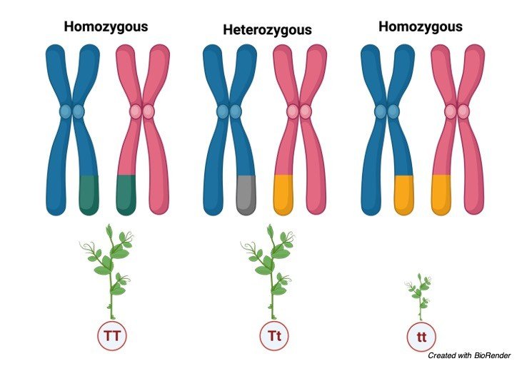
Homozygous refers to the state of the genes or genetic condition in which an individual has inherited the similar DNA sequence for a particular gene from both their biological parents and it is generally used with reference to the disease.
For instance, if an individual inherited a mutated allele in the DNA sequence from maternal side and similar gene from the paternal part as well, then the mutation is explained as homozygous for that individual.
Thus, after receiving 2 identical copies of the gene, detrimental effects will express phenotypically for which the genes are coded.
The term “heterozygous” attribute to a pair of alleles so heterozygous is when you have 2 dissimilar alleles that means you have received dissimilar copies from each parent.
In a heterozygous genotype, the allele which is dominant dominates over recessive and hence the dominant feature will be expressed while the recessive will not show but you will be still a carrier.
This will be passed it on to your children. Homozygous is the opposite of the heterozygous where the feature of the similar alleles — either dominant or recessive — is manifested.
There are three possible alleles for A, B, O blood type. However, the contribution from each parent’s gene makes a difference an individual exhibit with phenotypic characteristic.
Sometimes, it may be possible that both the parents pass on the same allele for a gene, making it a homozygous trait.
While in certain conditions, different set of alleles are triggered resulting in heterozygous trait.
A homozygous trait is mainly responsible for phenotypic appearance in the organism. A pea plant will definitely exhibit long stem, if it receives two alleles associated with tallness. In contrast, if it gets two “short” alleles, then plant with stunted growth will be evident.
Thus, for a specific characteristic, the genotype may be either homozygous or heterozygous, however, the way it is expressed it is identified as phenotype.
Features of Heterozygous
Heterozygous is a genetic condition where an individual inherits different alleles of a gene from the two parents.
Heterozygosity is observed in a diploid organism where a gene contains two different alleles at a gene locus.
In heterozygous chromosomes, the two alleles are different, and the heterozygous genotypes are represented by a capital letter, which indicates the dominant allele and a lowercase letter, which indicates the recessive allele, like Bb for eye color.
In heterozygous chromosomes with genes having traits that are expressed via complete dominance, only the trait coded by the dominant allele is expressed.
In complex dominance schemes, however, the expression of genes is more complicated. In incomplete dominance, the phenotypic trait observed is somewhere between dominant and recessive phenotypes.
Similarly, in co-dominance, the phenotypes are expressed by individual alleles in different parts of the body.
The heterozygous genotype has relatively higher fitness than the homozygous-dominant or homozygous-recessive genotypes. This fitness is termed as ‘hybrid vigour’.
Organisms reproducing by sexual breeding mechanisms are usually heterozygous for the traits that are varied.
Methods of sexual reproduction result in heterozygous chromosome formation. This ensures that the phenotypic characteristic in the offsprings is different from that in the parents.
Mostly, heterozygosity is defined at the specific locus where copies of the gene affecting a trait present on the two reciprocal homologous chromosomes are different.
Heterozygous cells or organisms are also called heterozygotes. As with homozygous genotypes, some heterozygous genotypes are also often associated with genetic conditions.
If the mutated allele is dominant, only the mutated copy is capable of causing the disease. This condition is called dominant-disease.
If the mutated allele is recessive, then the disease will not appear, and the organism will act as a carrier.
Diseases like Huntington’s disease, Marfan’s syndrome, and familial hypercholesterolemia are associated with heterozygous genotypes.
Homozygous vs Heterozygous
Comparison Homozygous Heterozygous Definition Homozygous is a hereditary condition where an individual acquires similar alleles of a quality from both the parents. Heterozygous is a hereditary condition where an individual acquires various alleles of a quality from the two guardians. Genotype representation Homozygous genotypes are addressed as AA or aa for homozygous-prevailing or homozygous-latent conditions, respectively. Heterozygous genotypes are addressed by Aa genotypes. Phenotypes Two various aggregates are conceivable with prevailing or latent homozygous conditions. The aggregate is for the most part because of the predominant allele in the heterozygous condition. Gametes Homozygous genotypes bring about a solitary sort of gamete. Heterozygous genotypes bring about two distinct kinds of gametes. Traits Homozygous genotypes produce similar qualities over various generations. Heterozygous genotypes produce various characteristics over various ages. Half and half vigour The homozygous condition doesn’t show mixture vigour. Heterozygous condition shows cross breed energy. Types Homozygous-prevailing and homozygous-passive are two kinds of homozygous conditions. The heterozygous condition can be communicated in three unique manners; co-strength, deficient predominance, and complete predominance. Additionally called Organisms or cells with the homozygous condition are named as homozygotes. Organisms or cells with the heterozygous condition are named as heterozygotes. Noticed in Homozygous genotypes are seen in creatures repeating by agamic means. Heterozygous genotypes are generally found in creatures duplicating by sexual means. Diseases Common illnesses related with the homozygous condition incorporate fibrosis, sickle cell frailty, and phenylketonuria. Common sicknesses related with the heterozygous condition incorporate Huntington’s infection, Marfan’s disorder, and familial hypercholesterolemia. Examples of Homozygous vs Heterozygous
Instances of heterozygous genotypes: Sickle-cell anaemia: The attribute for sickle-cell disorder is a passive characteristic that makes the platelets be shaped inaccurately.
Accordingly, in people with heterozygous genotype, the prevailing characteristic is communicated, forestalling the sickle-cell pale condition.
In sickle-cell paleness, the red platelets change its design into sickle-morphologic characteristics, which makes it little and lacking to convey sufficient oxygen.
Along these lines, the heterozygous genotype of the quality liable for sickle-cell iron deficiency gives a benefit to such people.
Wavy hair: The predominant attribute for hair type is wavy; subsequently, just individuals with homozygous-passive alleles have straight hair.
This quality codes for the protein that makes the hair be wavy. Heterozygous genotype makes the hair in such people be wavy, which is among wavy and straight hair.
This marvel is additionally called fragmented predominance, where the aggregate communicated is between the prevailing and passive ones.
In complete strength, the wavy hair straight is seen in people with the heterozygous condition.
Homozygous vs Heterozygous Citations
- Do heterozygous HTRA1 mutations carriers form a distinct clinical entity?CNS Neurosci Ther . 2018 Dec;24(12):1299-1300.
- Selective effects of heterozygous protein-truncating variants. Nat Genet . 2019 Jan;51(1):2.
- Managing Patients With Homozygous Familial Hypercholesterolemia. J Am Coll Cardiol . 2017 Aug 29;70(9):1171-1172.
- Multisystem presentation of a homozygous POLG2 variant. Eur J Med Genet . 2020 May;63(5):103899.
Share
Similar Post:
-

Hardy Weinberg Equilibrium: Definition, and Examples
Continue ReadingWhat is Hardy Weinberg Equilibrium?
The Hardy Weinberg equilibrium is a rule expressing that the hereditary variety in a populace will stay consistent starting with one age then onto the next without upsetting elements.
When mating is arbitrary in a huge populace with no problematic conditions, the law predicts that both genotype and allele frequencies will stay consistent on the grounds that they are in equilibrium.
Moreover, the genotype frequencies are identified with the allele frequencies by the square development of those allele frequencies.
As such, the Hardy Weinberg Law expresses that under a prohibitive series of expectations, it is feasible to ascertain the normal frequencies of genotypes in a populace if the recurrence of the various alleles in a populace is known.
In populace hereditary qualities, the Hardy–Weinberg standard, otherwise called the Hardy Weinberg equilibrium, model, hypothesis, or law, expresses that allele and genotype frequencies in a populace will stay steady from one age to another without other developmental impacts.
These impacts incorporate hereditary float, mate decision, assortative mating, regular choice, sexual determination, transformation, quality stream, meiotic drive, hereditary catching a ride, populace bottleneck, originator impact and inbreeding.
In the least complex instance of a solitary locus with two alleles indicated An and a with frequencies f(A) = p and f(a) = q, individually, the normal genotype frequencies under arbitrary mating are f(AA) = p2 for the AA homozygotes, f(aa) = q2 for the aa homozygotes, and f(Aa) = 2pq for the heterozygotes.
Without choice, transformation, hereditary float, or different powers, allele frequencies p and q are steady between ages, so equilibrium is reached.
About Hardy Weinberg Equilibrium?
The rule is named after G. H. Hardy and Wilhelm Weinberg, who initially exhibited it numerically. Hardy’s paper was centered around exposing the view that a prevailing allele would naturally will in general expansion in recurrence (a view perhaps dependent on a misconstrued question at a lecture.
Today, tests for Hardy–Weinberg genotype frequencies are utilized essentially to test for populace separation and different types of non-arbitrary mating.
The Hardy-Weinberg equilibrium can be down by various powers, including changes, normal choice, non-random mating, hereditary float, and quality stream.
For example, transformations upset the equilibrium of allele frequencies by bringing new alleles into a populace. Also, regular choice and non-random mating upset the Hardy Weinberg equilibrium since they bring about changes in quality frequencies.
This happens in light of the fact that specific alleles help or mischief the regenerative achievement of the organic entities that convey them.
Another factor that can disturb this equilibrium is hereditary float, which happens whenever allele frequencies become higher or lower by some coincidence and commonly happens in little populaces.
Quality stream, which happens when rearing between two populaces moves new alleles into a populace, can likewise change the Hardy Weinberg equilibrium.
Since these problematic powers normally happen in nature, the Hardy Weinberg equilibrium seldom applies actually.
Subsequently, the Hardy-Weinberg equilibrium portrays an admired state, and hereditary varieties in nature can be estimated as changes from this equilibrium state.
Use of Hardy Weinberg Equilibrium
The hereditary variety of regular populaces is continually changing from the hereditary float, transformation, relocation, and normal and sexual choice.
The Hardy Weinberg standard gives researchers a numerical pattern of a non-developing populace to which they can look at advancing populaces.
In the event that researchers record allele frequencies after some time and compute the normal frequencies dependent on Hardy-Weinberg esteems, the researchers can estimate the systems driving the populace’s advancement.
Hardy Weinberg Equilibrium, Equations, and Analysis
As per the Hardy-Weinberg rule, the variable p regularly addresses the recurrence of a specific allele, typically a dominant one.
For instance, expect that p addresses the recurrence of the dominant allele, Y, for yellow pea pods.
The variable q addresses the recurrence of the recessive allele, y, for green pea pods.
In the event that p and q are the lone two potential alleles for this trademark, then, at that point the amount of the frequencies should amount to 1, or 100%.
We can likewise compose this as p + q = 1.If the recurrence of the Y allele in the populace is 0.6, then, at that point we realize that the recurrence of the y allele is 0.4.
From the Hardy-Weinberg standard and the known allele frequencies, we can likewise deduce the frequencies of the genotypes.
Since every individual conveys two alleles for each quality (Y or y), we can anticipate the frequencies of these genotypes with a chi square.
In the event that two alleles are drawn indiscriminately from the genetic stock, we can decide the likelihood of every genotype. In the model, our three genotype prospects are:
pp (YY), delivering yellow peas; pq (Yy), likewise yellow; or qq (yy), creating green peas. The recurrence of homozygous pp people is p2; the recurrence of heterozygous pq people is 2pq; and the recurrence of homozygous qq people is q2.
In the event that p and q are the lone two potential alleles for a given attribute in the populace, these genotypes frequencies will whole to one: p2 + 2pq + q2 = 1.
n our model, the potential genotypes are homozygous dominant (YY), heterozygous (Yy), and homozygous recessive (yy).
In the event that we can just notice the aggregates in the populace, we know just the recessive aggregate (yy).
For instance, in a nursery of 100 pea plants, 86 may have yellow peas and 16 have green peas. We don’t have a clue the number of are homozygous dominant (Yy) or heterozygous (Yy), ergo we do realize that 16 of them are homozygous recessive (yy).
Consequently, by knowing the recessive aggregate and, in this way, the recurrence of that genotype (16 out of 100 people or 0.16), we can compute the number of different genotypes.
Assuming q2 addresses the recurrence of homozygous recessive plants, q2 = 0.16. In this way, q = 0.4. Because p + q = 1, then, at that point 1 – 0.4 = p, and we realize that p = 0.6.
The recurrence of homozygous dominant plants (p2) is (0.6)2 = 0.36. Out of 100 people, there are 36 homozygous dominants (YY) plants. The recurrence of heterozygous plants (2pq) is 2(0.6)(0.4) = 0.48. Thusly, 48 out of 100 plants are heterozygous yellow (Yy).
Summary of Hardy Weinberg Equilibrium
The Hardy-Weinberg guideline expects that in a given populace, the populace is huge and isn’t encountering change, movement, normal determination, or sexual choice.
The recurrence of alleles in a populace can be addressed by p + q = 1, with p equivalent to the recurrence of the predominant allele and q equivalent to the recurrence of the recessive allele.
The recurrence of genotypes in a populace can be addressed by p2+2pq+q2= 1, with p2 equivalent to the recurrence of the homozygous predominant genotype, 2pq equivalent to the recurrence of the heterozygous genotype, and q2 equivalent to the recurrence of the recessive genotype.
The recurrence of alleles can be assessed by ascertaining the recurrence of the recessive genotype, then, at that point figuring the square base of that recurrence to decide the recurrence of the recessive allele.
Hardy Weinberg Equilibrium Citations
- A century of Hardy Weinberg equilibrium. Twin Res Hum Genet . 2008 Jun;11(3):249-56.
- Hardy Weinberg Equilibrium Disturbances in Case-Control Studies Lead to Non-Conclusive Results. Cell J . 2021 Jan;22(4):572-574.
- A note on exact tests of Hardy Weinberg equilibrium. Am J Hum Genet . 2005 May;76(5):887-93.
- Recursive Test of Hardy Weinberg Equilibrium in Tetraploids. Trends Genet . 2021 Jun;37(6):504-513.
- Hardy Weinberg Equilibrium in the Large Scale Genomic Sequencing Era.Front Genet . 2020 Mar 13;11:210.
Share
Similar Post:
-

Pedigree Analysis: Definition, Pedigree Chart, and Examples
Continue ReadingAbout Pedigree Analysis
It is easy to represent a thing in a diagram manner than in terms of theory, which also helps in easy understanding.
Pedigree analysis is used to represent traits of an organism or an individual so that we can identify the genotypes and phenotypes of the future generation.
It also helps us to predict the characteristics of future generation by assuming with the present generation characteristics, which is very helpful for identifying the genetic diseases and thus we can provide counseling for the parents in such condition which helps them to take a better decision.
Pedigree Chart
In human beings-controlled crosses cannot be made, so genetists discovered the method for undergoing crosses to find an individual with established mating’s.
The information that has obtained by established mating’s is by a chance or a hope. This method of identifying the possibility is referred to as pedigree analysis.
The individual in a family who comes first in attention of a genitists is known as propositus.
Generally, the propositus phenotype sometimes causes exceptional, where dwarf character appears in such cases the investigator traces the history of a character and analyses the change by tracing the family tree or pedigree chart.
Pedigree chart is drawn by using certain symbols. A pedigree helps us to represent the traits and it also gives an understanding of genotypes and phenotypes that will be passing to through the inheritance.
It also relates the relationship between the parents, offspring and their siblings.
It also helps in better understanding of the inheritance within the family of a particular individuals.
How to Read a Pedigree
Before knowing how to relate the traits in pedigree it is important to know the specific terms. And few of them are listed below
Thus, these symbols are commonly used to indicate the traits in a pedigree.

A family pedigree chart usually has circles to indicate the female individuals, squares to indicate the male individuals and perpendicular line drawn to the marriage bar indicates the off springs.
The symbols which are present on the same line are considered as they are from the same generation and the generations are denoted by roman numerical as I, II etc.
The unshaded symbols shows that they are unaffected individuals and the shaded symbols shows they are partially or fully affected depending upon their shaded area, the arrow indicates the specific person who is carrying the defective trait throughout his generations.
Considering an example of polydactyl, which means that some individuals have extra fingers in their limbs.
When the pedigree illustration is done for this, a marriage has been done between polydactyl man and a normal woman they reproduce three children’s where the first generation has a polydactyl daughter and a son and the other normal son.
In second generation when these polydactyl individuals marry a normal person their reproductive off springs are shown in the graph below.

However, from this polydactyl pedigree chart it is clear that polydactyl condition is playing a dominant role on recessive i.e., normal trait.
Determination of Particular Trait
If the trait is dominant then it will be sure that one of the traits will be obtained from the parents because dominant genes don’t skip the generation.
However, neither trait will be obtained from the parents if it is heterozygous.
Determination of Autosomal and Sex-linked Trait
Males are usually most affected than females in a sex-linked recessive inheritance. Where as in autosomal recessive conditions male send female are equally affected.
The other terms to be taken into consideration while reading a pedigree chart is;
Genotype: Genetic characteristics of an organism., where TT denotes homozygous tall.
Phenotype: Phenotype indicates the physical characters of an individual., where Tt heterozygous tall also considered as a tall.
Dominant Allele: An allele which is phenotypically dominant over the other allele.
Recessive Allele: This allele is expressed only in cases where the dominant alleles are absent or unable to express.
How to Read a Pedigree Chart?
Mendelian recessive alleles are very helpful in determining the human disease and other exceptional condition.
The pedigree chart can be read easily with above sorted clues. When the diseased condition appears in the progeny of the unaffected parents.
Also, at the same time the affected parents cannot have the unaffected individuals these chances are more when recessive alleles are revealed by consanguineous marriages where the best example is cousin marriages.
This also suits the conditions where the rate of mattings of heterozygotes are expected to be rare in cases of albinism, Tay- Sachs disease, cystic fibrosis and phenylketonuria.
There are also some conditions which are not caused by dominant alleles. This condition often occurs in every generations, where unaffected individual transmit the condition to their offspring in case if they are a carrier.
In other case when two parents are affected, they are also chancing of being a child to be unaffected where they get their trait from their grandparents and these unaffected individuals passes a diseased condition to their children’s.
Examples of abnormal conditions caused by dominant alleles are Huntington’s chorea and brachydactyly which means these individuals have very short fingers.
This pedigree analysis also helps in identifying the phenotypical and genotypical traits in plants as well as in insects as in humans to identify the inheritance passed to the upcoming generations.
Pedigree Analysis Citations
- Peditree: pedigree database analysis and visualization for breeding and science. J Hered . Jul-Aug 2005;96(4):465-8.
- Pedigree analysis for conservation of genetic diversity and purging. Genet Res (Camb) . 2009 Jun;91(3):209-19.
- Pedigree analysis of Czech Holstein calves with schistosoma reflexum. Acta Vet Scand . 2012 Apr 2;54(1):22.
- Enabling Pedigree Visualization and Analysis in tranSMART. Stud Health Technol Inform . 2018;253:75-79.
Share
Similar Post:
-

Sex Chromosomes: Definition, Examples, and Facts
Continue ReadingAbout Sex Chromosomes
We all know our body is made up of types of chromosomes, one is autosomes and the other is allosomes or most commonly known as sex chromosomes.
Autosomes are commonly known as somatic or body chromosomes which does not have any practice in taking the memory to the next generation.
Whereas allosomes play an important role in taking the inheritance from present to future generations.
What are Sex Chromosomes?
Sex chromosomes are one of the type of chromosomes which play an important role in determination of sex of an individual.
Most of mammals including humans consists of two sex chromosomes.
Where as in males the sex chromosomes are named as X and Y and in females two chromosomes are named as X.
Hence male has X and Y chromosomes in their cells, eggs contain X chromosome and sperm, cells contain Y chromosome.
This is the reason why male parent determine the sex of the offspring. As sex chromosomes control the gender of an organism.
Considering in human beings we can consider xx as female and XY as male. But in other mammals these chromosomes have their namely differently.
The interesting thing about these chromosomes is that there is a large discrepancy in size of these chromosomes.
Where X chromosomes are larger than the Y chromosomes. These two chromosomes carry different genes for different functions.
But we cannot say appropriately that which chromosome performs this function as two chromosomes work together in making a gender and other functions properly, the defect in any of these chromosomes results in any deficiency or syndromes in that particular individual.
Before knowing about the syndromes caused by allosomes it is important to know about the morphology and structure of chromosomes.
Morphology of Chromosomes
Size: The chromosome size is generally measured during the phases of metaphase in mitotic cell division.
Usually, it measures 0.25µm in birds and fungi, 30µm in certain plants like Trillium and 8 to 12µm inn maize, 3µm in drosophila and 5µm in humans.
While the organisms which are having lesser number of chromosomes have their chromosomes with larger size than the one which is containing higher number of chromosomes.
Where the dicotyledons contains smaller chromosomes comparing with that of monocotyledons.
The animals contain smaller chromosomes on comparing with plants.
The chromosomes are also named depending upon their structure and on which organism it is present as lamp brush chromosomes which is present in few vertebrates and polytene and oocyte chromosomes which are found in certain insects.
Shape: The chromosomes change its shape at different phases during their cell division process.
Where as in the interchange phase the chromosomes look like a thin coiled thread like structures, and on passing to the metaphase and anaphase they become thick and filamented.
These chromosomes also have a centromere (often referred to as clear zone), and kinetochore which forms the length of the chromosome.
The two arms arise from the centromere and they are generally called as chromosomal arms.
However, the position varies accordingly, which results in various shape of the chromosomes as telocentric (centromere is situated at the proximal end), acrocentric (centromere at one end forming long and short arm which form a rod like appearance), metacentric (V- shaped chromosome, where centromere is at the centre), sub-metacentric (j shaped or L-shape where centromere is towards the median position)
Chromosome Structure
Chromosomes are generally the thread like structures present in the nucleus of the cell containing proteins along with one molecule of Deoxy ribo nucleic acid in each chromosome.
The structure of chromosomes also varies accordingly depending upon their structures, where as in metaphase two chromosomes consists of a symmetrical structure which is known as chromatids.

During some stages of interphase a bead like structures are found in the chromatin material which is known as chromomeres.
In some cases, the chromosomes contain a knob like structures which are referred to as satellites, along with all these chromosomes generally have the primary and secondary constrictions, nuclear organisers, centromere, kinetochore and telomeres which make up the perfect chromosomal structure.
Differentiation of Sex Chromosomes
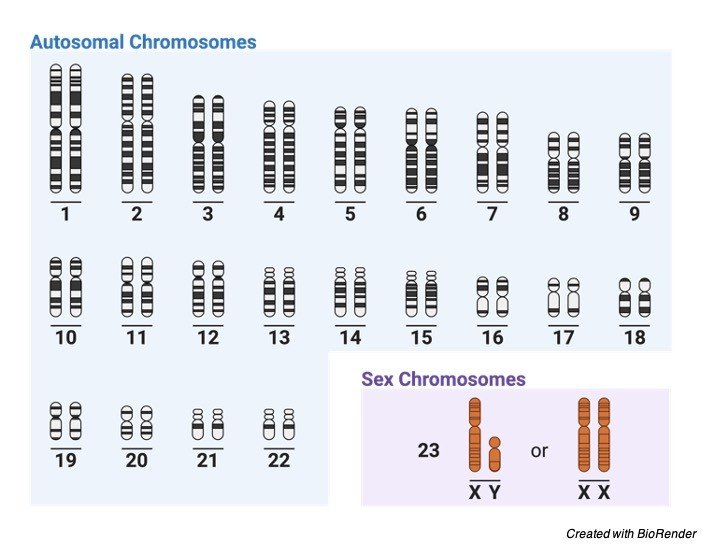
Each cell of a human consists of 23 pair of chromosomes which are 46 in number.
The first 22 sets are defining as autosomes and the remaining one set is known as sex chromosomes or allosomes.
Autosomes are generally known as homologous chromosomes because they contain the same genes in the same order along the entire length of the chromosomes i.e., in chromosomal arms.
Thus, females have all the 23 pairs as homologous pairs where as in males the lass pair is formed by X and Y chromosomes which are heterozygous in condition.
The X chromosome present in the 23rd pair is always an ovum where the other X and Y varies accordingly as sperm or ovum depending upon their gender.
In females during an early embryonic development the X chromosomes present in other cells neither than egg cells are deactivated partially and permanently.
In some cells the X chromosomes from female i.e. from mother is inherited where as in other cells the X chromosomes from father is deactivated.
This is the only reason why our body has only one functional X chromosome. Whereas the deactivated X chromosomes are repressed by heterochromatin and prevents the activation of more genes
Sex Determination
All diploid organism get fifty percentage of allosomes from their mother and father equally.
Since females have only two X chromosomes they can pass only the X chromosome, where as male passes either X or Y chromosome.
The individual which get X chromosome from the father is determined as female and the individual which gets Y chromosomes from the father are consider as male.
This is the reason why male sperm cells decide the sex of the individual.
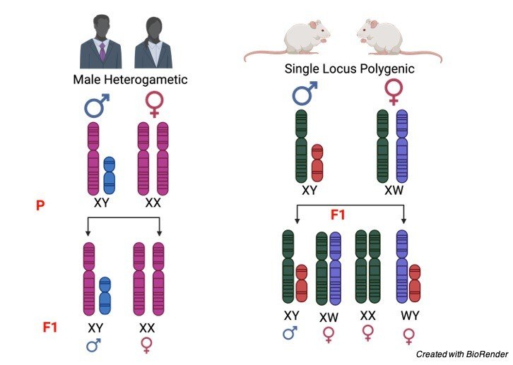
However few people rarely have a intersex due to the divergent form of sexual development.
This results when the allosomes are formed neither XX nor XY.
At sometimes the embryo may fuse and it also results in intersex. It can also be due to the exposure of chemicals which results in mutation of a particular gene.
Sex Chromosomes Disorders
Sex chromosome is not only involved in determining the gender of an individual but also in determining or carrying other genes which are responsible for other characteristics.
Genes which are being carried by sex chromosomes are commonly called as sex linked.
Sex linked genes are the ones which are passed down from their ancestors.
There is also a possibility that these genes will carry the diseases along with the genes to their offsprings if their ancestors have any one of them.
As only males carry y chromosomes there are the who transmits Y linked inherited diseases.
X linked diseases are transmitted either by female or male to their offspring.
Sex Chromosomes Citations
- Sex chromosomes manipulate mate choice. Nature . 2019 Jun;570(7761):311-312.
- Why Do Sex Chromosomes Stop Recombining?. Trends Genet . 2018 Jul;34(7):492-503.
- Genetic Diversity on the Sex Chromosomes. Genome Biol Evol . 2018 Apr 1;10(4):1064-1078.
- Young sex chromosomes in plants and animals. New Phytol . 2019 Nov;224(3):1095-1107.
- How to identify sex chromosomes and their turnover. Mol Ecol . 2019 Nov;28(21):4709-4724.
Share
Similar Post:
-

Autosomes: Definition, Facts, and Example I Research...
Continue ReadingAbout Autosomes
We all know our body is made up of types of chromosomes, one is autosomes and the other is allosomes or most commonly known as sex chromosomes.
Autosomes are commonly known as somatic or body chromosomes which does not have any practice in taking the memory to the next generation.
Whereas allosomes play an important role in taking the inheritance from present to future generations.
What are Autosomes
The autosomal members always pair in a diploid cell which often has the similar morphological features.
The DNA which is present in the autosomes are generally known as atDNA or auDNA.
Considering an example in human who have 22 pairs of chromosomes in their diploid genome and 1 pair of allosome, and total of 46 chromosomes which are 23 in pairs.
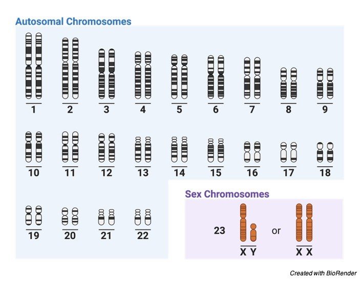
The autosomes are named as 1, 2, 3, 22 pair they are karyotyped according to their size and the base pairs present in them.
The change in chemical or physical form of the chromosomes leads to many syndromes and diseases, but it is not carried through generations.
But by research it has also be found that autosomes contain sex determination gene, which is also capable of deciding the characters of an individual by forming their outer appearance and male characters in male and feminine characters in female.
Taking an example of SRY gene, Y chromosome encodes a transcription factor which is known as TDF which is very important for the sex determination in males during their development.
TDF activates the SOX9 which is present on the chromosome 17 and this effect causes mutation in that particular gene and causes the Y chromosome to develop into a female.
Thus, it is also proven that autosomes also play an important role in determining the sex of an individual.
All the members of a species of a plant or the animal are characterized by a set of chromosomes which have certain constant characteristics.
These characteristics include the number of Karyotyping helps in identifying the size, position of centromere length of the arm, secondary constrictions and satellites.
This technique is used to find the characteristic of the chromosomes in a particular group.
A diagrammatic representation of this particular set of chromosomes to understand its morphological characteristics is referred to as idiogram.
This technique also has an advantage, which helps in sorting out the missing pair of chromosomes or any abnormalities found.
It is important to know about the structure and morphology of chromosomes before discussing about their abnormalities
Chromosomes Morphology
Size: The chromosome size is generally measured during the phases of metaphase in mitotic cell division.
Usually, it measures 0.25µm in birds and fungi, 30µm in certain plants like Trillium and 8 to 12µm in maize, 3µm in drosophila and 5µm in humans.
While the organisms which are having lesser number of chromosomes, have their chromosomes with larger size than the one which is containing higher number of chromosomes.
Where the dicotyledons contains smaller chromosomes comparing with that of monocotyledons.
The animals contain smaller chromosomes on comparing with plants.
The chromosomes are also named depending upon their structure and on which organism it is present as lamp brush chromosomes which is present in few vertebrates and polytene and oocyte chromosomes which are found in certain insects.
Shape: The chromosomes change its shape at different phases during their cell division process.
Where as in the interchange phase the chromosomes look like a thin coiled thread like structures, and on passing to the metaphase and anaphase they become thick and filamented.
These chromosomes also have a centromere (often referred to as clear zone), and kinetochore which forms the length of the chromosome.
The two arms arise from the centromere and they are generally called as chromosomal arms.
However, the position varies accordingly, which results in various shape of the chromosomes as telocentric (centromere is situated at the proximal end), acrocentric (centromere at one end forming long and short arm which form a rod like appearance), metacentric (V- shaped chromosome, where centromere is at the centre), sub-metacentric (j shaped or L-shape where centromere is towards the median position).
Chromosome Structure
Chromosomes are generally the thread like structures present in the nucleus of the cell containing proteins along with one molecule of Deoxy ribo nucleic acid in each chromosome.
The structure of chromosomes also varies accordingly depending upon their structures, where as in metaphase two chromosomes consists of a symmetrical structure which is known as chromatids.
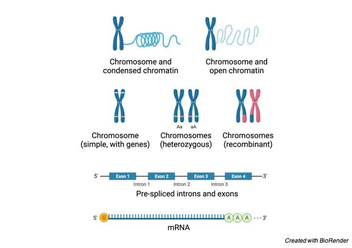
During some stages of interphase a bead like structures are found in the chromatin material which is known as chromomeres.
In some cases, the chromosomes contain a knob like structures which are referred to as satellites, along with all these; chromosomes generally have the primary and secondary constrictions, nuclear organisers, centromere, kinetochore and telomeres which make up the perfect chromosomal structure.
Autosome Chromosomal Abnormalities
The autosomal genetic disorders occur due to different causes; the most common thing often occurs is non-disjunction of chromosomes in the parental germ cells.
Autosomal genetic disorders which exhibit the characteristics of a mendelian inheritance gets inherited in an autosomal dominant or recessive condition.

These disorders pass from either of their parents with the same frequency. Autosomal dominant disorders are mostly present in both parents as well as their children.
If both the parents had a defective allele there is a chance where child is also being affected by the same, if only one parent i.e., in heterozygous manner then the child may be a carrier or an affected one.
In some cases, aneuploidy of the chromosomes also leads to the deletion of half a part of the chromosome which results partial monosomies.
It occurs mostly due to unbalanced translocations. Unbalanced translocations also lead to partial dislocation or translocations.
Autosomal translocations lead to a number of diseases apart from gene disorders like cancer and schizophrenia.
Where as the diseases caused by aneuploidy are due to improper gene dosage or non- functional gene product.
Autosomes Citations
- The different levels of genetic diversity in sex chromosomes and autosomes. Trends Genet . 2009 Jun;25(6):278-84.
- Local adaptation and the evolution of inversions on sex chromosomes and autosomes. Philos Trans R Soc Lond B Biol Sci . 2018 Oct 5;373(1757):20170423.
- Signatures of replication timing, recombination, and sex in the spectrum of rare variants on the human X chromosome and autosomes. Proc Natl Acad Sci U S A . 2019 Sep 3;116(36):17916-17924.
- Pseudodicentric Chromosome Originating from Autosomes 9 and 21 in a Male Patient with Oligozoospermia. Cytogenet Genome Res . 2019;159(4):201-207.
- A balancing act between the X chromosome and the autosomes. J Biol . 2006;5(1):2.
- The probability of fusions joining sex chromosomes and autosomes. Biol Lett . 2020 Nov;16(11):20200648.
Share
Similar Post:
-

Incomplete Dominance vs Codominance: Definition & Examples
Continue ReadingIncomplete Dominance vs Codominance
The laws of inheritance proposed by Mendel characterized the predominance factors in legacy and the impacts of alleles on the aggregates.
Codominance and inadequate strength are various sorts of legacy (explicitly hereditary). Be that as it may, both fragmented strength and codominance sorts of predominance were not distinguished by Mendel.
Nonetheless, his work prompts their distinguishing proof. A few botanists worked in the legacy field and tracked down these particular predominance types.
The deficient strength and codominance are regularly stirred up. Consequently, see the essential factors that lead to varying from one another.
Incomplete Dominance
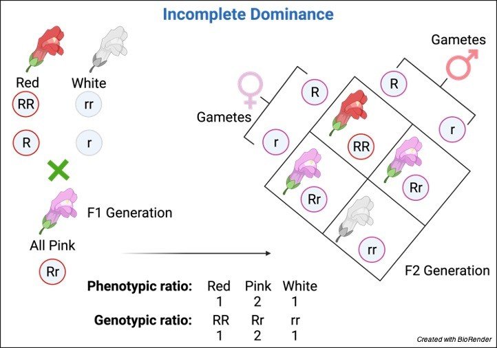
The incomplete dominance is a halfway dominance, which means the aggregate is in the middle of the genotype predominant and latent alleles.
In the above model, the subsequent posterity has a pink shading attribute notwithstanding the prevailing red tone and white shading characteristic because of incomplete dominance.
The prevailing allele doesn’t veil the passive allele bringing about an aggregate not quite the same as the two alleles, i.e., pink tone.
The incomplete dominance conveys hereditary significance since it clarifies the reality of the moderate presence of aggregate from two unique alleles.
Additionally, Mendel clarifies the Law of dominance that only one allele is prevailing over the other, and that allele can be one from both.
The overwhelming allele will diminish the impact of the latent allele.
While in incomplete dominance, the two alleles stay inside the delivered aggregate, yet the posterity have an entirely unexpected characteristic.
Mendel didn’t examine incomplete dominance on the grounds that the pea plant doesn’t show any incomplete dominance (middle of the road attributes).
Notwithstanding, the Mendel proposed proportion 1:2:1 will in general be exact for incomplete dominance, as found in the case of the four o’clock blossom, where the F1 age brings about red, pink, and white blossoms genotypic proportion of 1:2:1, separately.
These outcomes show the Law of Inheritance where alleles are acquired from guardians to posterity actually happens in the incomplete dominance portrayed by Mendel.
In research on quantitative hereditary qualities, the opportunities for incomplete dominance requires the subsequent aggregate to be in part identified with any of the genotypes (homozygotes); in any case, there will be no dominance.
Codominance
Codominance alludes to the dominance wherein the two alleles or qualities of the genotypes (of the two homozygotes) are communicated together in posterity (aggregate).
There is neither a predominant nor latent allele in cross-rearing.
Maybe the two alleles stay present and shaped as a combination of both of the alleles (that every allele tends to add phenotypic articulation during the rearing interaction).
Now and again, the codominance is additionally alluded to as no dominance because of the presence of the two alleles (of homozygotes) in the posterity (heterozygote).
Subsequently, the aggregate delivered is unmistakable from the genotypes of the homozygotes.
The capitalized letters are utilized with a few superscripts to recognize the codominant alleles while communicating them in compositions.
This composing style demonstrates that every allele can communicate even within the sight of different alleles (elective).

The case of codominance can be found in plants with white tone as passive allele and red tone as predominant allele produce blossoms with pink and white tone (spots) after cross-reproducing.
Additionally, Mendel likewise didn’t consider the codominance factor because of the pea plant’s restricted attributes.
In any case, further examination uncovered the codominance in plants and the other way around.
The genotypic proportion was equivalent to Mendel depicted. They delivered posterity that outcomes in the F1 age to incorporate red, spotted (white and pink), and white with a similar genotypic proportion.
Codominance can be effectively found in plants and creatures due to shading separation, just as in people to some terminated, for example, blood classification.
The incomplete dominance produces posterity with middle of the road qualities while the codominance includes the blending of allelic articulations.
In any case, in the two sorts of dominance, the parent alleles stay in the heterozygote. In any case, no allele is prevailing over the other.
Incomplete Dominance vs Codominance
Incomplete Dominance Codominance Incomplete dominance happens in the heterozygote, wherein the predominant allele doesn’t rule the passive allele totally; rather, a transitional quality shows up in the offspring. Codominance happens when the alleles don’t show any prevailing and passive allele relationship. Be that as it may, every allele from homozygote can add phenotypic articulations in the posterity or basically the “blend” of every allele. The posterity’s aggregate is a moderate of the guardians’ homozygous traits. The phenotypic articulation of homozygous in codominance is autonomous. The declaration of alleles in incomplete dominance is prominent, which means none of the alleles rules over the other. The articulation of alleles in codominance is consistently obvious, which means the two alleles have an equivalent possibility for communicating their belongings. The shaped characteristic (aggregate) is distinctive because of blending both parent’s aggregates and genotypes. The framed quality (aggregate) isn’t diverse because of the no blending of the two guardians’ aggregates and genotypes. The posterity don’t show the parental phenotype. The posterity shows both parental aggregates. The predominant allele doesn’t rule over the latent allele. None of the alleles is prevailing or passive, and the ruling relationship doesn’t happen. The predominant allele doesn’t overwhelm over the latent allele. The posterity aggregate created has the mix of two alleles and, subsequently, shows two aggregates together. The quantitative methodology can be utilized for the investigation of incomplete dominance in living beings (counting the examination of both non-ruling alleles). The quantitative methodology can be utilized for the examination of codominance in the living being (just including the investigation of quality articulations). Incomplete dominance models incorporate Pink blossoms of four o’clock blossoms (Mirabilis jalapa), and actual qualities in people, for example, hair tone, hand sizes, and height. Codominance can be found in people and just as in creatures. The blood classification (or gatherings A, B, and O) in people and the spots on quills or hairs of domesticated animals are instances of codominance. Incomplete Dominance vs Codominance Citations
- Defining codominance in plant communities. New Phytol . 2021 Jun;230(5):1716-1730.
- Incomplete dominance of deleterious alleles contributes substantially to trait variation and heterosis in maize. PLoS Genet . 2017 Sep 27;13(9):e1007019.
- Combined effects of dosage compensation and incomplete dominance on gene expression in triploid cyprinids. DNA Res . 2019 Dec 1;26(6):485-494.
- Codominance of Acer saccharum and Fagus grandifolia: the role of Fagus root sprouts along a slope gradient in an old-growth forest. J Plant Res . 2010 Sep;123(5):665-74.
Share
Similar Post:
-

Lactobacillus: Health Benefits, Uses, and Side Effects
Continue ReadingAbout Lactobacillus
Microorganisms comprises of varied microbes, fungi, protozoa. It also includes many microscopic plants viruses, viroid and also prions.
They can be seen anywhere on this planet anywhere where habitat is seen. It can flourish on nutritive media where researchers can do studies on it by cultivation and producing colonies and can help for research purpose.
Microorganisms gives rise to various diseases in living beings including plants and animals, although they cause diseases, they can be useful in many different and friendly ways.
One such type of bacteria is lactobacillus and there are many species of lactobacillus which are useful and can help in digestion, and on urinary and reproductive system as well and cause no harm.
They are also used in various consumable forms such as yogurts and probiotic drink or supplements.
It is most popular to treat diarrhea when patients are given antibiotics which may suppress the gut flora and this type of bacteria helps to protect and increase good gut flora which prevents it.
Lactobacillus contributes significantly in improving the overall health in living beings.

They are also used and prescribed for general digestion problems, large intestine disorders which causes stomach pain, irritable bowel syndrome (IBS), colic in infants, inflammation of bowel track or colon, etc.
It is a gram-positive, aerotolerant anaerobes or microaerophilic, rod-shaped, non-spore-forming bacteria under the genus Lactobacillus and has as many as 260 phylogenetically, metabolically varied species.
Its taxonomic update of the genus in 2020 put lactobacilli to 25 genera.
Metabolism of Lactobacillus
They are the only group of the lactic acid bacteria that comprises of homofermentative and heterofermentative organisms and in the Lactobacillaceae, either metabolism is jointly used by all strains of Lactobacillus.
In Lactobacilli hexoses are metabolised by glycolysis to lactate as higher end product which is termed as homofermentative.
It may be heterofermentative, which means that hexoses are broken down by the Phosphoketolase pathway to lactate, CO2 and acetate or ethanol as the end product.
They are aerotolerant which means it is manganese-dependent and has been seen in Lactiplantibacillus plantarum.
Few lactobacillus species respire only if heme and menaquinone are available in the growth medium but usually don’t need iron for development.
This species is homofermentative, do not exhibit pyruvate formate lyase, and manty don’t ferment pentoses example: in L. crispatus, pentose metabolism is strain selective and is gained by lateral gene transmit.
Genome of Lactobacillus
The genome is not constant and differs, size from 1.2 to 4.9 Mb (megabases) and the quantity of protein-coding genes vary from 1,267 to about 4,758 genes in Fructilactobacillus sanfranciscensis and Lentilactobacillus parakefiri.
Differences can be seen in one species as well for example in L. crispatus genome sizes varies in between 1.83 to 2.7 Mb, or 1,839 to 2,688.
Lactobacillus comprises a stock of compound microsatellites in the coding area that are not perfect and usually possess different motifs and they also comprises of many plasmids.
In many new research study they have delineated that the plasmids encode the genes which are necessary to acquittance of lactobacilli to the surrounding.
Types of Lactobacillus
L. delbrueckii is a type of species of the genus, is 0.5 to 0.8 μm and can be seen in single or in small groups in chains.
L. acidophilus, L. brevis, L. casei, and L. sanfranciscensis are some another types.
The quantity of lactic acid generated differs. In L. brevis and L. fermentum, glucose metabolism is heterofermentative, with lactic acid sums upto 50% of end products and ethanol, acetic acid, and carbon dioxide as the rest 50%.
The organisms that cannot metabolise glucose receives its energy organic components like galactose, malate, or fructose.
Health Benefits of Lactobacillus
Lactobacillus spp. are fundamental as purposeful and essential as deliberate or accidental component in many food available.
An excellent deal of attention has been given to its significant property as being probiotics.
Strains that are evaluated for this beneficial property comprises of L. acidophilus LA1, L. acidophilus NCFB 1748, Lactobacillus GG, L. casei Shirota, Lactobacillus gasseri ADH, and Lactobacillus reuteri.
Other beneficial property of Lactobacillus accommodates immune improvements, decreasing fecal enzyme process, inhibiting intestinal disorders, and decreasing incidence of viral diarrhea and this probiotic strains are said to own a capability to live in/habitat in the intestinal tract, and in a good way affecting the microflora and maybe excluding colonization by pathogens.
Albeit the positive effects from having of probiotics is critical, authentic records still remains in water and the clinical trials often have hurdles and costly to conduct specially in people within which compliance with experimental protocols and normalizing for other genetic and environmental factors are hard.
For now the major focus is onselective isolates, such as L. casei GG, and research is directed and is being built around these strains.
Ergo, variety of products are named as probiotics and are clubbed together with more traditional dairy products to be sold in the market. This are easily available for the normal people.
Lactobacillus spp. are important components in many food products which either we know or may be present unknowingly.
Significant amount of attention has been directed toward their potential benefits and importance as probiotics.
Strains that have been verified for their probiotic properties are L. acidophilus LA1, L. acidophilus NCFB 1748, Lactobacillus GG, Lactobacillus casei, L. Shirota, gasseri ADH, and Lactobacillus reuteri.
Many reported clinical effects attributed to the consumption of it consist of immune enhancement, lowering faecal enzyme activity, preventing intestinal problems, and reducing viral diarrhoea.
Most probiotic strains are accepted to have a capacity to colonize the intestinal lot, subsequently decidedly influencing the microflora and maybe barring colonization by microbes.
But the possible advantages from the utilization of probiotics are huge, research is as yet slacking.
Clinical preliminaries are troublesome and costly to perform particularly in people in which consistency with trial conventions and normalizing for other hereditary and natural components are troublesome.
Huge worth and spotlight are currently being set on specific confines, like L. casei GG, and protected innovation is being worked around these strains.
These are filled in milk and split it to frame curd. The LAB creates acids that coagulate and do not completely digest the milk proteins which upgrades the wholesome property by raising vit. B12.
Presently, various items are assigned as probiotics and are sold alongside more conventional dairy items.
Probiotics especially related to Bifidobacterium spp. and Lactobacillus spp. have studied and research shows that they exhibit anticarcinogenic, antimutagenic, and antitumorigenic activities ergo, more research needs to be executed to verify its authenticity.
Lactobacillus Citations
Share
Similar Post:
-

Proto-Oncogene: Definition, Function, and Examples
Continue ReadingWhat are Proto-Oncogene?
There are trillions of living cells in the body that develops, gap and bite the dust in a systematic design. This cycle is firmly directed by the qualities inside a cell’s core.
These qualities code for proteins that assist with controlling cell development. These significant qualities are called proto-oncogenes.
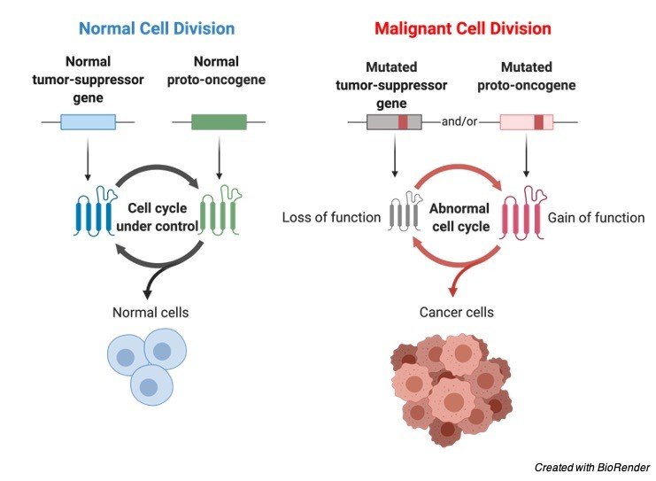
An adjustment of the DNA grouping of the proto-oncogene brings about an oncogene, which delivers an alternate protein and meddles with typical cell guidelines.
Features of Proto-Oncogene
Protooncogenes have numerous capacities in a cell however they regularly code for proteins that animate cell division, forestall cell separation, or manage customized cell demise (apoptosis).
These are for the most part fundamental cycles needed for ordinary development, improvement, and the support of solid organs and tissues.
Notwithstanding, a changed or blemished adaptation of a proto (oncogene) expands the creation of these proteins, subsequently prompting unregulated cell division, a slower pace of cell separation, and expanded hindrance of cell demise.
Combined this enhances and highlights properties of the cells that have gotten malignant.
Presently, scientists know about in excess of 40 unique sorts of proto-oncogenes in people.
A few hereditary components that cause a proto-oncogene to turn into an oncogene have additionally been clarified and models are given underneath:
• Point transformations, inclusions or erasures that lead to an overactive quality item.
• Point transformations, inclusions or cancellations that lead to an expansion in record.
• Quality intensification prompting extra duplicates of a proto-oncogene.
• Chromosomal movement that makes a proto-oncogene move to an alternate chromosomal site related with expanded articulation.
• Chromosomal movements that cause a protooncogene to intertwine with another quality to deliver a protein that has oncogenic action.
Proto-Oncogene Characteristic
A couple of malignancy conditions are brought about by acquired changes of proto-oncogenes that cause the oncogene to be turned on (initiated).
However, most disease-causing changes including oncogenes are obtained, not acquired.
They, by and large, actuate oncogenes by:
Chromosome modifications: Changes in chromosomes that put one quality close to another, which permits one quality to initiate the other.
Quality duplication: Having additional duplicates of a quality, which can prompt it making an over-the-top certain protein.
Investigation into oncogenes has worked on comprehension and information regarding why certain people are more helpless to malignant growth than others.
A few bunches are bound to have their proto-oncogenes changed over to oncogenes and foster malignancy.
The specialists that cause malignant growth like radiation, infections, and ecological poisons, for instance, are bound to cause disease in these people.
Example of Proto-Oncogene: HER2
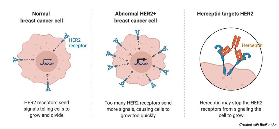
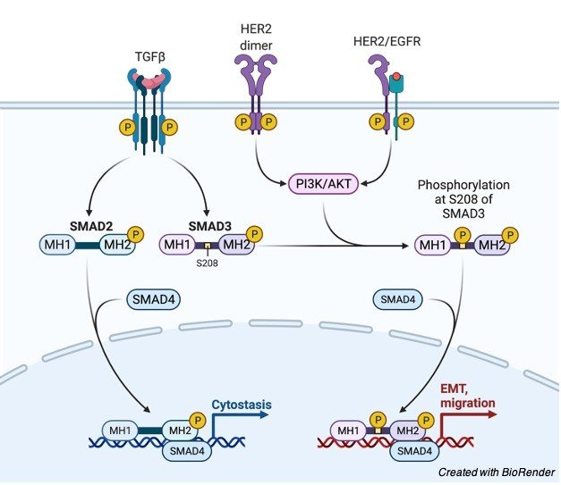
One illustration of a notable proto-oncogene is the HER2 quality. This quality codes for a transmembrane tyrosine kinase receptor called human epidermal development factor receptor 2.
HER2 Receptor and Breast Cancer
This protein receptor is associated with the development, fix, and division of cells in the bosom.
In a sound breast cell, there are two duplicates of HER2, yet in certain kinds of bosom malignant growth, the cells contain multiple duplicates, which prompts overabundance creation of the HER2 protein.
This makes the breast cells develop, partition and multiply considerably more rapidly than sound breast cells.
HER2/EGFR signaling pathway in Breast cancer
This hereditary deformity in the Breast cells isn’t acquired yet is well on the way to emerge because of maturing.
Specialists are as yet exploring whether natural factors, for example, smoke and contamination improve the probability of the imperfection happening.
In the event that a lady is analyzed as HER2 positive, she has a strange number of HER2 qualities and is creating a lot of the HER2 protein.
This implies cells in her Breast are filling in an upregulated way and shaping a carcinogenic tumor.
Assuming a lady is analyzed as HER2 negative, it’s anything but HER2 protein creation that is causing malignant growth.
Example of Proto-Oncogene: Myc
Another illustration of a proto-oncogene is the Myc quality, which codes for record factors.
At the point when the quality grouping of Myc is modified, these record factors are created at expanded rates and quality articulation is changed bringing about the arrangement of a tumor.
What is Oncogene?
On the contrary oncogenes are qualities that leads to malignancies.
As such, oncogenes can be characterized as destructive qualities. Oncogenes are transformed proto-oncogenes. At the point when the DNA succession of the proto-oncogene is changed or transformed, it’s anything but an oncogene.
Oncogene is coded with various proteins which impact the ordinary cell cycle.
Oncogenes produce inhibitors of the phone cycle which are equipped for proceeding with cell division in any event, during conditions that are not useful for cell division.
Oncogenes additionally produce positive controllers which are fit for keeping cells dynamic till the development of malignant growth.
Oncogenes run after disease development by advancing uncontrolled cell division, bringing down cell separation and restraining ordinary cell demise (apoptosis).
Proto-oncogenes become oncogenes because of a few hereditary changes or systems like transformations, quality intensifications, chromosomal movements.
They are rattled off as follows.
Creation of overactive quality items by point transformations, inclusions, or erasures.
Expanded record by point transformations, inclusions or erasures.
Creation of extra duplicates of proto-oncogenes by quality intensification.
Development of proto-oncogenes into various chromosomal site and cause for expanded articulation.
Combination of proto-oncogenes with different qualities which can cause oncogenic action.
Examples
HER2: Another notable protooncogene is HER2. This quality makes protein receptors that are engaged with the development and division of cells in the breast.
Numerous individuals with breast disease have a quality intensification transformation in their HER2 quality.
This kind of disease is frequently alluded to as HER2-positive breast malignant growth.
Myc : The Myc quality is related with a sort of malignant growth called Burkitt’s lymphoma.
It’s anything but a chromosomal movement moves a quality enhancer arrangement close to the Myc proto-oncogene.
Cyclin D: Cyclin D is another protooncogene. Its ordinary occupation is to make a protein called Rb tumor silencer protein dormant.
In certain diseases, similar to tumors of the parathyroid organ, Cyclin D is actuated because of a change.
Thus, it can presently don’t manage its work of making the tumor silencer protein inert.
This thus causes uncontrolled cell development.
Your cells contain numerous significant qualities that direct cell development and division.
The typical types of these qualities are called proto-oncogenes. The changed structures are called oncogenes. Oncogenes can prompt disease.
You can’t totally keep a change from occurring in a proto-oncogene, yet your way of life may have an effect.
You might have the option to bring down your danger of malignancy causing changes by opting for heathier lifestyle.
Citations
- Proto-oncogene fos: complex but versatile regulation. Cell . 1987 Nov 20;51(4):513-4.
- Proto-oncogene fos: an inducible gene. Princess Takamatsu Symp . 1986;17:279-90.
- A function for the lck proto-oncogene. Trends Biochem Sci . 1989 Oct;14(10):404-7.
- Role of proto-oncogene activation in carcinogenesis. Environ Health Perspect . 1992 Nov;98:13-24.
Share
Similar Post:
-

Genetic Disorders: Definition, Cause, Symptoms, and Examples
Continue ReadingWhat are Genetic Disorders?
Genetic disorders are type of biological inheritance which follows the principles that have been proposed by Gregor Mendel in the year 1865 and 1866.
Generally, in humans the Genetic disorders are type of genetic cause which happens due to abnormalities or defects in the genome of an individual.
These disorders are seen in an affected individual from birth and can be diagnosed and can be treatment to reduce the impact even though it cannot be cured purely.
It can also be identified the passages of traits by pedigree analysis or family tree.
This type of genetic disorders is rare, affecting one of thousand individuals.
These disorders may or may not be inherited. Inherited disorders are the ones which are passed through germ line cells, where as in non-inheritable conditions these disorders are caused due to mutations or some changes in the genetic material.
A best example for this condition is cancer, which is either caused by environmental conditions or lifestyle factors or though inherited conditions.
Principles of Genetic Disorders
The research carried out by Mendel on pea plants stands as a base for understanding all the inheritances which are characterized by a single gene disorders.
Mendelian genetic disorders are caused due to a mutation in a single gene locus, where the locus is present in autosome or allosome.
It occurs either as a dominant or a recessive allele. If the individual who is having this disorder then the pedigree analysis is followed to find whether this disorder is present in an autosome or allosome in large families then dominant and recessive condition can also be found through that.
Mendel also suggested two laws for knowing the inherited disorders better.
Law of Segregation
Mendel’s law of segregation states that at the time of formation of genes two genes are separated so that each gene will carry one of the alleles.
Law of Independence
Mendel’s law of independence states that during the time of separation of gametes, each gene pair separates independently with other pairs.
It can also be said that the allele received by the gamete for one pair is independent of the allele received for the other allele.
Types of Genetic Disorders
Genetic disorders are classified depending upon the Mendel’s law of inheritance.
It is of five types namely;
Autosomal dominant
Autosomal recessive
Sex-linked dominant
Sex-linked recessive
Mitochondrial
All these types of Mendelian disorders can be identified with the help of pedigree analysis.
Examples of Genetic Disorders
Examples of Mendelian Genetic disorders include;
Sickle cell anaemia
Muscular dystrophy
Cystic fibrosis
Thalassemia
Phenylketonuria
Colour blindness
Skeletal dysplasia
Haemophilia
Haemophilia
It is one of the types of sex-linked recessive disorder. Here, the unaffected carrier mother passes this condition to their sons.
This disorder is rare in females because they get only one gene through the parents at most of the times, so the probability will be as a carrier mother or as an unaffected one.
So, it is very rare that females will be an affected individual.
In this disorder the blood will not clot easily after an injury or any other accidents.
Males are most frequently affected to this type of disorder.
Sickle Cell Anaemia
It is one type of autosomal recessive disorder where the disorder is carried to the offspring’s if two parents are carriers of this disease.
This disorder is caused due to the mutation in the hemoglobin molecule where the glutamic acid present in the sixth position in a hemoglobin molecule in a beta chain is replaced by valine.
So, the hemoglobin molecules acquire a change in shape, by converting into a sickle shape from its normal biconcave shape which reduces the oxygen carrying capacity of the cells and this shape of molecules cannot be easily passed through the vascular system and it results in forming a block in small blood vessels such as capillaries.
Phenylketonuria
It is one of the autosomal recessive disorder caused due to the inborn error of phenylalanine metabolism which is one of the important amino acid needed for the body.
This disorder, affects the conversion of enzyme phenyl alanine into tyrosine as a result phenylalanine is being accumulated in the body which results in formation of many derivatives and causes mental retardation in most of the cases.
Thalassemia
It is one of the types of autosomal recessive disorder. In this type of disorder body makes abnormal amount of hemoglobin so it leads to a condition where higher amount of red blood cells is destroyed and causes anemia.
It causes symptoms such as abdominal swelling, dark urine excretion, deformities of facial bones etc.
Cystic Fibrosis
It is one of the types of autosomal recessive disorder where the lungs and digestive system gets affected completely and there is also a thick production or secretion of mucus which blocks the lungs and pancreas.
Usually, the person affected with this syndrome has very short life span.
Genetic Disorders Citations
- Genetic disorders. JEMS . 2014 Feb;39(2):64-71.
- Developmental Support for Infants With Genetic Disorders. Pediatrics . 2020 May;145(5):e20190629.
- Genetic disorders of nuclear receptors. J Clin Invest . 2017 Apr 3;127(4):1181-1192.
- Genetic Disorders of the Extracellular Matrix. Anat Rec (Hoboken) . 2020 Jun;303(6):1527-1542.
- Coatopathies: Genetic Disorders of Protein Coats. Annu Rev Cell Dev Biol . 2019 Oct 6;35:131-168.
- Maternal Genetic Disorders in Pregnancy. Obstet Gynecol Clin North Am . 2018 Jun;45(2):249-265.
Share
Similar Post:


