Category: Uncategorized

Category: Uncategorized

  • Cell Junctions: Definition, Types, and Function

    About PhD In Psychology

    o Certain cells need to be able to contact other nearby cells. Cell junctions adhere membranes of adjacent cells.

    Types of Cell Junctions

    There are three different types of cellular junctions:

    I. Gap Junctions

    o Connect the cytoplasm of adjacent cells, which allows for the movement of molecules between the cells.

    o Gap junctions are found in the heart muscle. Gap junctions allow for rapid spread of information between cells.

    o Ex. Electrical synapse

    Diagram Representing Types of cell Junctions

    Cell, Cell Structure, Cell Membrane Structure, Animal Cell, Plant cell,
    II. Tight Junctions

    o Provide waterproof seals between adjacent cells to prevent leakage of fluid.

    o Tissues held together by tight junctions may act as a complete fluid barrier.

    o Tight junctions also act as a barrier to protein movement between the apical and basolateral surface of a cell.

    o The portion of the cell exposed to the lumen is called its apical surface.

    o The rest of the cell (i.e., its sides and base) make up the basolateral surface.

    o The lumen is the space enclosed by an organ/organelle.

    o Tight junctions perform two vital functions:

    o They prevent the passage of molecules and ions through the space between cells.

    o So materials must actually enter the cells (by diffusion or active transport) in order to pass through the tissue.

    o This pathway provides control over what substances are allowed through.

    o They block the movement of integral membrane proteins (red and green ovals) between the apical and basolateral surfaces of the cell.

    o Thus the special functions of each surface, for example o receptor-mediated endocytosis at the apical surface o exocytosis at the basolateral surface can be preserved.

    III. Adhering Junctions (Desmosomes)

    o Join two cells at a single point and they directly attach the cytoskeleton of each cell.

    o They don’t prevent fluid from circulating around all sides of a cell.

    o Are the types of junctions found cells that experience a lot of stress and they often accompany tight junctions.

    o Ex. in epithelial cells which allow for stretching.

    o Desmosomes (adhering junctions) act like spot welds.

    Cell Junctions Citations

    Share

    Similar Post:

    Continue Reading
  • Cytoskeleton: Description, Structure, and Function

    Cytoskeleton

    o The cytoskeleton is a cellular “scaffolding” or “skeleton” contained within the cytoplasm.

    o The cytoskeleton is present in all cells; it was once thought this structure was unique to eukaryotes, but recent research has identified the prokaryotic cytoskeleton.

    o It is a dynamic structure that maintains cell shape, protects the cell, enables cellular motion (using structures such as flagella, cilia and lamellipodia), and plays important roles in both intracellular transport (the movement of vesicles and organelles, for example) and cellular division.

    Cytoskeleton Component

    Cytoskeleton, Cytoskeleton Description, Cytoskeleton Structure, Cytoskeleton Function 1

    Types of Cytoskeleton

    There are two types of filaments in the cytoskeleton;

    I. Microtubules

    o Microtubules are built from subunits-molecules of tubulin-each one of which is itself a dimer composed of two very similar globular proteins called a-tubulin and b-tubulin bound tightly together by noncovalent bonds.

    o Although tubulin is globular it can polymerize into long straight filaments.

    o Thirteen of these filaments lie alongside each other to form the microtubule.

    o In nine triplet sets (star-shaped), they form the centrioles.

    o Centrioles function in the production of flagella and cilia, but are NOT necessary for microtubule production.

    o The major portion of each flagellum and cilium, called the axoneme, contains nine pairs of microtubules forming a circle around two lone microtubules.

    o The latter formation is commonly referred to as a “9+2” arrangement, wherein each doublet is connected to another by the protein dynein.

    o The mitotic spindle is made from microtubules.

    o Mitotic spindle provides the machinery that will segregate the chromosomes equally into the two daughter cells.

    o Microtubules have a + and a – end.

    o This is because of the polarity of the a and b tubulin, thus making the microtubule polar.

    o The – end attaches to a microtubule-organizing center (MTOC) in the cell.

    o The major MTOC in animals is the centrosome.

    o A microtubule grows away from an MTOC at its + end.

    o Centrosomes are composed of two orthogonally arranged centrioles Centrosomes are often associated with the nuclear membrane during interphase of the cell cycle.

    o In mitosis the nuclear membrane breaks down and the centrosome nucleated microtubules can interact with the chromosomes to build the mitotic spindle.

    o The centrosome is copied only once per cell cycle so that each daughter cell inherits one centrosome, containing two centrioles.

    o The centrosome replicates during the S phase of the cell cycle.

    II. Microfilaments

    o Microfilaments are the thinnest filaments of the cytoskeleton found in the cytoplasm of all eukaryotic cells.

    o These linear polymers of made of actin subunits are flexible and relatively strong.

    o Microfilaments produce the contracting force in muscle as well as being active in cytoplasmic streaming, phagocytosis, and microvilli movement, cytokinesis, and muscle movement.

    o In fungi, the Septa is usually perforated to allow exchange of cytoplasm between cells, called cytoplasmic streaming.

    o Both microtubules and microfilaments are involved in intracellular movements in eukaryotic cells.

    o In both cases the movements are generated by motor proteins, which bind to microfilaments and microtubules and use the energy derived from repeated cycles of ATP hydrolysis to travel steadily along the microtubule or microfilaments in a single direction.

    o The motor proteins that move along cytoplasmic mircrotubules are: kinesins ( – ⇒ + ) and dyneins ( + ⇒ – ).

    o Actin uses myosin as its motor protein.

    Eukaryotic Flagella and Cilia

    o Eukaryotic flagella and cilia are specialized structures also made from microtubules.

    o The major portion of each flagellum and cilium, called the axoneme, is arranged in nine doublets oriented about two additional microtubules (wheel-shaped) they form cilia and flagella.

    o This formation is commonly referred to as a “9+2” arrangement, wherein each doublet is connected, via a crossbridge, to another by the protein dynein.

    o The cross bridges cause the microtubule pairs to slide along their neighbors creating a whip action in cilia causing fluid to move laterally, or a wiggle action in flagella causing fluid to move directly away from the cell.

    o Both cilia and flagella grow from a basal body.

    o In humans cilia are only found in the fallopian tubes and the respiratory tract and in cerebrospinal fluid in ependymal cells.

    o Bacterial flagella are long, hollow, rigid, helical cylinders made from a globular protein called flagellin, these shouldn’t be confused with eukaryotic flagella which are made up of microtubules.

    o They rotate counterclockwise. When they are rotated clockwise, the bacterium tumbles.

    o This tumbling acts to change the orientation of the bacterium allowing it to move forward in a new direction.

    o A basal body is an organelle formed from a centriole, a short cylindrical array of microtubules.

    o It is found at the base of a eukaryotic cilium or flagellum and serves as a nucleation site for the growth of the axoneme microtubules.

    Cytoskeleton Citations

    Share

    Similar Post:

    Continue Reading
  • Endoplasmic Reticulum: Definition, Function, and Structure

    Endoplasmic Reticulum

    o The endoplasmic reticulum (ER) by the nucleus has many ribosomes attached to it on the cytosolic side, giving its granular appearance, hence the name granular or rough ER.

    o Translation on the rough ER propels proteins into the ER lumen as they are created.

    o These proteins are tagged with a signal sequence of amino acids and sometimes glycosylated (carbohydrate chains are added – doesn’t occur in the cytosol).

    o A 20 amino acid sequence called a signal peptide near the front of the peptide is recognized by protein-RNA signal-recognition particles (SRPs) that carries the entire ribosome complex to a receptor on the ER.

    o The peptide is actually pulled through the membrane through an ATP driven process.

    o The signal peptide is also usually removed by an enzyme.

    o Some proteins have an ER retention signal which tells the ER that it is destined to function there, so if that protein escapes to the Golgi it will be returned to the ER.

    o Rough ER has ribosomes attached to its cytosol side, and it synthesizes virtually all proteins not used in the cytosol.

    o Proteins synthesized on the rough ER are pushed into the ER lumen and sent to the Golgi.

    o The ER lumen is contiguous in places with the space between the double bilayer of the nuclear envelope.

    Endoplasmic Reticulum Structure

    Endoplasmic Reticulum, Endoplasmic Reticulum Definition, Endoplasmic Reticulum Function, Endoplasmic Reticulum Structure 4

    Features of Endoplasmic Reticulum

    o The newly synthesized proteins are moved through the lumen toward the Golgi apparatus or Golgi complex.

    o The Golgi apparatus is a series of flattened, membrane bound sacs (cisterna) which are piled like stacks of plates.

    o The Golgi apparatus is usually located close to the nucleus, and in animal cell is often close to the centrosome.

    o Each Golgi stack has two distinct faces: an entry, or cis, face and an exit, or trans, face.

    o The cis face is adjacent to the ER and the trans face is facing the cytosol.

    o Small transport vesicles bud off from the ER and carry the proteins across the cytosol to the Golgi.

    o Proteins are distinguished based upon their signal sequence and carbohydrate chains.

    o Those proteins not possessing a signal sequence are packaged into secretory vesicles and expelled from the cell in a process known as bulk flow.

    o The Golgi may change proteins chemically by glycosylation or by removing amino acids.

    o The end product of the Golgi is a vesicle full of proteins.

    o These protein filled vesicles may either be expelled from the cell as secretory vesicles, released from the Golgi to mature into lysosomes, or transported to other parts of the cell.

    o The Golgi is like the post office it sorts and packages proteins.

    o It can also alter proteins.

    o Usually where proteins are modified and these modifications of the proteins determine where they will go.

    Secretory Vesicles

    o Secretory vesicles (sometimes called zymogen granules) may contain enzymes, growth factors, or extracellular matrix components.

    o Secretory vesicles release their contents through exocytosis.

    o Since exocytosis incorporates vesicle membranes into the cell membrane, secretory vesicles also act as the vehicle with which to supply the cell membrane with its integral proteins and lipids, and as the mechanism for membrane expansion.

    o Secretory vesicles are continuously released by most cells in a process called constitutive secretion.

    o Some specialized cells (ex. neuronal cells) can release secretory vesicles in response to a certain chemical (ex. Ca+2) or electrical stimulus in a process called regulated secretion.

    o Ex. Neural synapses

    o Some proteins are activated within secretory vesicles.

    o (Ex. Proinsulin ⇒ insulin).

    Lysosomes

    o Lysosomes contain acid hydrolases (hydrolytic enzymes) such as proteases, lipases, nucleases, and glycosidases.

    o Together, these enzymes are capable of breaking down EVERY major type of macromolecule within the cell, including old organelles.

    o Lysosomes generally have a pH of 5.

    o They fuse with endocytotic vesicles and digest their contents.

    o Any material not degraded by the lysosome is ejected from the cell through exocytosis.

    o Under certain conditions lysosomes will rupture and release their contents into the cytosol killing the cell in a process called autolysis.

    o This is useful in the formation of certain organs and tissues, like the destruction of the tissue between the digits of a human fetus in order to form fingers.

    o Contain hydrolytic enzymes that digest substances taken in by endocytosis. 

    Smooth Endoplasmic Reticulum

    o Smooth ER lacks ribosomes and contains an enzyme which is used in the liver, intestinal epithelial cells, and renal tubule epithelial cells, to make glucose from glycogen.

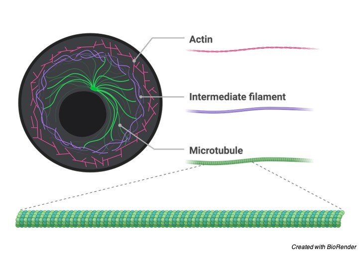
    o Rough ER tends to resemble flattened sacs, whereas smooth ER tends to be tubular.

    o Glucose ⇒ Glycogen = glycogenesis

    o Glycogen ⇒ Glucose = glycogenolysis

    o Triglycerides are produced in the smooth ER and stored as fat droplets.

    o Adipocytes are cells containing predominately fat droplets.

    o Such cells are important in energy storage and body temperature regulation.

    o Adipocytes, also called fat cells, are specialized cells whose cytoplasm contains almost nothing but triglycerides.

    o The Smooth ER and the cytosol share in the role of cholesterol formation and its conversion to various steroids.

    o Most of the phospholipids in a cell membrane are originally synthesized in the smooth ER.

    o The phospholipids are all synthesized on the cytosol side of the membrane and then some are flipped to the other side by proteins called phospholipid translocators located EXCLUSIVELY in the smooth ER.

    o Smooth ER oxidizes foreign substances, detoxifying drugs, pesticides, toxins, and pollutants.

    o The Smooth ER is the site of lipid synthesis, including steroids, detoxifying drugs, and useful for converting glycogen to glucose.

    o Peroxisomes are vesicles in the cytosol that grow by incorporating lipids and proteins from the cytosol.

    o Rather than budding off membranes like lysosomes from the Golgi, peroxisomes are self-replicating.

    o They are involved in the production and breakdown of hydrogen peroxide.

    o Peroxisomes inactivate toxic substances such as alcohol, regulate oxygen concentration, play a role in the synthesis and break of lipids, and in the metabolism of nitrogenous bases and carbohydrates.

    Endoplasmic Reticulum Citations

    Share

    Similar Post:

    Continue Reading
  • Endocytosis: Structure, Pinocytosis, and Phagocytosis

    Cell Membrane and Endocytosis

    o Besides transport across the membrane, cells can acquire substances from the extracellular environment through endocytosis.

    o Endocytosis/exocytosis requires ATP hydrolysis.

    o Prokaryotes’s membranes cant before endocytosis or exocytosis.

    o Viruses enter eukaryotic cells via endocytosis.

    Types of Endocytosis

    There are several types of endocytosis:

    I. Pinocytosis

    o Pinocytosis – (“cell-drinking”) is a form of endocytosis in which small particles are brought into the cell suspended within small vesicles.

    o It is primarily used for the absorption of extracellular fluids (ECF), and in contrast to phagocytosis, generates very small vesicles.

    o Unlike receptor-mediated endocytosis, pinocytosis is nonspecific in the substances that it transports.

    o The cell takes in surrounding fluids, including all solutes present.

    o A portion of the plasma membrane is invaginated and pinched off forming a membrane-bounded vesicle called an endosome.

    o This process, unlike phagocytosis, is nonselective.

    II. Phagocytosis

    o Phagocytosis is the cellular process of phagocytes (white blood cells) and protists of engulfing solid particles by the cell membrane to form an internal phagosome, which is a food vacuole.

    o Phagocytosis is a specific form of endocytosis involving the vesicular internalization of solid particles, such as bacteria, and is therefore distinct from other forms of endocytosis such as pinocytosis, the vesicular internalization of various liquids. 

    Cell Membrane, Cell Membrane Structure, Pinocytosis, Phagocytosis, Receptor mediated endocytosis 2

    o Phagocytosis is involved in the acquisition of nutrients for some cells, and in the immune system it is a major mechanism used to remove pathogens and cell debris.

    o The impetus for phagocytosis is the binding of proteins on the particulate matter to protein receptors on the phagocytic cell.

    o Organisms “eat” by stretching out pseudopodia and encircling any food particles they find in their paths.

    o In humans, antibodies or complement proteins bind to particles and stimulate receptor proteins on macrophages and neutrophils to initiate phagocytosis.

    III. Receptor Mediated Endocytosis

    o Receptor mediated endocytosis refers to specific uptake of macromolecules such as hormones and nutrients.

    o In this process, the ligand binds to a receptor protein on the cell membrane, and is then moved into a clathrin coated pit.

    o Clatherin is a protein that forms a polymer adding structure to the underside of the coated pit.

    o The coated pit invaginates to form a coated vesicle.

    o One way this process differs from phagocytosis is that its purpose is to absorb the ligands, whereas the ligands in phagocytosis exist only to act as signals to initiate phagocytosis of other particles.

    IV. Exocytosis

    o Exocytosis is simply the reverse of endocytosis.

    o In Eukaryotic cells, unlike prokaryotic, the membrane invaginates and separates to form individual, membrane bound compartments and organelles.

    Endocytosis Citations

    Share

    Similar Post:

    Continue Reading
  • Cell Nucleus: Definition, Function, Structure, and Facts

    Cell Nucleus

    o The major feature distinguishing eukaryotic cells from prokaryotic cells is the nucleus of the eukaryote.

    o The aqueous ‘soup’ inside the nucleus is called the nucleoplasm.

    o DNA is found in the nucleus and the mitochondria in eukaryotes.

    o The nucleus is wrapped in a double phospholipid bilayer (two membranes) said the nuclear envelope or membrane.

    Structure of Cell Nucleus

    Cell Nucleus, Cell Nucleus Definition, Cell Nucleus Function, Cell Nucleus Structure 1

    "DNA is found in the nucleus and the mitochondria in eukaryotes"

    o The nuclear envelope is perforated with large holes called nuclear pores, through which all molecules enter or leave.

    o Small molecules pass right though but larger molecules (such as RNAs and proteins) can’t pass through unless they carry an appropriate sorting signal.

    o RNA can exit the nucleus through the nuclear pores, but DNA cannot.

    o Proteins are transported in the their fully folded form in the nuclear pore and is actively transported in via GTP hydrolysis, proteins usually have to unfold to cross the membranes of other organelles.

    o Within the nucleus is an area called the nucleolus where rRNA is transcribed and the subunits of the ribosomes are assembled.

    o The nucleolus is not separated from the nucleus by a membrane.

    o Both the nucleus and the nucleolus disappears during prophase of mitosis and meiosis.

    "Both the nucleus and the nucleolus disappears during prophase of mitosis and meiosis"

    o The eukaryotic cell nucleus.

    o Visible in this diagram are the ribosome-studded double membranes of the nuclear envelope, the DNA (complexed as chromatin), and the nucleolus.

    o Within the cell nucleus is a viscous liquid called nucleoplasm, similar to the cytoplasm found outside the nucleus.

    o Chromatin is the complex combination of DNA, RNA, and protein that makes up chromosomes.

    o It is found inside the nuclei of eukaryotic cells, and within the nucleoid in prokaryotic cells.

    Cell Nucleus Citations

    Share

    Similar Post:

    Continue Reading
  • Fungi: Definition, Characteristics, Types, and Facts

    What are Fungi?

    o Fungi represent a distinct kingdom of organisms with tremendous diversity and are eukaryotes but have characteristics of both prokaryotes and eukaryotes (such as a cell wall).

    o Three divisions exist within this kingdom: Zygomycota, Ascomycota and Basidiomycota.

    o Fungi, like plants, are separated into divisions not phyla.

    o If something contains “mycota” in its division then it’s a fungus.

    o All cells of Zygomycota are haploid, except for the zygospore and don’t have cell walls except in their sexual structures.

    o Oomycota, which are slime molds and water molds, are not true fungus but are part of the Protista kingdom.

    o All fungi are eukaryotic heterotrophs that obtain their food by absorption rather than by ingestion: they secrete their digestive enzymes outside their bodies and then absorb the products of digestion.

    o Although most fungi are considered saprophytic (live off dead matter), many fungi do not distinguish between living and dead matter, and thus can be potential pathogens.

    o Fungi are larger than bacteria.

    Characteristics of Fungi

    o With the exception of yeasts, fungi are multicellular.

    o Yeasts are unicellular fungi.

    o Most fungi possess cell walls, called septa, made up of the polysaccharide, chitin.

    o Chitin is more resistant to microbial attack than is cellulose.

    o Arthopods’s (insects and crustaceans) exoskeleton is made up of chitin as well.

    o Septa is usually perforated to allow exchange of cytoplasm between cells, called cytoplasmic streaming.

    o This allows for rapid growth.

    o A fungal cell can contain multiple nucle.

    o The nuclei, in a single cell, may or may not be identical.

    o Fungi lack centrioles (as well as prokaryotes), mitosis takes place entirely within the nucleus, and their nuclear membranes don’t disappear during mitosis.

    o In the growth state, fungi consist of a tangle mass (called mycelium) of multiply branched thread-like structures called hyphae (haploid).

    Fungi Structure

    Fungi, Fungi Definition, Fungi Characteristics, Fungi Types, Fungi Facts 1

    Fungi Reproduction and Life Cycle

    o Like most organisms, fungi alternate between haploid and diploid stages in their life cycle; however, the haploid stage predominates, and is their growth stage.

    o Hyphae are haploid and some hyphae may form reproductive structures called sporangiophores.

    o By far the most important type of asexual reproduction is that of spore formation.

    o Asexual reproduction is extremely important to fungi.

    o It is responsible for the production of large numbers of spores throughout the year.

    o These structures release haploid spores that give rise to new mycelia in asexual reproduction.

    o Spore formation is NOT always via asexual reproduction.

    o Haploid spores can form and spread faster and more efficiently than diploid zygotes because they don’t undergo meiosis.

    o Yeasts rarely reproduce sexually by producing spores.

    o More often in yeasts, asexual reproduction occurs via budding.

    o Also called cell fission, in which a smaller cell pinches off from the single parent cell.

    "Sporangiophores release haploid spores that give rise to new mycelia in asexual reproduction"

    o When sexual reproduction occurs it is between hyphae from two mycelia of different mating types.

    o These two hyphae (haploid) grow towards one another eventually touching and forming a conjugation bridge, and becomes a gamete producing cell, called a gametangium.

    o In Zygomycota, the gametangia remain attached to the parent hyphae and the nuclei fuse with one another to produce a diploid zygote, called a zygospore.

    o After its formed it usually goes dormant, but when it is activated, the zygospore undergoes meiosis to produce haploid cells, one of which immediately grows a short sporangiophore to asexually reproduce many spores.

    o Sporangiophores release haploid spores that give rise to new mycelia in asexual reproduction.

    o Except for the zygospore all cell in Zygomycota are haploid, so they undergo mitosis.

    "When sexual reproduction occurs it is between hyphae from two mycelia of different mating"

    o The important thing to understand about fungal reproduction is that asexual reproduction.

    o Ex. Budding or mitosis normally occur when conditions are good.

    o Sexual reproduction (meiosis) occur when conditions are tough.

    Haploid state ⇒ asexual reproduction (mitsosis/budding (cell fission)) ⇒ conditions are good

    o Diploid state (zygospore) ⇒ sexual reproduction (meiosis) ⇒ conditions are bad.

    o Homologous chromosomes are chromosomes in a biological cell that pair (synapse) during meiosis.

    o The pair are non-identical chromosomes that both contain information for the same biological features and contain the same genes at the same loci but possibly each have different alleles (that is, different genetic information) at those genes.

    o Any cell that contains homologous chromosomes are called diploid.

    o Any cell that doesn’t contain homologous chromosomes are called haploid.

    o Lichens are composite organisms consisting of a symbiotic association of a fungus with a photosynthetic partner usually either a green alga or cyanobacterium.

    o The mixture of organisms regularly found at any anatomical site is referred to as the normal flora.

    Fungi Citations

    Share

    Similar Post:

    Continue Reading
  • Endospores: Definition, Function, and Diagram

    What is Endospore Formation?

    o An endospore is a dormant, tough, and non-reproductive structure produced by Gram-positive bacteria which forms when a bacterium produces a thick internal wall that encloses its DNA and part of its cytoplasm.

    o Remember that Gram + bacteria produce endospores.

    o The primary function of most endospores is to ensure the survival of a bacterium through periods of environmental stress.

    o They are therefore resistant to ultraviolet and gamma radiation, desiccation, lysozyme, temperature, starvation, and chemical disinfectants.

    Endospore Formation

    Endospore, Endospore Definition, Endospore Function, Endospore Diagram 1

    Endospore Formation Steps

    o In endospore formation, the bacterium divides within its cell wall.

    o One side then engulfs the other side.

    o The chemistry of the cell wall of the engulfed bacterium changes slightly to form the cortex of the endospore.

    o Several protein layers lie over the cortex to form the resistant structure called the spore coat.

    o A delicate covering called the exosporium, sometimes surrounds the spore coat.

    oThe outer cell then lyses, releasing the dormant endospore.

    o The endospore must be activated before it can be germinated and grow.

    o Activation usually involves heat.

    o Germination is triggered by nutrients.

    Endospore Formation Citations

    Share

    Similar Post:

    Continue Reading
  • Bacterial Growth Curve: Definition, Stages, and Graph

    Bacterial Growth Curve

    Bacterial growth in batch culture can be modeled with four different phases: lag phase, exponential or log phase, stationary phase, and death phase.

    Bacterial Growth Curve

    Bacterial Growth, Bacterial Growth Curve, Bacterial Growth Definition, Bacterial Growth Stages, Bacterial Growth Graph 1

    I. Lag Phase

    o During lag phase, bacteria adapt themselves to growth conditions.

    o It is the period where the individual bacteria are maturing and not yet able to divide.

    o During the lag phase of the bacterial growth cycle, synthesis of RNA, enzymes and other molecules occurs.

    II. Exponential Phase

    o Exponential phase (sometimes called the log phase) is a period characterized by cell doubling.

    o The number of new bacteria appearing per unit time is proportional to the present population.

    o If growth is not limited, doubling will continue at a constant rate so both the number of cells and the rate of population increase doubles with each consecutive time period.

    o For this type of exponential growth, plotting the natural logarithm of cell number against time produces a straight line.

    o The slope of this line is the specific growth rate of the organism, which is a measure of the number of divisions per cell per unit time.

    o The actual rate of this growth (i.e. the slope of the line in the figure) depends upon the growth conditions, which affect the frequency of cell division events and the probability of both daughter cells surviving.

    o Exponential growth cannot continue indefinitely, however, because the medium is soon depleted of nutrients and enriched with wastes.

    III. Stationary Phase

    o During stationary phase, the growth rate slows as a result of nutrient depletion and accumulation of toxic products.

    o This phase is reached as the bacteria begin to exhaust the resources that are available to them.

    o This phase is a constant value as the rate of bacterial growth is equal to the rate of bacterial death.

    IV. Death Phase

    At death phase, bacteria run out of nutrients and die.

    Bacterial Growth Curve Citations

    Share

    Similar Post:

    Continue Reading
  • Bacterial Conjugation: Steps, Definition, and Diagram

    Bacterial Reproduction: Bacterial Conjugation

    o Bacteria (prokaryotes) CAN’T undergo meiosis or mitosis, don’t have centrioles, and they can ONLY undergo Asexual Reproduction.

    o The type of cell division they are capable of undergoing is called binary fission, which exhibits exponential growth.

    o In binary fission, the circular DNA is replicated in a process similar to replication in eukaryotes.

    o Two DNA polymerases begin at the same point on the circle (ORI) and move in opposite directions making complementary single strands that combine with their template strands to form two complete DNA double stranded circles.

    o The cell then divides, leaving one circular chromosome in each daughter cell.

    o The daughter cells are genetically identical.

    Genetic Recombination

    There are three types of genetic recombination that can occur in bacterium:

    I. Bacterial Conjugation

    o Bacterial Conjugation requires that one of the bacterium have a plasmid with the gene that codes for the sex pilus.

    o If the plasmid can integrate into the chromosome of the host it’s called an episome.

    o In order for a bacterium to initiate conjugation, it must contain a conjugative plasmid.

    o Conjugative plasmids possess the gene for the sex pilus.

    o The sex pilus a hollow protein tube that connects the two bacteria to allow the passage of DNA.

    o The plasmid replicates differently than the circular chromosome.

    o One strand is nicked, and one end of this strand begins to separate from its complement as its replacement is replicated.

    o The loose strand is then replicated and fed through the pilus.

    o Two plasmids of interest are: the F plasmid and the R plasmid.

    o The F plasmid is called the fertility factor or F factor.

    o It allows the production of the sex pilus.

    o A bacterium with an F factor is designated as F+, and one without it is designated F-.

    o The F plasmid can be in the form of an episome and if the sex pilus is made while the F factor is integrated into the chromosome, then some or the entire chromosome may be replicated and transferred.

    Bacterial Conjugation Diagram

    Bacterial Conjugation, Bacterial Conjugation Steps, Bacterial Conjugation Definition, Bacterial Conjugation Diagram 1

    o R-plasmids often contain resistance genes coding for multiple antibiotic resistance.

    o As well as resistance transfer genes, they also code for the production of a conjugation (sex) pilus.

    o The conjugation pilus enables the donor bacterium to transfer a copy of the R-plasmid to a recipient bacterium, making it also multiple antibiotic resistant and able to produce a conjugation pilus.

    o Conjugation is conservative because the donor retains a complete original copy of the plasmid after the transfer is complete.

    II. Bacterial Transformation

    Transformation is the process by which bacteria may incorporate DNA from the external environment into their genome.

    III. Bacterial Transduction

    o Sometimes the capsid of a bacteriophage will mistakenly encapsulate a DNA fragment of the host cell.

    o When these new virions infect a new bacterium, they inject harmless bacterial DNA fragments instead of virulent viral DNA fragments.

    o This type of genetic recombination is called transduction.

    o The virus mediates that mediates transduction is called the vector.

    o This can be done artificially in a lab.

    o Mnemonic for transduction: the bacteriophage induces a change.

    Bacterial Conjugation Citations

    Share

    Similar Post:

    Continue Reading
  • Bacterial Cell Wall Structure: Gram Positive vs...

    Bacterial Cell Wall

    o The bacterial plasma membrane and everything inside of it is called the protoplast.

    o A protoplast isn’t a complete bacterium.

    o Protoplast: Plant, bacterial or fungal cell with the cell wall removed using either mechanical or enzymatic means.

    o Surrounding the protoplast is the bacterial envelope.

    o The component of the envelope, adjacent to the plasma membrane is the cell wall.

    o One of the functions of the cell wall is to prevent the protoplast from bursting.

    o Most bacteria (prokaryote) are hypertonic (hypotonic solution) to their environment (this means that the aqueous solution of their cytosol contains more particles than the aqueous solution surrounding them.

    o The resulting osmotic pressure causes a net movement of water into of the cell).

    o Compare isotonic where the cytosol contains the same amount of particles and hypotonic (hypertonic solution) where the cytosol contains less particles.

    o The cell wall is strong and able to withstand high pressure.

    o As the cell fills with water and the hydrostatic pressure builds, it eventually equals the osmotic pressure and the filling stops.

    o If the cell wall is removed, the plasma membrane cannot withstand the pressure.

    o Osmosis describes the movement of water.

    Peptidoglycan

    o Peptidoglycan, also known as murein, is a polymer consisting of sugars and amino acids that forms a mesh-like layer outside the plasma membrane of bacteria, forming the cell wall.

    o The polymers are crosslinked by an interbridge of more amino acids.

    o It is porous, so it allows large molecules to pass through.

    Structure and Synthesis of Peptidoglycan

    Peptidoglycan Cell wall, Peptidoglycan Structure, Peptidoglycan Synthesis, Gram Positive vs Gram Negative

    o Archaea don’t have peptidoglycan cell walls.

    o Peptidoglycan is more elastic than cellulose, which as we recall is carbohydrate formed by plants and contains Beta linkages (only bacteria eat Beta linkages and is part of the plants cell wall).

    o Some antibacterial drugs such as penicillin interfere with the production of peptidoglycan by binding to bacterial enzymes which make the crosslinks.

    "One of the functions of the cell wall is to prevent the protoplast from bursting"

    o Lysozyme, an enzyme produced naturally by humans, attacks the crosslinks as well.

    o In both cases the cell wall is disrupted and the cell lyses killing the bacterium.

    o Don’t confuse this with a lysosome which are organelles containing digestive enzymes

    o Capsules: This type of surface layer is composed primary of polysaccharides.

    o If the layer is strongly adhered to the cell wall, it is called a capsule; if not, it is called a slime layer.

    o Not all bacteria have this.

    o These layers provide resistance to phagocytosis and serve as antigenic determinants.

    o One method of classification of bacteria is according to the type of cell wall that they possess.

    o A staining technique, called gram staining, used to prepare bacteria for viewing under the light microscope, stains two major cell walls differently

    Gram Positive Bacteria

    o The first type is called gram-positive bacteria.

    o Gram-positive bacteria are those that are stained dark blue or violet by Gram staining.

    o This is in contrast to Gram- negative bacteria, which cannot retain the crystal violet stain, instead taking up the counterstain and appearing red or pink.

    o Gram-positive organisms are able to retain the crystal violet stain because of the high amount of peptidoglycan in the cell wall.

    o Thick peptidoglycan layer.

    Gram Positive Cell Wall
    Gram Positive Bacteria - Gram Positive Bacteria Cell wall - Gram Positive Bacteria Diagram - Gram Positive Bacteria Definition

    o Stain blue/violet.

    o The space between the peptidoglycan layer and the plasma membrane is known as the periplasmic space and it contains proteins that help the bacteria acquire nutrition.

    Gram Negative Bacteria

    o Gram-negative bacteria appear red or pink in color when gram stained.

    o Many species of Gram-negative bacteria are pathogenic, meaning that they can cause disease in a host organism.

    o This pathogenic capability is usually associated with certain components of Gram-negative cell walls, in particular the lipopolysaccharide (also known as LPS or endotoxin) layer.

    Gram Negative Cell Wall
    Gram Negative Bacteria - Gram Negative Bacteria Cell wall - Gram Negative Bacteria Diagram - Gram Negative Bacteria Definition

    o The following characteristics are displayed by Gram-negative bacteria:

    1. Thin peptidoglycan layer (which is present in much higher levels in Gram-positive bacteria)

    2. Outer membrane containing lipopolysaccharide (LPS) (can form a protective barrier from antibodies and many antibiotics) outside the peptidoglycan layer, this outer membrane is also more permeable than the inner, even allowing molecules the size of glucose to pass through

    3. Porins exist in the outer membrane, which act like pores for particular molecules

    4. A lipoprotein in the outer membrane called Braun’s lipoprotein points inward toward the cell wall and attaches covalently to the peptidoglycan

    5. There are two spaces between the layer of peptidoglycan and the two membranes

    6. Stain red/pink

    7. The periplasmic space is the space between the two membranes.

    o Some gram-negative bacteria possess fimbrie or pili (not to be confused with the sex pilus discussed below).

    o Fimbriae are short tentacles that can attach a bacterium to a solid surface.

    o They are NOT involved in cell motility.

    "Outer membrane is also more permeable than the inner, even allowing molecules the size of glucose to pass through"

    o Bacterial flagella are long, hollow, rigid, helical cylinders made from a globular protein called flagellin; these shouldn’t be confused with eukaryotic flagella which are made up of microtubules.

    o They rotate counterclockwise.

    o When they are rotated clockwise, the bacterium tumbles.

    o This tumbling acts to change the orientation of the bacterium allowing it to move forward in a new direction.

    o The movement of a bacterium toward or away from a particular stimulus is called taxis.

    o Such stimuli include chemicals (chemotaxis) and light (phototaxis).

    o The flagellum is propelled using the energy from a proton gradient rather than by ATP.

    Bacterial Cell Wall Citations

    Share

    Similar Post:

    Continue Reading