Author: Admin

Author: Admin

  • Phenylketonuria: Types, Symptoms, Causes, and Treatment

    What is Phenylketonuria

    Phenylketonuria is an inborn error of phenylalanine metabolism which is due to the decreased metabolism of the amino acid.

    If left untreated the conditions may lead to seizures, intellectual disability, mental problems and behavioral disorders.

    It also leads to lighter and musty smell skin.

    A baby which was born to a mother who is heterozygous carrier of this disease also gets affected with small head and low weight at birth.

    Cause of Phenylketonuria

    Phenylketonuria is an inherited genetic disorder due to a mutation in a PAH gene which causes low levels of the secretion of an enzyme phenylalanine hydrolase.

    This is generally an autosomal recessive condition where both the copies of gene have to be defected to acquire this condition.

    Types of Phenylketonuria

    There are two types of phenylketonuria namely classic PKU and variant PKU depending upon the function of the enzyme.

    However, the individual with only one copy of mutant gene do not have any symptoms. In many countries the screening test is done to the new born to protect them from severe symptoms.

    It affects 1 in 10,000 people every year. However early screening can help to start the treatments quickly and helps in avoiding death of an individual in early ages.

    Symptoms of Phenylketonuria

    Phenylketonuria causes mild to severe ranges. The severe form of phenylalanine is known as classic variant.

    If the baby is suffering from this classic variant it shows only mild symptoms and does show severe symptoms. 

    If it left untreated it leads to severe symptoms such as seizures, tumors, trembling and shaking of the limbs, stunted growth, hyperactivity, and also eczema in the skin and musty like odor in skin, urine and even in breath of an affected individual.

    If Phenylketonuria is not diagnosed at an early stage it can also leads to irreversible brain damage and disabilities of intellectual abilities and also other behavioral problems in the adult children.

    The other type of Phenylketonuria is also known as variant Phenylketonuria which does not cause any severe symptoms or Non Phenylketonuria hyperphenylalaninemia.

    This occurs mostly in babies who has too much phenylalanine but they cause only mild symptoms in infants.

    On following a proper diet from their Early stages Phenylketonuria can be prevented from becoming severe and also can be diminished.

    Pedigree Analysis of Phenylketonuria

    As we know this condition is caused due to an autosomal recessive condition which means that where the parents are in a heterozygous condition of being a carrier, it affects the child.

    The graph for this is listed below.

    Phenylketonuria - research tweet 1

    Frequency of Getting Phenylketonuria

    The frequency of obtaining this disease ranges according to the geographical regions. Phenylketonuria occurs 1 in 10,000 to 15,000 babies; who are new born.

    They are mostly detected at an early stage with the help of screening tests and treatments are started early, so the severity of this diseases can be prevented. So that the classic Phenylketonuria can be prevented from obtaining and variant form of this disease can be diminished.

    Cause of Phenylketonuria

    The genetic mutation that is happening in the PAH gene results in phenylketonuria. As PAH gene only instructs the body cells to synthesise an enzyme known as phenylketonuria; which helps in the conversion of amino acid phenylalanine to form other vital components that are needed for the body.

    Due to this gene mutated condition, the activity of phenylalanine hydrolase is reduced which effects the activity of processing phenylalanine in diets.

    This leads to a condition of building toxic levels in blood cells and other tissues of the body.

    This condition occurs because the nerve cells in the brain are sensitive particularly to the phenylalanine levels where excessive production can cause damage to the brain cells.

    Classic PKU happens when a phenylalanine activity is reduced to the extent levels or it may be absent at most severe cases.

    People suffering from this condition if left untreated it causes severe brain damage and other serious health problems.

    Mutation in this gene retains the activity of some enzymes and results in milder variant PKU.

    Phenylketonuria Diagnosis

    Screening test is one of the most popular test. It is performed in babies whose parents have this condition of inheritance.

    Through this test it can be identified that whether the gene is inherited from parents to their children and if found in early stages it can be treated and cured before its s becoming severe.

    These tests are done after 6 weeks of birth of a child.

    Treatment of Phenylketonuria

    The babies found out with this type of disorders can be provided or treated with proper nutritious diet which limits the intake of phenylalanine and they are led to breast fed to get the essential nutrients.

    Physicians also provide these kinds of children a special formula of diet known as Lofenalac which is very high in protein.

    Phenylketonuria Citations

    Share

    Similar Post:

    Continue Reading
  • Sickle Cell Anemia: Types, Symptoms, Causes, and...

    What is Sickle Cell Anemia

    We come across many disorders in our day-to-day life, some are acquired and some are transmitted through genes from our ancestors, which is known as genetic disorder.

    Sickle cell anemia is one such inherited disease which is caused through genetic disorder or through mutations occurring due to environmental factors, which leads to a defect in a DNA molecule.

    Sickle cell anemia - research tweet 2

    Cause of Sickle Cell Anemia

    It is one of the abnormal inherited disorder caused by mutation in an autosomal gene.

    Here the red blood cells of an affected individual is of sickle shaped instead of having a biconcave shape. Where these sickle shaped blood cells do not provide a capacity to carry enough oxygen and nutrients all over the body which results in anemia as well as other diseases.

    The people with this type of red blood cells are called as sickle cell traits.

    The sickle shaped cells cannot pass through the blood vessels easily as they gets struck in between the small blood vessels such as capillaries which results in haemolytic anemia and leads to death of an individual.

    This is caused due to the mutation in the single-base, the sixth amino acid in the chain is valine, rather than glutamic acid

    Characteristics of Sickle Cell Anemia

    Linus Pauling was the first one to identify this type of mutation in an individual where the genotype for this mutation is Hbs1 Hbs1which is a condition that our body does not have enough haemoglobin, because Hbs1 have negative charge compared to that of HbA1.

    If the individual is a carrier of this syndrome, they have their genotype as HbA1 Hbs1 which leads to a condition of mild sickle shaped anemia.

    This is considered by Pauling as one of the gene molecular disease.

    Effects of Sickle Cell Anemia

    The sickle cell anemia leads to the increased fragility of the red blood cell which leads to haemolytic anemia and it also increases the viscosity of the blood and it also lets the blood cells to stag in the small blood vessels which forms a thrombus and infracts.

    This phenomenon also causes damage to the spleen which is often said to be autopsleenectomy where the spleen ends as a non-functional unit and it also leads to other side effects such as pneumococcal meningitis and other infectious diseases.

    Sickle Cell Anemia Epidemiology

    The Sickle cell anemia is most common in central Africa where almost 20 percent of the people are having this trait and are affected with this disease.

    About 2% of these die in an early stage of their life.

    It has also been distributed widely in USA where this trait Hbs has found in Arab racial groups and it is also rarely seen among India and American Negros.

    How Sickle Cell Anemia Affects?

    The people who are having sickle cell trait as Sis/Sis are affected by this disease but they don’t show symptoms in certain cases.

    But in some cases, in high altitudes, it affects the respiratory tract and bleeding in the renal papillae.

    The persons who are having homozygous trait of sickle cell anemia do not get infected by malaria. But they die at the early stages of life.

    The normal trait persons are not resistant to malaria. The persons who have the heterozygous condition of causing sickle cell anemia is also resistant to malarial infections and they lead almost a normal life style and anemia is not severe in such cases.

    Types of Sickle Cell Anemia

    There are many types of sickle cell anemia depending upon the various changes in the traits.

    Some of the types are discussed as follows;

    HbSS: In this case the person gets two inherited genes from each of their parents This type is most severe.

    HBSC: Here the defected gene is inherited from a single parent and a normal gene from the other parent it also results in abnormal haemoglobin and results in a carrier offspring but it does not cause any serious symptoms.

    HbS- Beta thalassemia: A individual suffering from this disorder get one of allele from an infected sickle cell parent and the other from the Beta thalassemia affected person.

    Symptoms of Sickle Cell Anemia

    In general, if our body does not get any enough oxygen it causes many complications and symptoms as the outcome of an infection.

    These symptoms vary according to the age and life style factors of the individual. It causes early symptoms such as fatigue, jaundice; which leads to yellowing of the skin and eyes, swelling of hands and feet; which causes severe pain.

    Later the symptoms are little complicated such as acute chest syndrome, damage in a normal vision, enlarging of spleen, ulcers in the leg, Heart attack, stroke, deep thrombosis in the veins, infections in the heart, kidney or liver.

    Formation of gallstones, infertility in males and causes of malnutrition in kids, high blood pressure in the lungs which is also known as Pulmonary hypertension, bone and joint inflammation etc.

    Treatment of Sickle Cell Anemia

    Currently the sickle anemia has no cure but it is possible to overcome the difficulty of this syndrome through blood transfusions or through stem cell transplants the disease can be controlled.

    Sickle Cell Anemia Citations

    Share

    Similar Post:

    Continue Reading
  • Chromosomal Instability: Definition, Types, and Examples

    What is Chromosomal Disorder or Chromosomal Instability?

    Many types of disorders are arising in humans, each are due to various reasons such as genetic, acquired or through mutated conditions.

    These mutated conditions are mostly due to any abnormality or defect in the chromosome of an individual.

    Humans usually have 23 pairs of chromosomes in that 22 pairs are autosomes and the last pair is known as allosome or sex chromosome.

    Chromosomal disorders occurs if there is any addition of chromosomes to the particular number or reduction in this particular set of numbers.

    Any change in the number of chromosomes is known as ploidy. And this disorder occurs when there is any change in the structure of the chromosome.

    Why Chromosomal Disorder or Chromosomal Instability?

    Chromosomal disorders are due to any abnormality or change in structure or function of a chromosome.

    A change in a structure of any one of the chromosomes or missing of a part of a chromosome or a whole chromosome or mutation or any defects in a particular gene in a chromosome may be due to chromosomal disorders.

    These disorders are detected with the help of banding patterns which helps us to identify the abnormalities in the chromosome by staining the chromosomes with colour stains.

    Karyotyping also helps us to detect the missing pair of chromosomes. In karyotyping the chromosomes are arranged in a definite manner and seen under a light microscope.

    Idiogram also helps in karyographing of chromosomes. Any small change in the chromosome can lead to vigorous effects in the genes and affect the enzyme producing capacity; which leads to various diseases.

    These chromosomal abnormality happening in an allosomes are definitely passed to the next generations, where as the abnormalities of autosomes may or may not pass to the next generation.

    Types of Chromosomal Instability

    Generally, there are two types of chromosomal instability which leads to chromosomal disorders.

    1. Structural Chromosomal Instability

    2. Numerical Chromosomal Instability

    1. Structural Chromosomal Instability

    The structural chromosomal instability are due to a change in structure of a chromosome or missing of any part of the chromosome.

    As said, there are about 23 pair of chromosomes which are 46 in number.

    It also occurs when there is a change in structure or missing of any part of the chromosome or when the missing parts are translocated to any other areas.

    This condition causes many birth effects due to addition or deletion of a part of a chromosome.

    Chromosomal Instability - research tweet 2

    All chromosomes are made up of many segments which are commonly divided into short and long arm, where the short arm forms upper half of the chromosome and the long arm forms a lower part of the chromosome.

    The short arm is known as “p” arm and the long arm is known as “q” arm.

    i. Chromosomal Instability: Deletion of Chromosome

    In deletion of chromosomes the part of a chromosome is deleted or went missing.

    Even a small piece of a missing chromosome can lead to vigorous disorders or misleading of gene encoding, as a small part contains various information for coding different genes and enzymes.

    When these conditions happen in a pregnant mother this condition is being inherited to an offspring.

    The best example for deletion is Cri-du-chat syndrome where there is the damage or missing of chromosome number five.

    Cri-du-chat Syndrome

    Cri-du-chat syndrome is usually seen among one in 20,0000 people. It occurs mostly in the children of America.

    It is caused due to a defect or deletion in the chromosome number five. Which occur especially in the p arm and it is said as 5p.

    The characteristic feature of this syndrome is that the children’s have a high pitch cry, poor tone of muscles and the heads of these children are very small.

    They are usually underweight. They also face the problem while speaking, they express their emotions with the help of signs.

    Other related health problems are with feeding, hyperactivity of an individual, and disability in their intellectual activities.

    Mostly the people affected with this Cri-du-chat syndrome have a normal life span in most of the cases.

    They result in series condition only when they have organ defects along with this syndrome, which leads to short life span of an individual.

    The children with this type of syndrome are involved in language and physical therapy to improve themselves.

    ii. Chromosomal Instability: Duplication of Chromosome

    The term itself provides a complete meaning for this syndrome, here the duplication of chromosomes occurs where the same chromosome forms a copy of the other, which results in two copies of the same chromosome, as a result two copies of the same genetic material are formed.

    Even if the genes are present in many copies it leads to non-functioning of the chromosomes.

    Or by giving extra instruction to the same enzyme production which leads to errors in developing a baby.

    This duplication leads to a Pallister-Killian syndrome.

    Pallister-Killian Syndrome

    In this syndrome the extra copy of chromosome 12 is present. Which leads to mosaicism.

    Infants with this condition has many serious problems such as intellectual disability, poor muscle tone, poor feeding, stiffness of joints, cataracts in eye.

    Usually, a people in this syndrome have a very short life span. These people have only their life span till 40 years.

    iii. Chromosomal Instability: Inversion of Chromosome

    This occurs when the part of the chromosome results in breakage and reuniting by combining by rotating at 180º with its own axis.

    This results in rearrangement of genes. But it is not severe as much as other structural abnormalities.

    iv. Chromosomal Instability: Translocation of Chromosome

    The transfer of chromosomes with a non-homologous pair is known as translocation. The addition or deletion or rearrangement of genes may occur in this condition.

    Which leads to a phenotypic change resulting in abnormalities in the development of egg, sperm or zygote. It often results in miscarriage of a pregnant women.

    2. Numerical Chromosomal Instability

    Any abnormalities in the chromosomal number leads to this condition. It is divided into two types as aneuploidy and euploidy

    i. Chromosomal Instability: Aneuploidy

    It is a condition where loss or gain of a chromosome occurs. It mostly occurs due to a non-disjunction in a chromosome, during cell division. Aneuploidy is further classified as Trisomy (2n+1) and monosomy (2n-1).

    The disorders caused due to aneuploidy is Turners syndrome, Klinefelter’s syndrome and Downs syndrome.

    ii. Chromosomal Instability: Euploidy

    Here the loss or gain of whole set of the chromosomes occurs. This condition mostly occurs in plants.

    Haploid: In this condition loss of one set of the chromosomes occurs.

    Polyploid: In this condition the loss of one or more sets occurs.

    Chromosomal Instability Citations

    Share

    Similar Post:

    Continue Reading
  • Metformin: Description, Properties, and Health Benefits

    History of Metformin

    Recently called metformin, Dimethylbiguanide, was initially integrated in 1929 after an investigation into a more harmful hypoglycaemic subsidiary guanidine; found in Galega officinalis which had been utilized to treat the sign and effects of diabetes since the seventeenth century.

    Metformin was not utilized in Europe to treat diabetes, because of the poisonousness of its subsidiaries and the creation of engineered insulin in 1922, until the last part of the 1950’s when French Physician Jean Sterne who made note of its antihyperglycaemic impacts during preliminaries into discovering new malarial medicines.

    Metformin and Type-II Diabetes

    Today, metformin is the principal line and most ordinarily endorsed prescription for the treatment of type-II diabetes mellitus (T2DM).

    This is as it is unfathomably protected, savvy, being one of the fewanti-hyperglycaemic prescriptions to not build weight and decrease cardiovascular sickness hazard.

    In spite of metformin’s fame, there is as yet a general absence of comprehension of its systems of activity.

    Metformin likewise has low power, with a maximum portion of 2500mg, and regular contraindications coming about in around 25% of medicines.

    Does this suggest the conversation starter; is metformin the best first-line treatment for type-II diabetes mellitus?

    Mechanism of Metformin

    Since the 1950’s there have been considerable endeavors into understanding the manners by which metformin can bring down hepatic glucose creation (HGP) and plasma glucose levels (PGL).

    Up to this point, the hypothesis was that metformin’s enemy of hyperglycemic impact was fundamentally intervened through a Liver Kinase B1 (LKB1) – 5′ AMP-enacted protein kinase (AMPK) subordinate pathway.

    Metformin - research tweet 1

    This has now been tested after examinations showing that hereditarily inadequate mice, lacking AMPK in the liver, kept up with tantamount blood glucose levels to that of the benchmark group of mice.

    This shows that metformin doesn’t intervene gluconeogenesis, essentially exclusively, in the liver through the LKB1-AMPK pathway.

    Subsequently further exploration has been embraced to decide the genuine instruments of activity of metformin.

    There are presently different working speculations concerning the objective for metformin’s capacity to oversee HGP notwithstanding almost certainly, it intervenes through various methods of activity.

    One late hypothesis is that, because of high measures of metformin should have been powerful, it adjusts the microbiota of the host, expanding Akkermansia muciniphila populaces.

    A. muciniphila was displayed to altogether expand glucose resistance in High-fat-diet (HFD) prompted diabetic mice.

    There is likewise mounting proof from contemplates showing the gut may assume a vital part in metformin’s intervention of HGP and PGL. One such examination showed that intraduodenal mixtures of metformin gave the main change in HGP.

    This examination likewise exhibited the dependence of glucagon-like peptide-1 receptors (GLP-1R) and protein kinase A (Pka) to bring down HGP, recommending an AMPK – 1.

    GLP-1R – Pka subordinate pathway. This signallling pathway doubtlessly brings down HGP through neuronal intercession of glucose homeostasis through the gut-cerebrum liver hub.

    Different investigations have likewise shown supplement detecting upper intestinal lipids setting off a gut-cerebrum liver pivot to direct gluconeogenesis exhibiting the body’s capacity to control through this pathway.

    Advantage of Metformin in Diabetes

    Metformin is used for treatment of Type 2 Diabetes Mellitus, this is to a great extent because of its expense, solid wellbeing record, minor incidental effects – discarding uncommon instances of lactic acidosis – and cardioprotective impacts.

    Metformin might be the best first-line treatment for overweight type II diabetic patients.

    Another advantage of metformin over other regular enemy of hyperglycaemic medicines is that it doesn’t bring about extra weight acquire and may advance less eating and weight reduction.

    This is probably because of its capacity to decrease HGP giving less glucose in the circulation system for adipocytes to change over into fat contrasted with insulin which would have the contrary impact and increment weight acquire because of expanded cell take-up.

    Is Metformin really Effective?

    Metformin tends to the manifestations of hyperglycaemia as opposed to effectively remedying the basic issues related with T2DM.

    Diabetes is regularly described to have 8 significant physiological disturbances known as the ominous octet.

    Along these lines thought to have a powerful treatment in which HbA1c is brought down and kept up with by turning around the fundamental dysfunctions related with the infection.

    One such proposed treatment is regulating the GLP-1R agonists (GLP-1RAs). GLP-1RAs, for example, liraglutide have exhibited the capacity to address 6 of the ominous octets.

    It has likewise been shown that liraglutide brings down HbA1c quicker and drastically more than metformin, a decrease of 1.14% (1.8mg-liraglutide) longer than a year, contrasted with sulfonylurea, which has comparative HbA1c decrease to metformin, lessening by 0.51%.

    Hence thought to have a powerful treatment in which HbA1c is brought down and kept up with by switching the fundamental dysfunctions related with the infection.

    One such proposed treatment is regulate GLP-1R agonists (GLP-1RAs). GLP-1RAs, for example, liraglutide have exhibited the capacity to address 6 of the ominous octet.

    It has additionally been shown that liraglutide brings down HbA1c quicker and drastically more than metformin, a decrease of 1.14% (1.8mg-liraglutide) longer than a year, contrasted with sulfonylurea, which has a comparative HbA1c decrease to metformin, lessening by 0.51%.

    Metformin has obviously solid advantages related with treatment. It is reliably viable in decreasing danger factors related with T2DM and is adequate treatment in lessening both HGP and PLG.

    Nonetheless, because of the far and wide utilization of metformin and its evident adequacy different medicines, for example, GLP-1RAs are not being investigated as generally accessible choices for patients with T2DM.

    It ought to likewise be noticed that regardless of the clear suitability of GLP-1RAs, there is yet to be a moderate choice, liraglutide yearly expense is ~$9,300 contrasted with metformin <$50.

    Further exploration should likewise be done into the conceivable results of GLP-1RAs as there is still some debate encompassing expanded dangers of the thyroid malignant growth, pancreatitis and the kidney impairment.

    Considering the expenses and the vulnerability encompassing more current choices, for example, GLP-1RAs I accept that metformin ought to stay the main line treatment for type-II diabetes mellitus in any case, further investigation into new strategies following the very authoritative opinion as that of GLP-1RA medicines ought to be investigated in order to track down a more successful and designated treatment.

    Metformin Citations

    Share

    Similar Post:

    Continue Reading
  • DNA vs RNA: Definition, Differences, and Similarities

    What is DNA

    It is defined as a long polymer that has a deoxyribose and phosphate foundation having 4 definite bases namely, thymine, adenine, cytosine and guanine.

    It is primarily located in the nucleus of a cell and in the mitochondria.

    It has 2 de-oxyriboses.

    Its important function is to transmit genetic information to produce the various kinds of proteins and ultimately the whole body of the organism.

    Thus, it acts like a extended storage.

    The DNA is a double-stranded molecule that has an extended chain of nucleotides.

    DNA can reproduce on its own, thus, known as self-replicating.

    The base pair for the DNA is as follows;

    • C:G :: Cytosine pairs with Guanine

    • A:T :: Adenine pairs with Thymine

    In cells, DNA (Deoxyribonucleic corrosive) is the nucleic acid that capacities as the first outline for the synthesis of proteins.

    DNA contains the sugar deoxyribose, phosphates and a special sequence of the nitrogenous bases adenine (A), guanine (G), cytosine (C) and thymine (T).

    The DNA molecules contain directions a living element needs to develop, create and imitate.

    These directions are available inside every cell and are inherited from the parents to their offsprings.

    It is comprised of nucleotides which contain nitrogenous group, a phosphate bunch, and a sugar group.

    The request for the nitrogenous bases – thymine(T), guanine(G), cytosine(C), and adenine(A), is vital in deciding the hereditary code.

    Genes are created by the order of the nitrogenous bases available in the DNA which is important for protein synthesis.

    The RNA is another nucleic acid that translates hereditary data into proteins from DNA.

    DNA vs RNA - research tweet 1

    The nucleotides are connected together for the generation of two long strands which twist to create a structure known as the double-helix which takes after that of a ladder wherein the sugar and phosphate particles from the sides while the rungs are shaped by the bases.

    The bases situated on one strand clubbed with the bases on the other strand, as in – guanine sets with cytosine and adenine sets with thymine.

    The DNA molecule are extensively long and consequently without the right packaging, they can’t find a way into cells.

    In this manner, DNA is firmly coiled to deliver developments alluded to as chromosomes. Each chromosome has a solitary DNA molecule. In human, there are 23 sets of chromosomes that are available inside the core of the cells.

    Types of DNA

    A-DNA

    It is found at a humidity of 75%.

    In a climate where there is a raised salt concentration or ionic concentrations, like K+, Na+, Cs+ or in a condition of dehydration it’s anything but a structure that contains 11 nucleotide pairs with a rise of 2.56Aº in an upward direction for each base pair.

    It has the broadest helical diameter amongst all DNA structures – 23Aº DNA which is an ordinary helix that is right-handed with a rotation of 32.7º per base pair.

    B-DNA

    It is seen at a humidity of 9.25 and a low salt concentration or ionic strength. It has 10 base pairs for each turn which from the helix axis.

    There is a distance of 3.4Aº with a helical width of 20Aº.

    The Watson-Crick’s double helix model is characterized as a B-type of DNA.

    C-DNA

    It is seen at a humidity of 66% and in the inhabitance of a couple of ions like Lithium(Li+).

    It intently has 9.33 base pairs for each turn.

    The distance across the helix is about 19Aº and the upward ascents for each base pair for the right-handed helix is 3.32º.

    D-DNA

    It is noticed infrequently as an outrageous variation.

    The 8 base pairs are tilted negatively from the helix axis with an axial rise of about 3.03Aº.

    Z-DNA

    It is found in a climate with an extremely high salt concentration. In contrast to the A, B, and C types of DNA, it is a left-handed helical structure.

    The foundation is arranged in a crisscross shaped made up of the sugar-phosphate linkage wherein the recurrent monomer is the dinucleotide rather than the mononucleotide, which is seen in substitute structures.

    RNA

    It is defined as a polymer with a ribose and phosphate foundation with 4 diverse bases namely, uracil, cytocine, adenine and guanine.

    It is usually found in the cytoplasm, nucleus and in the ribosome.

    It has riboses. The RNA is a single-stranded molecule that has a miniature chain oof nucleotides.

    RNA cannot reproduce on its own, it needs DNA to synthesize its molecule.

    The base pair for the RNA is as follows;

    • C:G :: Cytosine pairs with Guanine 

    • A:U :: Adenine pairs with Uracil

    Ribonucleic corrosive (RNA) is a nucleic acid which is straightforwardly engaged with protein combination.

    Ribonucleic acid is a significant nucleotide with long chains of nucleic corrosive present in every living cell.

    Its primary job is to go about as a courier passing on guidelines from DNA for controlling the proteins synthesis.

    RNA contains the sugar ribose, phosphates, and the nitrogenous bases adenine (A), guanine (G), cytosine (C), and uracil (U). DNA and RNA share the nitrogenous bases A, G, and C.

    Thymine is normally just present in DNA and uracil is typically just present in RNA.

    Types of RNA

    Just a portion of the genes in cells are expressed into RNA.

    Coming up next are the sorts of RNA wherein each type is encoded by its own kind of gene:

    • tRNA: also known as the transfer RNA or the tRNA translate amino acids to ribosomes while translation.

    DNA vs RNA - research tweet 2

    • mRNA:  also known as the messenger RNA or the mRNA encodes amino acid sequences of a polypeptide.

    • rRNA:  also known as the ribosomal RNA or the rRNA produces ribosomes with the ribosomal proteins that are organelles accountable for the translation of the mRNA.

    • snRNA:  The small nuclear RNA creates the complexes alongside proteins which are used in RNA processing in the eukaryotes.

    DNA vs RNA Citations

    Share

    Similar Post:

    Continue Reading
  • DNA Replication: Steps, Process, and Mechanism I...

    What is DNA Replication?

    In atomic science, DNA replication is the organic cycle of delivering two indistinguishable replicas of DNA from one unique DNA particle.

    DNA replication happens in all living beings going about as the most fundamental part for natural legacy.

    It is perused in the 3 ‘to 5’ direction by the DNA polymerase, which implies that the subsequent strand is combined in the 5 ‘to 3’ direction.

    Replication includes the creation of indistinguishable DNA helices from a double stranded DNA molecule.

    Catalysts are basic to DNA replication as they promote vital strides in this cycle.

    The whole DNA replication measure is critical for both cell development and growth in organisms. It’s likewise significant in cell fix.

    Beginning of DNA Replication

    Replication consistently starts at a particular point on the DNA, which is known as the origin of replication and is perceived by its sequence.

    DNA Replication Steps DNA Replication - research tweet 2

    Steps of DNA Replication

    A) Initiation: Preparatory step

    • Step 1: Replication fork formation.

    B) Elongation: DNA Synthesis Begins

    • Step 2: Primer binding

    • Step 3: Synthesis of leading and lagging strands

    • Step 4: Remove primer and gap fill

    • Step 5: Proofreading

    C) Termination:

    • Step 6: End of the replication

    Step 1: Replication Fork Formation

    Before DNA can be duplicated, the double stranded molecule should be “unzipped” into two solo strands.

    DNA has four bases called adenine (A), thymine (T), cytosine (C), and guanine (G) that form pair between the two strands. Adenine just combines with thymine and cytosine just ties to guanine.

    To loosen up DNA, these base-pair interactions should be broken. This is finished by a protein known as DNA helicase.

    Notwithstanding, there is a unique initiator protein that is needed to sets off DNA replication, to be specific DnaA.

    It ties areas at the oriC site all through the cell cycle.

    To start the replication, notwithstanding, the DnaA protein should tie to a couple of explicit oriC groupings that have five repeats of the 9 bp arrangement also known as the R site.

    At the point when DnaA ties to the oriC site, it enlists a helicase catalyst (DnaB helicase). Presently the DNA helicase breaks the hydrogen bond that holds reciprocal DNA bases together.

    The detachment of the two single strands of DNA makes a two Y-formed design called a replication fork.

    Together they structure a bubble-like design called a replication bubble. These two separate strands fill in as a layout for the creation of the new DNA strands.

    Helicase is the main replication compound to be stacked at the beginning of replication. Helicase’s responsibility is to just move the replication forks forward by “unwinding” the DNA.

    As we probably are aware, DNA is entirely unstable as a single strand. Along these lines, cells can keep them from returning together in a double helix.

    To do this, a particular protein called single-stranded DNA binding proteins (SSBs) covers and keeps the isolated strands of DNA close to the replication fork.

    When the helicase rapidly unwinds the double helix. It raises the tension on the remaining DNA particle.

    Topoisomerase plays a significant support part during DNA replication. This protein forestalls the DNA double helix in front of the replication fork from turning out to be too tight when the DNA is opened.

    It does this by making impermanent nicks in the helix to release tension and afterward fixing the nicks to forestall perpetual harm.

    Elongation of DNA Replication: DNA Synthesis Begins

    Step 2: Primer binding

    Another enzyme was presented in this progression, which assumes the main part in the fabrication of DNA, called as DNA polymerase.

    It can just add nucleotides at the 3 ‘end of a current DNA strand. Primase forms the RNA primer, or short nucleic acid strand, that finishes the format, providing a 3 ‘end for working on DNA polymerase.

    A commonplace primer has around five to ten nucleotides. The primer starts the synthesis of DNA. When the RNA primer is set up, DNA polymerase “extends” it and consecutively adds nucleotides to make another strand of DNA that is complementary to the template strand.

    DNA Replication Steps DNA Replication - research tweet 1
    Step 3: Synthesis of Leading and Lagging Strands

    One of the strands is arranged in the 3′ to 5′ bearing (towards the replication fork), this is the leading strand.

    The other strand is situated in the 5′ to 3’direction (away from the replication fork), this is the trailing strand. Due to their diverse direction.

    Leading Strand Synthesis

    A short piece of RNA called a primer (made by enzyme known as primase) comes by and attach to the terminal of the leading strand.

    The primer fills in as the beginning stage for DNA synthesis.

    The DNA polymerase ties to the leading strand and afterward strolls alongside it, adding new complementary nucleotide bases (A, C, G, and T) in the 5′ to 3′ direction to the DNA strand.

    This sort of replication is known as continuous.

    Lagging Strand Synthesis

    The DNA polymerase consistently runs in the 5 ‘to 3’ direction. At the point when the two strands have been incorporated consistently while the replication fork is moving.

    One strand would consequently must be exposed to a 3 to 5 synthesis. Okazaki tracked down that one of the new strands of DNA was integrated in short pieces known as Okazaki fragments.

    This work eventually prompted to concluded that one strand is synthesized persistently and others irregularly.

    DNA polymerase III uses one bunch of its core subunits (the core polymerase) to continuously synthesize the leading strand. While the other two sets of the core subunit lie starting with one Okazaki fragment then onto the next on the looped duct.

    In vitro, there are just two arrangements of core subunits containing DNA polymerase III holoenzymes that can combine both the leading and lagging strand.

    Nonetheless, the third arrangement of core subunits expands the productivity of delayed strand synthesis just as the processivity of the in general replisome.

    At the point when DnaB helicase ties before DNA polymerase III. It starts by unwinding the DNA on the replication fork as it moves alongside the trailing strand template in the 5 ‘to 3’ direction.

    Primase every so often connects with DnaB helicase and synthesize a short RNA primer.

    Another slide clamp is currently situated on the primer through the clamp loading complex of DNA polymerase III. At the point when the union of the Okazaki fragment is complete.

    Replication stops and the core subunits of DNA polymerase III separate from their slide clamps and associate with the new clamp.

    This starts the synthesis of another Okazaki fragment. Two sets of core subunits can be engaged with the union of two unique Okazaki fragments simultaneously.

    When an Okazaki piece is complete, its RNA primers are taken out by DNA polymerase I or RNase H1. What’s more, that space is supplanted by DNA by the polymerase. The leftover nick has now been fixed by the DNA ligase.

    Step 4: Remove Primer and Gap Fill

    When both the continuous and discontinuous strands are framed, an enzyme called an exonuclease (DNA polymerase I or RNase H1) eliminates all RNA primers from the first strands.

    These primers were supplanted by appropriate DNA bases. The excess nick was fixed by the DNA ligase.

    DNA ligase catalyzes the development of a phosphodiester bond between a 3′-hydroxyl group on the end of one strand of DNA and a 5′-phosphate on the end of another strand.

    Step 5: Proofreading

    The DNA replication happens with high constancy. It contains some unacceptable nucleotide once for each 104–105 polymerized nucleotides.

    The exactness of replication relies upon the capacity of replicative DNA polymerases to choose the right nucleotide for the polymerization reaction.

    This high devotion isn’t accomplished in a single step yet rather is produced through the activity of a few progressive error avoidance and processing steps.

    These steps incorporate the selection of the right DNA base by the DNA polymerase, the editing of polymerase miss addition mistakes by exonucleolytic proofreading, lastly, the post-replicative DNA crisscross repair, which recognizes DNA mismatch and corrects recently replicated DNA.

    Termination of DNA Replication

    DNA replication stops when two forks of replication meet on a similar stretch of DNA, and the accompanying occasions happen, however not really in a specific order: forks converge until the entirety of the mediating DNA is loosened up; remaining gaps are filled and tied; Catenans are removed, and replication proteins are discharged.

    Step 6: End of DNA Replication

    At last, the parent strand and its complementary DNA strand coils into the recognizable double helix shape. The outcome is two DNA atoms comprising of one new and one old chain of nucleotides.

    Every one of these two little daughter helices is an almost precise of the parental helix.

    DNA Replication Citations

    Share

    Similar Post:

    Continue Reading
  • Homozygous vs Heterozygous: Definition and Differences

    Homozygous vs Heterozygous

    Features of Homozygous

    Homozygous refers to the state of the genes or genetic condition in which an individual has inherited the similar DNA sequence for a particular gene from both their biological parents and it is generally used with reference to the disease.

    For instance, if an individual inherited a mutated allele in the DNA sequence from maternal side and similar gene from the paternal part as well, then the mutation is explained as homozygous for that individual.

    Thus, after receiving 2 identical copies of the gene, detrimental effects will express phenotypically for which the genes are coded.

    The term “heterozygous” attribute to a pair of alleles so heterozygous is when you have 2 dissimilar alleles that means you have received dissimilar copies from each parent.

    In a heterozygous genotype, the allele which is dominant dominates over recessive and hence the dominant feature will be expressed while the recessive will not show but you will be still a carrier.

    This will be passed it on to your children. Homozygous is the opposite of the heterozygous where the feature of the similar alleles — either dominant or recessive — is manifested.

    There are three possible alleles for A, B, O blood type. However, the contribution from each parent’s gene makes a difference an individual exhibit with phenotypic characteristic.

    Sometimes, it may be possible that both the parents pass on the same allele for a gene, making it a homozygous trait.

    While in certain conditions, different set of alleles are triggered resulting in heterozygous trait.

    A homozygous trait is mainly responsible for phenotypic appearance in the organism. A pea plant will definitely exhibit long stem, if it receives two alleles associated with tallness. In contrast, if it gets two “short” alleles, then plant with stunted growth will be evident.

    Thus, for a specific characteristic, the genotype may be either homozygous or heterozygous, however, the way it is expressed it is identified as phenotype.

    Homozygous vs Heterozygous - research tweet 1
    Features of Heterozygous

    Heterozygous is a genetic condition where an individual inherits different alleles of a gene from the two parents.

    Heterozygosity is observed in a diploid organism where a gene contains two different alleles at a gene locus.

    In heterozygous chromosomes, the two alleles are different, and the heterozygous genotypes are represented by a capital letter, which indicates the dominant allele and a lowercase letter, which indicates the recessive allele, like Bb for eye color.

    In heterozygous chromosomes with genes having traits that are expressed via complete dominance, only the trait coded by the dominant allele is expressed.

    In complex dominance schemes, however, the expression of genes is more complicated. In incomplete dominance, the phenotypic trait observed is somewhere between dominant and recessive phenotypes.

    Similarly, in co-dominance, the phenotypes are expressed by individual alleles in different parts of the body.

    The heterozygous genotype has relatively higher fitness than the homozygous-dominant or homozygous-recessive genotypes. This fitness is termed as ‘hybrid vigour’.

    Organisms reproducing by sexual breeding mechanisms are usually heterozygous for the traits that are varied.

    Methods of sexual reproduction result in heterozygous chromosome formation. This ensures that the phenotypic characteristic in the offsprings is different from that in the parents.

    Mostly, heterozygosity is defined at the specific locus where copies of the gene affecting a trait present on the two reciprocal homologous chromosomes are different.

    Heterozygous cells or organisms are also called heterozygotes. As with homozygous genotypes, some heterozygous genotypes are also often associated with genetic conditions.

    If the mutated allele is dominant, only the mutated copy is capable of causing the disease. This condition is called dominant-disease.

    If the mutated allele is recessive, then the disease will not appear, and the organism will act as a carrier.

    Diseases like Huntington’s disease, Marfan’s syndrome, and familial hypercholesterolemia are associated with heterozygous genotypes.

    Homozygous vs Heterozygous

    ComparisonHomozygousHeterozygous
    DefinitionHomozygous is a hereditary condition where an individual acquires similar alleles of a quality from both the parents.Heterozygous is a hereditary condition where an individual acquires various alleles of a quality from the two guardians.
    Genotype representationHomozygous genotypes are addressed as AA or aa for homozygous-prevailing or homozygous-latent conditions, respectively.Heterozygous genotypes are addressed by Aa genotypes.
    PhenotypesTwo various aggregates are conceivable with prevailing or latent homozygous conditions.The aggregate is for the most part because of the predominant allele in the heterozygous condition.
    GametesHomozygous genotypes bring about a solitary sort of gamete.Heterozygous genotypes bring about two distinct kinds of gametes.
    TraitsHomozygous genotypes produce similar qualities over various generations.Heterozygous genotypes produce various characteristics over various ages.
    Half and half vigourThe homozygous condition doesn’t show mixture vigour.Heterozygous condition shows cross breed energy.
    TypesHomozygous-prevailing and homozygous-passive are two kinds of homozygous conditions.The heterozygous condition can be communicated in three unique manners; co-strength, deficient predominance, and complete predominance.
    Additionally calledOrganisms or cells with the homozygous condition are named as homozygotes.Organisms or cells with the heterozygous condition are named as heterozygotes.
    Noticed inHomozygous genotypes are seen in creatures repeating by agamic means.Heterozygous genotypes are generally found in creatures duplicating by sexual means.
    DiseasesCommon illnesses related with the homozygous condition incorporate fibrosis, sickle cell frailty, and phenylketonuria.Common sicknesses related with the heterozygous condition incorporate Huntington’s infection, Marfan’s disorder, and familial hypercholesterolemia.

    Examples of Homozygous vs Heterozygous

    Instances of heterozygous genotypes: Sickle-cell anaemia: The attribute for sickle-cell disorder is a passive characteristic that makes the platelets be shaped inaccurately.

    Accordingly, in people with heterozygous genotype, the prevailing characteristic is communicated, forestalling the sickle-cell pale condition.

    In sickle-cell paleness, the red platelets change its design into sickle-morphologic characteristics, which makes it little and lacking to convey sufficient oxygen.

    Along these lines, the heterozygous genotype of the quality liable for sickle-cell iron deficiency gives a benefit to such people.

    Wavy hair: The predominant attribute for hair type is wavy; subsequently, just individuals with homozygous-passive alleles have straight hair.

    This quality codes for the protein that makes the hair be wavy. Heterozygous genotype makes the hair in such people be wavy, which is among wavy and straight hair.

    This marvel is additionally called fragmented predominance, where the aggregate communicated is between the prevailing and passive ones.

    In complete strength, the wavy hair straight is seen in people with the heterozygous condition.

    Homozygous vs Heterozygous Citations

    Share

    Similar Post:

    Continue Reading
  • Hardy Weinberg Equilibrium: Definition, and Examples

    What is Hardy Weinberg Equilibrium?

    The Hardy Weinberg equilibrium is a rule expressing that the hereditary variety in a populace will stay consistent starting with one age then onto the next without upsetting elements.

    When mating is arbitrary in a huge populace with no problematic conditions, the law predicts that both genotype and allele frequencies will stay consistent on the grounds that they are in equilibrium.

    Moreover, the genotype frequencies are identified with the allele frequencies by the square development of those allele frequencies.

    As such, the Hardy Weinberg Law expresses that under a prohibitive series of expectations, it is feasible to ascertain the normal frequencies of genotypes in a populace if the recurrence of the various alleles in a populace is known.

    In populace hereditary qualities, the Hardy–Weinberg standard, otherwise called the Hardy Weinberg equilibrium, model, hypothesis, or law, expresses that allele and genotype frequencies in a populace will stay steady from one age to another without other developmental impacts.

    These impacts incorporate hereditary float, mate decision, assortative mating, regular choice, sexual determination, transformation, quality stream, meiotic drive, hereditary catching a ride, populace bottleneck, originator impact and inbreeding.

    In the least complex instance of a solitary locus with two alleles indicated An and a with frequencies f(A) = p and f(a) = q, individually, the normal genotype frequencies under arbitrary mating are f(AA) = p2 for the AA homozygotes, f(aa) = q2 for the aa homozygotes, and f(Aa) = 2pq for the heterozygotes.

    Without choice, transformation, hereditary float, or different powers, allele frequencies p and q are steady between ages, so equilibrium is reached.

    About Hardy Weinberg Equilibrium?

    The rule is named after G. H. Hardy and Wilhelm Weinberg, who initially exhibited it numerically. Hardy’s paper was centered around exposing the view that a prevailing allele would naturally will in general expansion in recurrence (a view perhaps dependent on a misconstrued question at a lecture.

    Today, tests for Hardy–Weinberg genotype frequencies are utilized essentially to test for populace separation and different types of non-arbitrary mating.

    The Hardy-Weinberg equilibrium can be down by various powers, including changes, normal choice, non-random mating, hereditary float, and quality stream.

    For example, transformations upset the equilibrium of allele frequencies by bringing new alleles into a populace. Also, regular choice and non-random mating upset the Hardy Weinberg equilibrium since they bring about changes in quality frequencies.

    This happens in light of the fact that specific alleles help or mischief the regenerative achievement of the organic entities that convey them.

    Another factor that can disturb this equilibrium is hereditary float, which happens whenever allele frequencies become higher or lower by some coincidence and commonly happens in little populaces.

    Quality stream, which happens when rearing between two populaces moves new alleles into a populace, can likewise change the Hardy Weinberg equilibrium.

    Since these problematic powers normally happen in nature, the Hardy Weinberg equilibrium seldom applies actually.

    Subsequently, the Hardy-Weinberg equilibrium portrays an admired state, and hereditary varieties in nature can be estimated as changes from this equilibrium state.

    Use of Hardy Weinberg Equilibrium

    The hereditary variety of regular populaces is continually changing from the hereditary float, transformation, relocation, and normal and sexual choice.

    The Hardy Weinberg standard gives researchers a numerical pattern of a non-developing populace to which they can look at advancing populaces.

    In the event that researchers record allele frequencies after some time and compute the normal frequencies dependent on Hardy-Weinberg esteems, the researchers can estimate the systems driving the populace’s advancement.

    Hardy Weinberg Equilibrium, Equations, and Analysis

    As per the Hardy-Weinberg rule, the variable p regularly addresses the recurrence of a specific allele, typically a dominant one.

    For instance, expect that p addresses the recurrence of the dominant allele, Y, for yellow pea pods.

    The variable q addresses the recurrence of the recessive allele, y, for green pea pods.

    In the event that p and q are the lone two potential alleles for this trademark, then, at that point the amount of the frequencies should amount to 1, or 100%.

    We can likewise compose this as p + q = 1.If the recurrence of the Y allele in the populace is 0.6, then, at that point we realize that the recurrence of the y allele is 0.4.

    From the Hardy-Weinberg standard and the known allele frequencies, we can likewise deduce the frequencies of the genotypes.

    Since every individual conveys two alleles for each quality (Y or y), we can anticipate the frequencies of these genotypes with a chi square.

    In the event that two alleles are drawn indiscriminately from the genetic stock, we can decide the likelihood of every genotype. In the model, our three genotype prospects are:

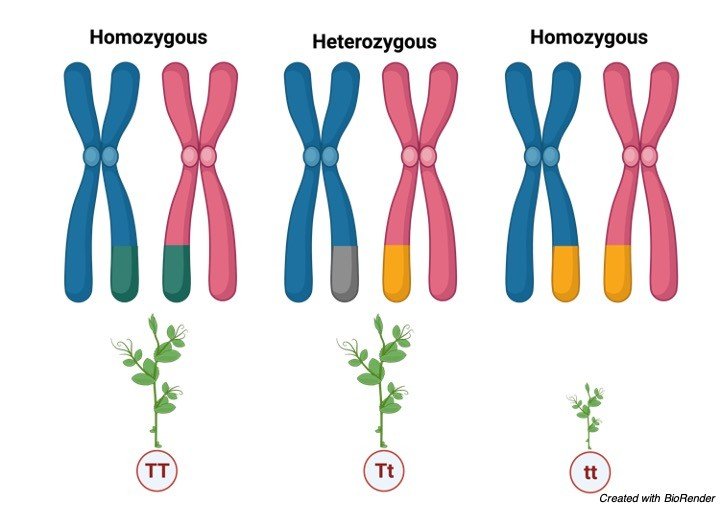
    pp (YY), delivering yellow peas; pq (Yy), likewise yellow; or qq (yy), creating green peas. The recurrence of homozygous pp people is p2; the recurrence of heterozygous pq people is 2pq; and the recurrence of homozygous qq people is q2.

    In the event that p and q are the lone two potential alleles for a given attribute in the populace, these genotypes frequencies will whole to one: p2 + 2pq + q2 = 1.

    n our model, the potential genotypes are homozygous dominant (YY), heterozygous (Yy), and homozygous recessive (yy).

    In the event that we can just notice the aggregates in the populace, we know just the recessive aggregate (yy).

    For instance, in a nursery of 100 pea plants, 86 may have yellow peas and 16 have green peas. We don’t have a clue the number of are homozygous dominant (Yy) or heterozygous (Yy), ergo we do realize that 16 of them are homozygous recessive (yy).

    Consequently, by knowing the recessive aggregate and, in this way, the recurrence of that genotype (16 out of 100 people or 0.16), we can compute the number of different genotypes.

    Assuming q2 addresses the recurrence of homozygous recessive plants, q2 = 0.16. In this way, q = 0.4. Because p + q = 1, then, at that point 1 – 0.4 = p, and we realize that p = 0.6.

    The recurrence of homozygous dominant plants (p2) is (0.6)2 = 0.36. Out of 100 people, there are 36 homozygous dominants (YY) plants. The recurrence of heterozygous plants (2pq) is 2(0.6)(0.4) = 0.48. Thusly, 48 out of 100 plants are heterozygous yellow (Yy).

    Summary of Hardy Weinberg Equilibrium

    The Hardy-Weinberg guideline expects that in a given populace, the populace is huge and isn’t encountering change, movement, normal determination, or sexual choice.

    The recurrence of alleles in a populace can be addressed by p + q = 1, with p equivalent to the recurrence of the predominant allele and q equivalent to the recurrence of the recessive allele.

    The recurrence of genotypes in a populace can be addressed by p2+2pq+q2= 1, with p2 equivalent to the recurrence of the homozygous predominant genotype, 2pq equivalent to the recurrence of the heterozygous genotype, and q2 equivalent to the recurrence of the recessive genotype.

    The recurrence of alleles can be assessed by ascertaining the recurrence of the recessive genotype, then, at that point figuring the square base of that recurrence to decide the recurrence of the recessive allele.

    Hardy Weinberg Equilibrium Citations

    Share

    Similar Post:

    Continue Reading
  • Pedigree Analysis: Definition, Pedigree Chart, and Examples

    About Pedigree Analysis

    It is easy to represent a thing in a diagram manner than in terms of theory, which also helps in easy understanding.

    Pedigree analysis is used to represent traits of an organism or an individual so that we can identify the genotypes and phenotypes of the future generation.

    It also helps us to predict the characteristics of future generation by assuming with the present generation characteristics, which is very helpful for identifying the genetic diseases and thus we can provide counseling for the parents in such condition which helps them to take a better decision.

    Pedigree Chart

    In human beings-controlled crosses cannot be made, so genetists discovered the method for undergoing crosses to find an individual with established mating’s.

    The information that has obtained by established mating’s is by a chance or a hope. This method of identifying the possibility is referred to as pedigree analysis.

    The individual in a family who comes first in attention of a genitists is known as propositus.

    Generally, the propositus phenotype sometimes causes exceptional, where dwarf character appears in such cases the investigator traces the history of a character and analyses the change by tracing the family tree or pedigree chart.

    Pedigree chart is drawn by using certain symbols. A pedigree helps us to represent the traits and it also gives an understanding of genotypes and phenotypes that will be passing to through the inheritance.

    It also relates the relationship between the parents, offspring and their siblings.

    It also helps in better understanding of the inheritance within the family of a particular individuals.

    How to Read a Pedigree

    Before knowing how to relate the traits in pedigree it is important to know the specific terms. And few of them are listed below

    Thus, these symbols are commonly used to indicate the traits in a pedigree.

    Pedigree Analysis - research tweet 2

    A family pedigree chart usually has circles to indicate the female individuals, squares to indicate the male individuals and perpendicular line drawn to the marriage bar indicates the off springs.

    The symbols which are present on the same line are considered as they are from the same generation and the generations are denoted by roman numerical as I, II etc.

    The unshaded symbols shows that they are unaffected individuals and the shaded symbols shows they are partially or fully affected depending upon their shaded area, the arrow indicates the specific person who is carrying the defective trait throughout his generations.

    Considering an example of polydactyl, which means that some individuals have extra fingers in their limbs.

    When the pedigree illustration is done for this, a marriage has been done between polydactyl man and a normal woman they reproduce three children’s where the first generation has a polydactyl daughter and a son and the other normal son.

    In second generation when these polydactyl individuals marry a normal person their reproductive off springs are shown in the graph below.

    Pedigree Analysis - research tweet

    However, from this polydactyl pedigree chart it is clear that polydactyl condition is playing a dominant role on recessive i.e., normal trait.

    Determination of Particular Trait

    If the trait is dominant then it will be sure that one of the traits will be obtained from the parents because dominant genes don’t skip the generation.

    However, neither trait will be obtained from the parents if it is heterozygous.

    Determination of Autosomal and Sex-linked Trait

    Males are usually most affected than females in a sex-linked recessive inheritance. Where as in autosomal recessive conditions male send female are equally affected.

    The other terms to be taken into consideration while reading a pedigree chart is;

    Genotype: Genetic characteristics of an organism., where TT denotes homozygous tall.

    Phenotype: Phenotype indicates the physical characters of an individual., where Tt heterozygous tall also considered as a tall.

    Dominant Allele: An allele which is phenotypically dominant over the other allele.

    Recessive Allele: This allele is expressed only in cases where the dominant alleles are absent or unable to express.

    How to Read a Pedigree Chart?

    Mendelian recessive alleles are very helpful in determining the human disease and other exceptional condition.

    The pedigree chart can be read easily with above sorted clues. When the diseased condition appears in the progeny of the unaffected parents.

    Also, at the same time the affected parents cannot have the unaffected individuals these chances are more when recessive alleles are revealed by consanguineous marriages where the best example is cousin marriages.

    This also suits the conditions where the rate of mattings of heterozygotes are expected to be rare in cases of albinism, Tay- Sachs disease, cystic fibrosis and phenylketonuria.

    There are also some conditions which are not caused by dominant alleles. This condition often occurs in every generations, where unaffected individual transmit the condition to their offspring in case if they are a carrier.

    In other case when two parents are affected, they are also chancing of being a child to be unaffected where they get their trait from their grandparents and these unaffected individuals passes a diseased condition to their children’s.

    Examples of abnormal conditions caused by dominant alleles are Huntington’s chorea and brachydactyly which means these individuals have very short fingers.

    This pedigree analysis also helps in identifying the phenotypical and genotypical traits in plants as well as in insects as in humans to identify the inheritance passed to the upcoming generations.

    Pedigree Analysis Citations

    Share

    Similar Post:

    Continue Reading
  • Sex Chromosomes: Definition, Examples, and Facts

    About Sex Chromosomes

    We all know our body is made up of types of chromosomes, one is autosomes and the other is allosomes or most commonly known as sex chromosomes.

    Autosomes are commonly known as somatic or body chromosomes which does not have any practice in taking the memory to the next generation.

    Whereas allosomes play an important role in taking the inheritance from present to future generations.

    What are Sex Chromosomes?

    Sex chromosomes are one of the type of chromosomes which play an important role in determination of sex of an individual.

    Most of mammals including humans consists of two sex chromosomes.

    Where as in males the sex chromosomes are named as X and Y and in females two chromosomes are named as X.

    Hence male has X and Y chromosomes in their cells, eggs contain X chromosome and sperm, cells contain Y chromosome.

    sex chromosomes - research tweet 3

    This is the reason why male parent determine the sex of the offspring. As sex chromosomes control the gender of an organism.

    Considering in human beings we can consider xx as female and XY as male. But in other mammals these chromosomes have their namely differently. 

    The interesting thing about these chromosomes is that there is a large discrepancy in size of these chromosomes.

    Where X chromosomes are larger than the Y chromosomes. These two chromosomes carry different genes for different functions.

    But we cannot say appropriately that which chromosome performs this function as two chromosomes work together in making a gender and other functions properly, the defect in any of these chromosomes results in any deficiency or syndromes in that particular individual.

    Before knowing about the syndromes caused by allosomes it is important to know about the morphology and structure of chromosomes.

    Morphology of Chromosomes

    Size: The chromosome size is generally measured during the phases of metaphase in mitotic cell division.

    Usually, it measures 0.25µm in birds and fungi, 30µm in certain plants like Trillium and 8 to 12µm inn maize, 3µm in drosophila and 5µm in humans.

    While the organisms which are having lesser number of chromosomes have their chromosomes with larger size than the one which is containing higher number of chromosomes.

    Where the dicotyledons contains smaller chromosomes comparing with that of monocotyledons.

    The animals contain smaller chromosomes on comparing with plants.

    The chromosomes are also named depending upon their structure and on which organism it is present as lamp brush chromosomes which is present in few vertebrates and polytene and oocyte chromosomes which are found in certain insects.

    Shape: The chromosomes change its shape at different phases during their cell division process.

    Where as in the interchange phase the chromosomes look like a thin coiled thread like structures, and on passing to the metaphase and anaphase they become thick and filamented.

    These chromosomes also have a centromere (often referred to as clear zone), and kinetochore which forms the length of the chromosome.

    The two arms arise from the centromere and they are generally called as chromosomal arms.

    However, the position varies accordingly, which results in various shape of the chromosomes as telocentric (centromere is situated at the proximal end), acrocentric (centromere at one end forming long and short arm which form a rod like appearance), metacentric (V- shaped chromosome, where centromere is at the centre), sub-metacentric (j shaped or L-shape where centromere is towards the median position)

    Chromosome Structure

    Chromosomes are generally the thread like structures present in the nucleus of the cell containing proteins along with one molecule of Deoxy ribo nucleic acid in each chromosome.

    The structure of chromosomes also varies accordingly depending upon their structures, where as in metaphase two chromosomes consists of a symmetrical structure which is known as chromatids.

    Chromosomes, Chromatids, Chromosomes vs Chromatids

    During some stages of interphase a bead like structures are found in the chromatin material which is known as chromomeres.

    In some cases, the chromosomes contain a knob like structures which are referred to as satellites, along with all these chromosomes generally have the primary and secondary constrictions, nuclear organisers, centromere, kinetochore and telomeres which make up the perfect chromosomal structure.

    Differentiation of Sex Chromosomes

    Each cell of a human consists of 23 pair of chromosomes which are 46 in number.

    The first 22 sets are defining as autosomes and the remaining one set is known as sex chromosomes or allosomes.

    Autosomes are generally known as homologous chromosomes because they contain the same genes in the same order along the entire length of the chromosomes i.e., in chromosomal arms.

    Thus, females have all the 23 pairs as homologous pairs where as in males the lass pair is formed by X and Y chromosomes which are heterozygous in condition.

    The X chromosome present in the 23rd pair is always an ovum where the other X and Y varies accordingly as sperm or ovum depending upon their gender.

    In females during an early embryonic development the X chromosomes present in other cells neither than egg cells are deactivated partially and permanently.

    In some cells the X chromosomes from female i.e. from mother is inherited where as in other cells the X chromosomes from father is deactivated.

    This is the only reason why our body has only one functional X chromosome. Whereas the deactivated X chromosomes are repressed by heterochromatin and prevents the activation of more genes

    Sex Determination

    All diploid organism get fifty percentage of allosomes from their mother and father equally.

    Since females have only two X chromosomes they can pass only the X chromosome, where as male passes either X or Y chromosome.

    The individual which get X chromosome from the father is determined as female and the individual which gets Y chromosomes from the father are consider as male.

    This is the reason why male sperm cells decide the sex of the individual.

    sex chromosomes - research tweet 5

    However few people rarely have a intersex due to the divergent form of sexual development.

    This results when the allosomes are formed neither XX nor XY.

    At sometimes the embryo may fuse and it also results in intersex. It can also be due to the exposure of chemicals which results in mutation of a particular gene.

    Sex Chromosomes Disorders

    Sex chromosome is not only involved in determining the gender of an individual but also in determining or carrying other genes which are responsible for other characteristics.

    Genes which are being carried by sex chromosomes are commonly called as sex linked.

    Sex linked genes are the ones which are passed down from their ancestors.

    There is also a possibility that these genes will carry the diseases along with the genes to their offsprings if their ancestors have any one of them.

    As only males carry y chromosomes there are the who transmits Y linked inherited diseases.

    X linked diseases are transmitted either by female or male to their offspring.

    Sex Chromosomes Citations

    Share

    Similar Post:

    Continue Reading