-

Taxonomy: Definition, Level, and Examples
Continue ReadingTaxonomy
Taxonomy originates from a Greek word and can be splitted into two words; “taxis” which means arrangement and “nomia” means method. It involves, identifying, classifying and providing a name to the organism. Nomenclature is important as it help to identify put them into various classes and sub-classes.
Taxonomy Definition
Scientifically, taxonomy can be described as allocating names and classifying both biotic and dead organism. Thus, a person who studies taxonomy is called as taxonomist. He has studied the organism in depth so that he can identify and classify it into proper groups. Ex; although human and whales are mammals, yet they are very un-identical from each other.
Other Definition of Taxonomy
According to Enghoff and Seberg; taxonomy has 7 stages starting from describing, naming, recognizing, comparing, classification of taxa, genetic variations, species identification and in the ecosystem defining the taxa.
According to Judd, taxonomy is grouping organism into a class and then classifying them into a larger group by allocating a name.
According to Simpsons; taxonomy is identifying, classifying and placing the organism into its group.
According to Lawrence; it is the classification of organism on the basis of its traits, it possess.
According to Walker; taxonomy is naming the biotic entities and their species.
According to Kirk; it is organizing and classifying the organism.
Systematics: It involves studying the data of the organism so that it can properly classified, identified and place into its family.
Taxonomy Overview
It is a branch of science which deals with classification of biological entities. It begins with grouping them into classes called taxa and then providing a taxonomic rank, thus, forming a level from high to low taxa.
Taxonomist
They are scientist, who study more about the organism’s characteristic and its behavior, lifestyle and on the basis of that, they are classified into groups and sub-groups. Each taxonomist would study about a particular kingdom such as plants, birds, insects, animals and etc.
History of Taxonomy
Since the 18th century, taxonomy has been to mankind. However, at that time the classification was on the basis of the appearance, its medicinal and agricultural importance. Thus, the classification system was called artificial. Charles Darwin’s theory made it further easier to classify organism into natural system, the scientific classification.
Before the Linnaean Era: It has work of all the taxonomist from 1500 BC, which is the pre-Linnaean era.
Early Egyptian Taxonomist: The various Egyptian taxonomist, who identified and classified the organism are:
a) Aristotle: When Aristotle was residing on an, island, he started observing and studying them and classified into plants and animals. Animals were further classified on the basis of warm blooded, number of legs, eggs laying pattern, birth pattern and etc. animals were classified into further groups such as cetaceans, enhaima, anhaima and others. Enhaima are animals containing blood such as vertebrates, whereas animals with no blood are called as anhaima, also referred as invertebrates. His work was continued by his students where his students classified around 500 types of plants.
b) Medieval Thinkers: At the time of Aristotle, microscopes weren’t present, thus a classification system was developed. A natural ladder called Scala Naturae was used to classify organism into one class. The other scholars were Thomas Aquinas, Procopius and Demetrios.
c) Andrea Cesalpino: She was an Italian taxonomist, infact the very first taxonomist. She came up with two plants family; they are Brassicaceae and Asteraceae which are yet used. In her book she has described around 1500 plants species.
d) John Ray: John came up with various classifications and he brought a change in the field of taxonomy as his book “Methodus Plantarum Nova” contained information about 1800 plant species.
e) Joseph Pitton de Tournefort: Around 9000 species were identified which he further classified into 698 genera.
f) Carl Linnaeus: Linnaeus is the one who came up with Linnaeus classifications system. His work is highly acknowledged. Some of his books were Systema Naturae, Species Plantarum, Systema Naturae were his works. The binomial system of classification was also given by him, which contained class, species, order, genus and others. His work was so popular, that before his findings the work done was called as Pre- Linnaean and his Linnaean system was the classification system for taxonomy.
Classification System
To classify an organism, they were placed into two kingdoms. Kingdom Plantae and kingdom Animalia. The further, after modification came the three-kingdom classification and finally five kingdom classification.
I. Two Kingdom Classification
In this classification system, classification was on the basis of two kingdom; kingdom Plantae and Kingdom Animalia. In kingdom plantae, organism can make food for themselves through photosynthesis and are referred by the name Autotrophs. Those who cannot synthesize food on their own are called as Heterotrophs belonging to the Kingdom Animalia.
In kingdom plantae lies bacteria, algae and fungi. However, they had confusion for organism like euglena which has plants and animals like properties. Thus, where does it reside is a major problem. Thus, came the three-kingdom classification.
II. Three Kingdom Classification
The problem faced to categorize euglena into which kingdom was taken care by Ernst Hackel in 1866, when the third kingdom arose which is Kingdom Protista. Bacteria was also placed in this kingdom, but fungi remained in kingdom plantae and scientist were not okay with it because unlike plants it cannot prepare its own food. Thus, the five-kingdom classification came into picture.
III. FiveKingdom Classification
Robert Whittaker introduced the five-kingdom classification in 1967. He first categorized organism into prokaryotes and eukaryotes. Prokaryotes consists of bacteria, whereas eukaryotes consist of plants and animals. On the basis of unicellular, multicellular, food uptake and other characteristics, organism were classified into five kingdom; Monera, Protista, Fungi, Plantae and Animalia.
Taxonomic Hierarchy
Taxonomic hierarchy is organizing organism into various levels, thus forming a pyramid structure. From the higher taxonomy to the lower taxonomy organisms are placed i.e., domain to species.
Taxonomic Ranks
Organism from higher taxonomy to lower taxonomy are given ranks which can be in any direction. The ranks pf taxonomy are as follows:
a) Domain: There are 3 domains
b) Kingdom: There are 5 kingdoms
c) Phylum: These are the classes
d) Class: These are the orders
e) Order: These are the families
f) Family: It consist of genera
g) Genus: It consist of species
h) Species: It consist of identical organisms.
a) Domain
It is the highest level of taxonomy which was added 250 years later the Linnaeus taxonomy in the year 1990. The three domains in taxonomy are Bacteria, Archaea and Eukaryota. The first two are prokaryotes, the latter is eukaryote.
b) Kingdom
There are five kingdoms and they are:
i. Kingdom Plantae
Also called by the kingdom Metaphyta. It consist of eukaryotic multicellular plants. Through the process of photosynthesis, they prepare food and produce oxygen and take up carbon dioxide. There are more than 250,000 species of plants on earth. Examples are flowering plants, mosses as well as fern belonging to kingdom plantae.
ii. Kingdom Animalia
It consist of eukaryotic multicellular organisms, lacking cell wall. All kinds of animals are placed into this kingdom. This kingdom also include humans. Animals move from one place to other. Few animals have symmetrical body such as fishes, echinoderms, mollusks and etc.
iii. Kingdom Monera
c) These are unicellular prokaryotes. Majority of the organism in this kingdom are heterotrophs, only few are autotrophs that can prepare their own food. Examples are bacteria and cyanobacteria.
iv. Kingdom Fungi
These are multicellular eukaryotes. They cannot prepare their own food, however some fungi can. Examples of fungi are molds, mushroom, yeast, smut, rust and others. Fungi are said to be the decomposers. They obtain food through absorption. There are around 2- 4 million species of fungi.
v. Kingdom Protozoa
It includes parasites which are unicellular eukaryotes. They obtain nutrition from dead matter; hence they are heterotrophs.
vi. Kingdom Archaea
They are assumed to be the ones that were to be first found. They are also unicellular prokaryotes, lacking nucleus and cell organelles. They were first included in the kingdom bacteria, but further research proved that they are quite versatile from bacteria.
c) Phylum
It was added to taxonomy in 19th century. There are overall 34 phylum; five belonging to archaea, in bacteria there are 29, protist has 20 phylum, plants have 13 and 8 phylum in fungi. Other animal phylum are Arthropoda, Porifera and Chordata.
d) Class
From the Linnaean system, the class has emerged, although the classes described by Linnaeus are not in function. 108 classes are found in animalia such as Mammalia, Aves and Reptilia.
e) Order
This was also given by Linnaeus and few of his orders are yet used. There are around 420-450 orders. Example of mammalian order are primates, carnivora, chiroptera and etc.
f) Family
It is more specific than order. Examples are Felidae, Mephitidae and Canidae.
g) Genus
It is the scientific name of the organism, which is written in Italian and is always in capital form. Example Homo sapiens, where homo is the genus name.
h) Species
There are around 9 million species on Earth, although many species have not been identified as well as classified. These are also written in Italian, but not capitalized. The genus is always followed by the species.
Taxonomy Evolution
Due to natural selection, variation and genetic drift leads to evolution. Evolution is what makes the organism adaptable to various unfavorable condition, thus surviving, producing and adapting to changes so that a new type of species arises which is well adapted. This new species formation is called as speciation, which is of various types; such as Sympatric speciation, Allopatric speciation, Parapatric speciation and Peripatric speciation.
Taxonomy Example: Dog
Domain: Eukarya
Kingdom: Animalia
Phylum: Chordata
Class: Mammalia
Order: Carnivora
Family: Canidae
Genus: Canis
Species: lupus
Taxonomy Citations
Share
Similar Post:
-

Analogous Structures: Definition, Examples, and Structure
Continue ReadingAnalogous Structures Definition
Analogous structures in evolutionary biology are biological structures with comparable or related functions but not from the same evolutionary origin. In other words, these biological features are used by species for the same function, despite the fact that these species come from different evolutionary lines.
Convergent evolution is the name given to this type of evolution. Convergent evolution occurs when animals evolve structures or functions that are comparable (analogous) to those of their evolutionary forebears, despite their evolutionary ancestors being highly distinct or unrelated. As a result, even if they developed from distinct evolutionary origins, homologous structures in unrelated species might have similar or matching functions.
Analogous Structures Examples
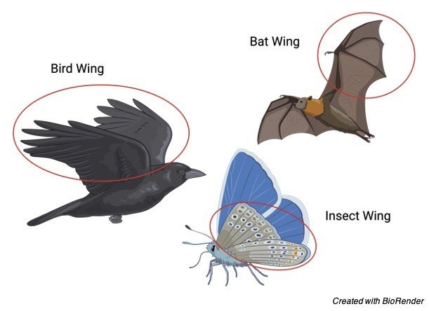
A bird’s wing and an insect’s wing are similar organs. Both of these animals have wings and use them to fly, yet their wings have different evolutionary origins. Therapod dinosaurs, especially Maniraptora members, are thought to be the evolutionary origins of birds. In the case of insects, their evolutionary history is unknown.
The origins of insect flying are unknown. The paranotal hypothesis proposes that their wings evolved from the paranotal lobes of the thoracic terga, according to one prevalent theory. Winged insects may have originated from a terrestrial rather than an aquatic progenitor, according to phylogenomic studies.
Analogous structures may not have to be of the same structure, as seen in the example. Birds’ wings are modified forelimbs in general. The humerus, ulna, and radius are the bones that make up their forelimbs. These bones, like the rest of the skeleton of a bird, are light. This is, in fact, one of the most important characteristics of birds that can fly. They have air sacs in them and are mostly hollow bones to save weight.
The existence of flying feathers at the tips of the wings is another modification that allows them to fly. The structure of the bird’s feathers gives the bird its initial lift and later its real powered flight. In the case of insects, the wing is an extension of the exoskeleton seen in the mesothorax and metathorax. The forewings are the paired wings that emerge from the mesothorax, whereas the hindwings emerge from the metathorax. Their wings do not have the same bone structure as birds’ wings.
Rather, the insect wings are made up of crisscrossing veins, with a nerve, a trachea, and hemolymph running through each of the main veins. The veins are surrounded by a cuticle, which thickens and supports the insect wing structurally. Instead of feathers, the insect wing possesses two types of hairy structures: microtrichia (finer) and macrotrichia (bigger) (larger).
Aside from the bird wings and insect wings mentioned above, here are some additional structures that are similar:
1. Vertebrates, cephalopods (squid and octopus), cubozoan jellyfish, and arthropods (insects, spiders, and crustaceans) developed their sophisticated eyes independently. Their eyes, on the other hand, are mostly used for vision.
2. The shells of brachiopods and bivalve mollusks are remarkably similar.
3. Plant hormones such as gibberellin and abscisic acid, which are produced by both plants and fungus.
4. Analogous organs include the sense of smell organs. An analogous organ is one that operates similarly to an organ from a different species but evolved at a different time. The sensilla of insects are comparable to the smelling organs of the terrestrial coconut crab. They both evolved organs that can sense odours in the air and flip antennas for better reception.
Analogous Structures vs Homologous Structures
Homologous structures are those that have a common ancestor or have the same evolutionary or developmental origin. Homologous structures may or may not serve the same purpose. Human and bat forelimbs are examples of homologous structures.
Humans and bats share the same core skeletal structure, which originated from the same components and is derived from the same embryonic origin. They are both mammalian in origin. Their forelimbs, on the other hand, are employed in a different way. The bats fly by using their forelimbs. The radius, like that of other mammals, is the most important component of the bat forelimb.
The digits, on the other hand, are extended and webbed, extending around the wrist. The patagium membrane, which extends between the arm and finger bones, is a fragile membrane. Muscles, nerves, blood arteries, and connective tissues make up the membrane, which is an extension of the skin.
Analogous structures, in contrast to homologous structures, have comparable functions but evolve separately. The fins of fish and the flippers of whales (mammals) are similar features of evolutionarily unrelated creatures that utilise them for swimming but do not have a common ancestor.
The anatomical features of analogous and homologous structures may differ, but the anatomical features of homologous structures may be identical. Homologous structures have comparable tendencies in terms of development, whereas analogous structures do not.
One might look at their common ancestors to see if the structure is similar to that of another species. As previously stated, analogous structures are structures that have a comparable or equivalent function but do not have the same evolutionary origin as the two species under investigation.
The Evolutionary Process
Convergent evolution is the process through which organisms evolve identical structures (or functions) despite their progenitors being substantially different or unrelated. The structures in the two species must have the same purpose, but they do not have to have the same physical characteristics to be called similar.
Analogous structures do not have a similar ancestral origin since they differ in anatomy and developmental origin. They evolve independently of the characteristics or qualities that they share.
The adaptation of the species to a comparable environment is linked to the production of equivalent structures. For example, the smelling organ of a coconut crab differs significantly from that of other crabs, resembling more like an insect sensilla. Their comparable terrestrial modes of existence have resulted in homologous structures.
Unlike crabs that live in water, the coconut crab spends more time on land, using its highly developed sense of smell to locate food sources (such as the scent of rotting flesh) over vast distances. By exposing the dwelling species to the same environmental variables and limitations, the same habitat and ecological niche might induce similar structures to develop amongst species of different lineages.
Analogous structures give evidence for evolution since they exist. This implies that species develop in reaction to their surroundings and as a measure of their ability to fit in and survive. They may have formed an anatomically comparable structure to those of their lineage, but the function may serve a different purpose that is more appropriate for their ecological environment. Analogous structures indicate that unrelated species evolve in ways that are both similar and distinct.
Analogous Structures Citations
Share
Similar Post:
-

Chemiosmosis: Definition, Mechanism, and Function
Continue ReadingChemiosmosis Definition
Chemiosmosis is a biological process that involves transporting ions (such as protons) to the other side of a membrane, resulting in an electrochemical gradient that may be utilised to drive ATP production.
What is Chemiosmosis?
Chemiosmosis is the process of diffusion of ions (usually H+ ions, also known as protons) across a selectively permeable membrane and thus proton gradient developed. In many cells, proton gradient provides the energy for the synthesis of ATP.
With the aid of the proteins buried in the membrane, the gradient also encourages the ions to return passively. By passively, we mean that the ions will migrate from a high-concentration location to a low-concentration area. Water molecules flow passively in this mechanism, comparable to osmosis.
Chemiosmosis, on the other hand, includes the movement of ions across the membrane, whereas osmosis involves the movement of water molecules. Both processes, however, need a gradient. This is known as an osmotic gradient in osmosis. Osmosis is caused by pressure variations between the two sides of the membrane.
In chemiosmosis, an electrochemical gradient, such as a proton gradient, drives the flow of ions. Chemiosmosis is not just comparable to osmosis. It’s also comparable to assisted diffusion and other kinds of passive transfer. It works on the same basis. Ions travel downward.
Membrane proteins also assist in the transport of molecules to the other side of the membrane. Membrane proteins aid ion movement across the membrane, which is not easily permeable to ions due to its bilipid structure. These proteins in the membrane function as a temporary shuttle, a conduit, or a tunnel, allowing them to move around more easily.
Membrane proteins are used in chemiosmosis to transport particular ions. Furthermore, unlike an active transport system, it does not require chemical energy (e.g., ATP). The development of an ion gradient in chemiosmosis results in the generation of potential energy, which is sufficient to drive the process.
Chemiosmosis can happen anywhere. It happens in eukaryotes during cellular respiration and photosynthesis in the mitochondria and chloroplasts. Chemiosmosis will occur in the cell membrane of prokaryotes since they lack these organelles.
Chemiosmotic Theory
Chemiosmosis is driven by an electrochemical proton gradient that is required for the synthesis of ATP, according to the chemiosmotic theory. Peter D. Mitchell (1920–1992), a British scientist, presented this idea. Mitchell’s idea, on the other hand, was not immediately accepted until a solid foundation for proton pumping was built. With the discovery of ATP synthase and the pH difference across the thylakoid, the bioenergetics field began to question his hypothesis’ validity.
He was aware of the phenomenon of membrane potential in the 1960s, in which the inner side of the membrane was negatively charged in relation to its surroundings. At the time, ATP was also recognised as the cell’s primary energy currency. However, the actual mechanism through which living creatures create ATP is unknown. The organelles responsible for ATP production have long been recognised as mitochondria.
However, it was unclear how these organelles produced ATP. It was previously thought to be related to phosphorylation at the substrate level (as what happens in glycolysis). Mitchell suggested that chemiosmosis might potentially create ATP.
He demonstrated that ATP production is linked to a proton gradient electrochemically. This laid the groundwork for understanding how oxidative phosphorylation leads to ATP production.
Chemiosmosis Model
Chemiosmosis is an ATP-producing energy-coupling process used by living organisms. It is one of the most important stages in cellular respiration in respiring cells.
Because mitochondria produces the majority of ATPs, it is known as the cell’s powerhouse. It is dedicated to the production of ATP. The organelle is a double-membraned structure, so keep that in mind. An outer membrane and an inner membrane make up the mitochondrial membrane. Both layers are made up of lipid layers that prevent ions from passing through them easily.
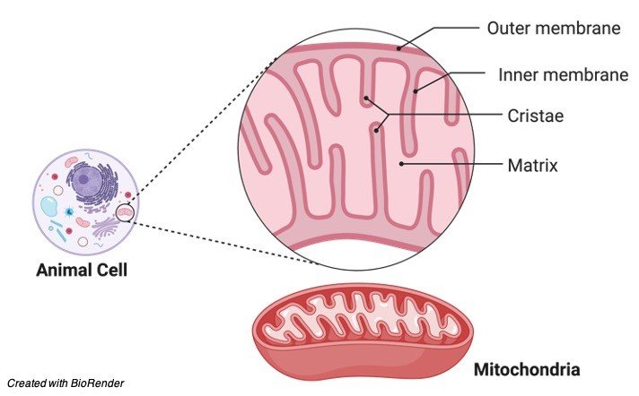
The intermembrane gap is the space between the two membranes. Cristae are many infoldings in the inner membrane. The mitochondrial matrix is the region within the inner membrane. The citric acid cycle, a cyclic metabolic mechanism in which food molecules are churned to create energy-rich phosphate compounds, takes place in the matrix.
The pyruvate produced by glycolysis is transformed into acetyl CoA, which is then transported to the mitochondrion where it is completely oxidised and degraded into carbon dioxide. The citric acid cycle generates one ATP for every pyruvate molecule via substrate phosphorylation.
The electron transport chain (ETC) and the enzyme ATP synthase are integrated into the mitochondrial membrane, so oxidative phosphorylation will provide the majority of the ATP.
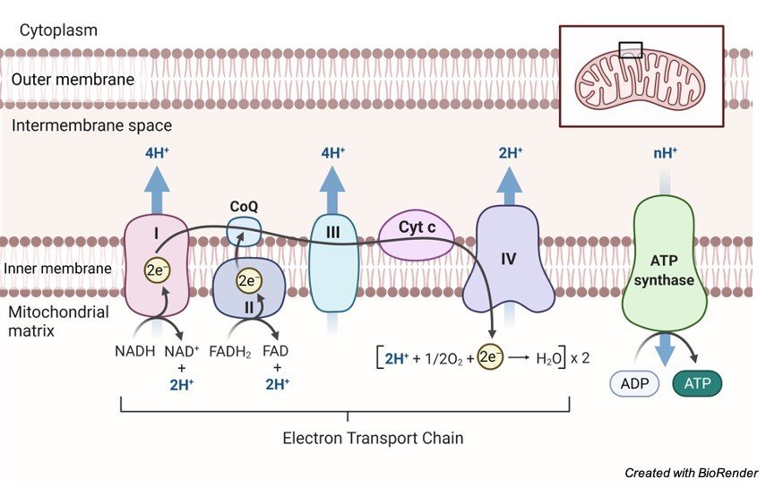
Most of the high-energy electrons are transported to NAD+ and FAD through redox processes, resulting in NADH (and H+) and FADH2, respectively. The electrons will be shuttled to the ETC for oxidative phosphorylation by these electron-carrying molecules. Every ETC member performs a redox reaction as the electrons move along the chain, receiving and giving electrons.
When electrons are transferred to the last electron acceptor, molecular oxygen, the electron flow will come to a halt. Water is formed as a result of the reaction: 2 H+ + 12 O2 H2O. ATP is not produced by ETC. Instead, as electrons pass through, ETC members push H+ (protons) into the intermembrane gap.
Protons accumulate on one side of the membrane as they are pushed across it. A proton (H+) gradient is created as a result of this. The proton-motive force was the name given to it by scientists. The energy created by the transport of protons (or electrons) through an energy-transducing membrane is defined by them.
Through the ATP synthase channel, protons will flow down to their gradient, i.e., from the intermembrane space to the matrix. When protons release energy as they traverse the ATP synthase, the hydrogen ion migration leads to ATP production. The energy causes the enzyme’s rotor and rod to revolve. The enzyme is then triggered to use this force to create an ATP molecule by forming a high-energy bond between the ADP molecule and the inorganic phosphate (Pi). ADP + Pi ATP is the reaction.
Chemiosmosis Fucntion
It’s all about energy coupling in chemiosmosis. The production of a proton motive force is the link between chemiosmosis and ATP synthesis. As previously stated, chemiosmosis is the process that drives ATP production via oxidative phosphorylation in cellular respiration.
The electrons from the citric acid cycle (which breaks down pyruvate-turned-acetyl coenzyme A into carbon dioxide) are transferred to electron carriers and transported to the ETC. The proton motive force generated by protons accumulating on one side of the membrane during energy transfer in the ETC via a series of redox reactions will be utilised to synthesise ATP from ADP and inorganic phosphate.
As a result, there will be no proton motive force for ATP synthase to employ during ATP synthesis if chemiosmosis is absent. As a result, fewer ATP end products will be produced without the need for chemiosmosis. In photosynthesis, where chemiosmosis is also a critical stage in ATP generation, a similar effect can be predicted.
Chemiosmosis in Chloroplast
Chemiosmosis occurs in the mitochondria of eukaryotes, as previously stated. Apart from the mitochondria, photosynthetic eukaryotes like plants have another organelle in which chemiosmosis occurs: the chloroplast.
The chloroplast is the organelle that plays the most important role in photosynthesis. It possesses a light-harvesting thylakoid system. As a result, it acts as the site of light responses (or light-dependent processes). The stroma is the stroma of the chloroplast matrix. The thick fluid comprises enzymes, chemicals, and other substrates that are engaged in dark reactions (or light-independent processes).
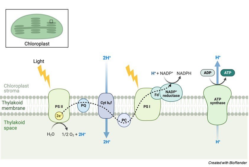
Chemiosmosis occurs in the thylakoid of chloroplasts. This membrane system contains its own ATP synthase and transport chain. The source of energy is one of the key distinctions between chemiosmosis in mitochondria and chloroplasts. The high-energy electrons in mitochondria are taken from the food molecules (through a redox reaction), whereas the source in the chloroplast is photons collected from the light source.
The proton (H+) gradient is formed by the accumulation of H+ ions in the thylakoid compartment (i.e., the space inside the thylakoid). The H+ ions can come from three sources:
(1) water splitting during light processes,
(2) protons translocated across the thylakoid membrane when electrons travel through the transport chain, and
(3) stromal H+ ions taken up by NADP+.
Because the quantity of H+ ions inside the thylakoid compartment (lumen) is higher, they will diffuse into the stroma via passing through the ATP synthases implanted in the thylakoid membrane.
Chemiosmosis in Prokaryotic Cells
Chemiosmosis occurs in the cell membrane of prokaryotes such as bacteria and archaea, which lack mitochondria and chloroplasts. When a proton gradient occurs on the other side of the membrane, hydrogen ions (protons) travel across the cellular membrane via ATP synthase (a transport protein).
During electron transport and redox processes, hydrogen ions accumulate as they are forcibly pushed to the other side, forming a proton gradient. As additional hydrogen ions accumulate on the other side of the membrane, they will return to the cell via passing through the ATP synthase. Energy is released when they pass through, and it is used to convert ADP to ATP via phosphorylation.
Chemiosmosis vs Oxidative Phosphorylation
Chemiosmosis is the method through which oxidative phosphorylation generates ATP directly. ATP synthase, on the other hand, will be unable to do so without the proton motive force generated by the ETC, which pushes protons (H+) to the other side of the membrane as electrons travel through the chain.
The metabolic process of oxidative phosphorylation creates ATP from the energy generated by a series of redox events in the ETC. As a result, electron transport-linked phosphorylation is another name for it. Because molecular oxygen is the ultimate electron acceptor, it is an aerobic process. This distinguishes it from the other type of phosphorylation, namely substrate-level phosphorylation, in which ATP is produced directly from an intermediary substrate. In contrast, oxidative phosphorylation is an indirect method of ATP synthesis. It is combined with chemiosmosis, which involves the movement of protons through the membrane.
Chemiosmosis Citations
- Quantum Mechanics predicts evolutionary biology. Prog Biophys Mol Biol . 2018 Jul;135:11-15.
- Mitochondrial proticity and ROS signaling: lessons from the uncoupling proteins. Trends Endocrinol Metab . 2012 Sep;23(9):451-8.
- What is the Role of Lipid Membrane-embedded Quinones in Mitochondria and Chloroplasts? Chemiosmotic Q-cycle versus Murburn Reaction Perspective. Cell Biochem Biophys . 2021 Mar;79(1):3-10.
Share
Similar Post:
-

Gamete: Definition, Formation, Examples, and Facts
Continue ReadingGamete Definition
A gamete is a mature reproductive or sex cell with a haploid number of chromosomes (i.e., just one set of dissimilar chromosomes) and the ability to fuse with another haploid reproductive cell to produce a diploid zygote. The zygote is created by fusing (or merging) two gametes, namely a male and a female gamete. Fertilization is the process of gametes coming together to form a zygote.
What are Gametes?
Gametes are an organism’s reproductive cells. Gametes are also referred to as sex cells. Female gametes are called ova or egg cells, and male gametes are called sperm.
A mature haploid reproductive cell generated by gametogenesis and that which unites with another from the opposing sex at fertilisation to form a zygote that grows into a new person.
Gamete Etymology
From the Ancient Greek (gamet), which means “woman.” sex cell; reproductive cell are synonyms. One of the gametes is generally bigger and non-motile, as is customary. It’s also called a female gamete, ovum, or egg cell.
The other gamete cell is smaller in size and motile. It’s also known as a sperm cell or a male gamete. Each human gamete has 23 chromosomes, and when they fuse, a diploid zygote with 46 chromosomes is formed.
These reproductive cells are generated in the male and female gonads, or reproductive organs, in mammals. Male gametes are pollen in seed-bearing plants, whereas female gametes are contained in the plant’s ovules. In plants, however, the gamete may or may not be a haploid cell.
Types of Gametes
The gametes used in fertilisation might be identical (known as isogamy) or distinct (known as polygamy) (referred to as anisogamy).
i. Isogamy
Isogamy refers to gametes that have identical morphology, such as size and form. Heterogamy is another name for this situation. These gametes aren’t classified as either male or female. These gametes are denoted by the letters “+” or “-“.
Unicellular algae gametes, Chlamydomonas reinhardtii, and Carteria palmata are examples.
ii. Anisogamy
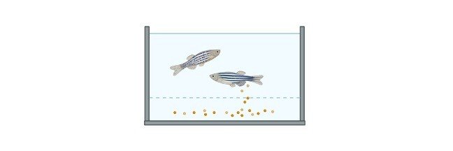
Anisogamy refers to gametes with morphological differences, such as size and form. Female and male gametes are the two kinds of gametes. The gamete with the smallest size is called a sperm or male gamete, whereas the gamete with the largest size is called an ova, egg, or female gamete. Additionally, these gametes can be both motile and non-motile.
In the instance of Polysiphonia, a red algae, both gametes are non-motile. A zygote is formed when a non-motile sperm joins a non-motile egg. Spermatia is a non-motile male gamete or sperm. This may also be found in flowering plants where both non-motile gametes are present in the gametophyte. Pollen is a non-motile male gamete found in plants.
iii. Oogamy
Oogamy occurs when one of the gametes, the male gamete or sperm, is motile while the other gamete, the egg or female gamete, is non-motile in humans and animals. Oogamy is a situation in which a big non-motile egg is fertilised or will fuse with a tiny motile sperm to produce a zygote.
Anisogamy (or heterogamy) is a type of sexual reproduction that involves female and male gametes of differing sizes. In contrast, isogamy is a kind of sexual reproduction in which both the male and female gametes are the same size.
Size of the Gametes
The size of the gametes can also be used to classify them. Gametes are classified as follows based on their size:
• Microgametes are tiny versions of gametes. These are motile, usually generated in vast numbers, and do not have nutrition storage. Consider sperm cells.
• Macrogametes are gametes that are larger than normal. These are non-motile, generated in small quantities, and contain a high amount of nutrient storage. Egg cells or ova are two examples.
Gamete Examples
Male and female gametes are created in their reproductive organs through a process known as “gametogenesis.” A diploid (2n) cell goes through meiosis to create four haploid (n) cells during gametogenesis.
In most cases, gametogonia is the first step in the gametogenesis process. The primordial germ cells give rise to gametogonia (PGCs). Mitosis is the mechanism by which these germ cells multiply. These cells are transported to the gonadal ridge in the late embryonic stage, where they are known as gametogonia. Following the development of gametogonia, subsequent gametogenesis results in the creation of an egg or sperm, depending on the individual’s sex.
Males and females have entirely distinct gametogenesis processes. Spermatogenesis is the gametogenesis that results in the creation of sperm, whereas oogenesis is the gametogenesis that results in the formation of an egg or ova.
What are some gamete examples?
A gamete is a haploid pair of chromosomes found in a reproductive cell or sex cell. It is generated by a germ cell that goes through gametogenesis, which is a gamete production process involving meiosis. Oogenesis is the gametogenesis that leads to the development of the female gamete. Spermatogenesis is the process of creating a male gamete.
Structure and Function: Sperm Cell
The sperm cell is the male reproductive cell or gamete. Anisogamy is a condition in which the structure of the male gamete differs from that of the female gamete. Sperm in animals, including humans, are tiny and motile. The flagellum is the motile organ found in sperm. Sperm cells have a finite lifespan and are unable to divide. Mammalian sperm have two different structures that are separated by a single membrane.

• The haploid nucleus, which is densely packed with DNA, is found in the head. A sperm cell also contains an acrosome, a thin, flattened sac-like structure, and a vacuole, in addition to the extremely compressed genetic material. The enzyme essential for breaching the ovary or egg cell is found in the acrosome. Exocytosis is the process through which the enzyme is released. The head is 5.1 m by 3.1 m in size.
• The tail directs the sperm towards the egg and finally enters it, ending up at the nucleus’s posterior end. This is also the sperm cell’s longest section. The tail is 50 metres in length. The tail travels at a rate of 1-3 millimetres each second.
The neck, which is rich in mitochondria, connects the two sections, namely the head and tail. Mitochondria are vital to the sperm cell because they supply all of the energy required for sperm motility. Mitochondria manufacture the ATP necessary for sperm motility. The neck also includes centrioles in addition to mitochondria. Sperm is a haploid gamete that has 23 chromosomes in humans.
Spermatogenesis
Spermatogenesis is a process that happens in male humans’ testes and begins only after they reach puberty. Spermatogenesis, on the other hand, is a continuous process that persists throughout a person’s lifespan once it begins (unlike oogenesis). Seminiferous tubules are tubular structures where spermatogenesis takes place.

Spermatogonia, or immature germ cells, are found at the basal lamina on the outside border of the seminiferous tubules. Mitosis is the mechanism by which these germ cells proliferate indefinitely. Some of these proliferating cells do not proliferate and become primary spermatocytes. These main spermatocytes then proceed through the first meiotic phase, in which each pair of homologous chromosomes contributes to cross-over, before going through division I of meiosis, which results in the production of two secondary spermatocytes with 22 duplicated autosomal chromosomes (it can be a duplicated X or a duplicated Y chromosome).
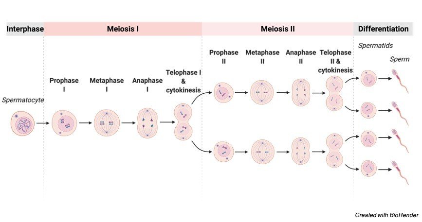
Phase II of meiosis occurs in these secondary spermatocytes, culminating in the creation of haploid spermatids, which are later differentiated to produce sperm. These sperm eventually make their way into the lumen of the seminiferous tubule. Later, sperm go to the epididymis, a coiled tube located above the testes where they develop and are stored in a liquid known as semen. This entire process takes around 70 days and may live in the female reproductive canal for over 5 days. Outside the body, however, they may only live for a few hours.
Sperm can be frozen for months or years and then thawed to preserve their ability to fertilise eggs. The fructose in the semen provides energy to the sperm for motility.
Sperm can’t swim backwards, which is an intriguing fact.
Ferns, cycads, and ginkgo trees all have flagellated sperm. Nematode sperm are amoeboid in nature. Rather than swimming, they move around by crawling.
Non-motile sperms, for example, non-motile sperms of Polysiphonia, a red alga, are dispersed by water currents after being discharged, relying on environmental circumstances for dispersal and finally reaching the egg cells. These non-motile sperm are carried by flies, butterflies, and insects.
Sperm have developed a number of important modifications that make them effective cells.
The following are some of these adaptations:
• Sperm have a streamlined shape and a tapered head, which aids in motility and agility.
• The energy for sperm movement is provided by condensed packing of mitochondria (almost 70 in number) in the neck region of the sperm.
• Sperm include certain basic amines that aid in the successful fertilisation of an egg by allowing them to establish an alkaline microenvironment even in the acidic vaginal canal.
• The sperm acrosome includes lysosomal enzymes (such as lysozyme) that aid sperm penetration into the egg during fertilisation.
Function of Sperm
The sperm’s job is to go to the egg and fuse or fertilise it to produce a zygote, transferring the male genetic material and centriole in the process (which eventually determines the microtubule cytoskeleton). The colour of the eyes, hair, and skin of the progeny is determined by the genetic content of the sperm. The X and Y chromosomes in sperm determine the sex of the offspring.
Structure and Function: Ovum (Egg Cell)
The egg cell, also known as the ovum, is a non-motile ovoid or spherical gamete generated in the female reproductive system’s ovaries. A fertilised egg is bigger than sperm. The average diameter of a human egg is around 0.1mm. It ranges from 1-2mm in fish and frogs. The biggest egg is the ostrich egg, which measures 170 x 135 mm. In humans, the egg or ova is a haploid gamete with 23 chromosomes.

The cytoplasm of an egg is called ooplasm. The egg’s cytoplasm is divided into two parts: the formative yolk and the nutritional yolk. Because the human egg has such a small amount of nutritious yolk, it is referred to as alecithal. The cytoplasm of avian eggs, on the other hand, is dense in nutritional yolk (which is made up of lipoproteins, pigment granules, and water).
The germinal vesicle, which houses the egg’s nucleus, and the germinal spot, a vacuole, are both found in the cytoplasm. The ovum’s nucleus is big, bloated with nucleoplasm, and positioned eccentrically. As a result, the human ovum has polarity, with animal and vegetable poles. The animal pole is the side of the ovum that contains the nucleus and polar body, whereas the vegetable pole is the opposite side. The cortex is a peripheral layer made up of microvilli and cortical granules that surround the cytoplasm.
Protective Membranes of Ovum
The zona striata or zona pellucida is a thick, translucent membrane that surrounds the ovum. The vitelline membrane is a thin layer that lies underneath the zona pellucida. The zona pellucida and the vitelline membrane are separated by a thin gap known as the perivitelline space. The corona radiata, which radiates from the egg surface, is the outermost layer, which lies above the zona pellucida.
Function of Ova
The major purpose of the ovum is to transport genetic material, which in a human ovum consists of 23 pairs of chromosomes, and to result in the creation of a zygote following fusion with the male gamete. It also offers the necessary conditions for sperm to fertilise the egg. The nutrients from the ovum are necessary for the zygote’s development after fertilisation.
Oogenesis
The female reproductive organ known as the ovaries undergoes oogenesis, which is the process of ovum differentiation. The oogenesis process varies from species to species. An ovarium is formed in humans from germ cells that are present at the time of a female child’s birth. As a result, there are two major phases of oogenesis:
Pre-natal Phase
The ova are made up of oogonia, which is a kind of germ cell. Mitosis allows the oogonia to multiply and produce primary oocytes. At the moment of birth, all of the germ cells multiply to produce a significant number of primary oocytes (roughly 2 million). A
fter birth, no primary oocytes are generated (this is in contrast with spermatogenesis, wherein the primary spermatocytes are continuously formed at puberty). The primordial follicle is formed when initial oocytes are surrounded by follicular cells. Prior to the birth of the female offspring, the main oocyte starts the first meiotic division. However, until adolescence, this meiotic division is not completed and halts at the prophase in the diplotene stage.
In summary, the embryo initiates the first phase of meiotic division, which then enters a stop period for around 12 years until puberty occurs. It transmits the signal to restart meiosis at that point. Some oocytes have been seen to remain in the meiotic prophase for over 50 years. In addition, only 400 primary oocytes develop in a female’s lifetime out of a population of millions.
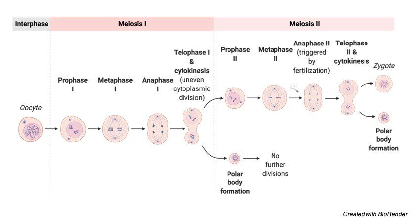
Post-natal Phase
The latent phase of the primary oocyte lasts until adolescence. The main oocyte undergoes maturation in the ovary after birth and before puberty. The primary oocyte remains inside the follicles during maturation, when it grows in size and forms the zona pellucida membrane.
The main oocyte restarts and completes meiosis before ovulation. The cytoplasm, on the other hand, is divided unequally, with the secondary oocyte acquiring the bulk of the cytoplasm. The initial polar body, on the other hand, only receives a small quantity of cytoplasm. The initial polar body is a non-functional cell that degenerates over time.
The nucleus of the secondary oocyte initiates the second meiotic division at the moment of ovulation. However, it only advances to metaphase before pausing the meiotic division process. In the event of fertilisation, the second meiotic division is restarted and finished.
The maturation of an oocyte happens with each menstrual cycle, resulting in the development of an ovum by division. This division produces cells of uneven size, such as secondary oocytes (120-150 mm and fertile) and polar bodies (120-150 mm and fertile) (not more than 10 mm and non-fertilizable).
In some species, such as humans, there are two types of gametes: the male gamete (i.e., sperm cell) and the female gamete (i.e., ovum). Male gametes are smaller and motile, but female gametes are many times larger and non-motile. The two gametes must be haploid in order for the chromosomal number to be maintained over generations during fertilisation during sexual reproduction.
Importance of Haploid
All animals are diploid genetically due to the fusing of two haploid gametes. The haploid gamete cells guarantee that the genetic content or number of chromosomes stays consistent from generation to generation. If the gamete cells are not haploid, each future generation will contain double the number of chromosomes or genetic material as the preceding one.
It’s crucial to keep in mind that cancer cells have a non-diploid condition. The presence or absence of a pair of chromosomes during cell replication might cause instability. This, in turn, can lead to the development of a disease, such as cancer. In humans and most animals, ploidy changes are typically deadly.
Aside from that, haploids are utilised for crop development (particularly in rice and tobacco) since they can be generated in a short period of time. As a result, haploids can aid crop development by shortening the breeding cycle and producing unique genetic compositions. Haploids are also an excellent cytological tool for researching mutations and genetic diseases.
Sex Determination in Humans and Animals
Primary sex determination in animals is decided by chromosomes rather than the environment, and is determined by the gonads. In placental animals, the presence of a Y chromosome determines sex. Female cells normally have two X chromosomes, i.e., XX, whereas male cells have an X and a Y chromosome, i.e., XY. As a result, each of the female eggs has a single X chromosome, whereas male sperm have two kinds, one with the X chromosome and the other with the Y chromosome.
As a result of the fusing of the X chromosome-containing egg with the X chromosome-containing sperm, female offspring with XX chromosomal makeup are produced. In contrast, when a male gamete carrying the Y chromosome fuses with an ovum holding the X chromosome, a zygote with sex chromosomes, XY, is formed, it develops into a male child. The testis-determining factor, which results in the development of testes in male progeny, is encoded by the SRY gene on the Y chromosome.
Sex Determination in Birds and Vertebrates
The sex determinants in birds are the Z and W chromosomes, with females being heterogametic (having ZW chromosomes) and males being homogametic (having just Z chromosomes) (i.e., with ZZ chromosomes). The Z chromosome is significantly bigger than the W chromosome. FET1 and ASW, two genes found on the W chromosomes, control the development of female birds. In chickens, the mechanism of determining sex is not well understood.
However, unlike mammals, chickens’ gonads differentiate into male or female reproductive organs after a period of time following birth. For sex determination, chickens require oestrogen. A male chicken can be turned into a female chicken by injecting oestrogen into the eggs during the development period. The ZW chromosomal sex determination is also seen in certain reptiles, fish, and amphibians.
Sex Determination in Insects and Invertebrates
Distinct insects have different sex determination patterns. Females are heterogametic in butterflies and moths (order Lepidoptera), whereas males are homogametic. In Lepidoptera, the sex is determined by the W and Z chromosomes. The W chromosome is linked to female traits. Males with ZZ chromosomal content grow into ZZ chromosomal males, whereas females with ZO chromosomal content develop into ZO chromosomal females.
In the absence of the W chromosome, a moth, Talaeporia tubulosa, uses ambient temperature to identify sex. Warmer temperatures result in the development of more female eggs, whereas cooler temperatures result in the formation of more male eggs. This is an excellent example of adaptation, in which certain conditions, such as warmth, encourage the development of more female offspring because warm conditions assure the availability of nutrients for later reproduction.
The XX/XO sex-determination system, which is a single-chromosome system, is employed in grasshoppers. Males have only one sex chromosome, XO (heterogametic), whereas females have two chromosomes, XX and homogametic.
Sex Determination in Drosophila
Drosophila melanogaster, a fruit fly, has been extensively researched to better understand genetics. The number of X chromosomes to the number of sets of autosomes, or the X: A ratio, is used to determine sex in Drosophila. The X chromosome encodes female-determining factors, whereas the autosomes encode male-determining factors.
The sex of a fruit fly is thus determined by the balance between X and A. Male flies have XY and XO chromosomes, whereas female flies have XX, XXY, and XXYY. Isogamy has evolved into anisogamy, which is the evolutionary successor of isogamy. Isogamous individuals, such as fungus, algae, and yeast, produce the same sort of gametes. ‘+’ and ‘-‘are used to denote isogamous gametes.
In anisogamy, the male and female gametes have physical differences and are referred to as male and female. According to popular belief, the genesis of anisogamy is based on the fact that the largest number of positive fusions of gametes happens when the population’s gametic material has been divided with a high degree of anisogamy. As a result, it is assumed that a set amount of reserve material is required for zygote formation, and that only disassortative fusions (between small and big gametes) occur. According to this idea, males generate a high number of sperm to enhance the chances of conception.
Evidence shows that sperm density in the female tract has a favourable effect on fertility (i.e., the proportion of fertilised ovum discharged). As a result, the more sperm in the sperm, the better the likelihood of conception. This is also due to the fact that a larger quantity of sperm increases sperm competition for fertilisation, resulting in increased fertility.
Furthermore, the sole purpose of male sperm is to transmit genetic material, and a large quantity of tiny sperm provides an evolutionary advantage. The ovum expends far more energy than a male gamete in generating a viable zygote. The egg supplies genetic material from its nucleus, mitochondrial genes, and vital nutrients for the zygote’s early growth in order to increase its chances of survival.
As a result, in order to deliver all of the essential materials, the ovum is big in size and contains a sufficient amount of all of the required substances. As a result, anisogamy is said to have evolved, in which eggs are non-motile, enormous in size, and restricted in quantity, whereas sperm are tiny, motile structures generated in huge quantities.
Chromosomal Anomalies in Gametes
The existence of an aberrant number of chromosomes is known as aneuploidy. In a typical human cell, there are 46 chromosomes. An aneuploid is a person who has 45 or 47 chromosomes. Because of the aberrant chromosomal number, a genetic imbalance occurs, resulting in a disease. The second most frequent type of mutation is somatic mutation.
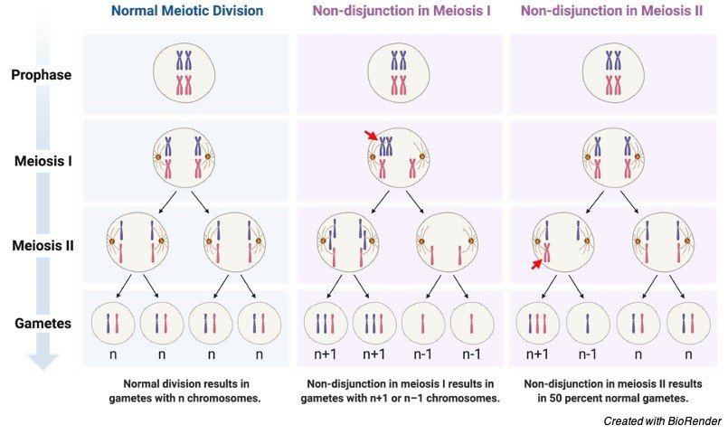
Nondisjunction, or the failure to separate chromosomes between two cells during cell division, causes aneuploidy. Miscarriage is caused by aneuploidy in the germline. A normal woman is a 46 XX, while a typical man is a 46 XY. Trisomy is the most prevalent kind of aneuploidy.
The following are some examples of aneuploidy:
• Loss or acquisition of a portion of a chromosome causes partial aneuploidy or trisomy.
• Monosomy refers to the absence of one of the typical complement’s chromosomes. Monosomy is exemplified by Turner’s syndrome. Females with Turner’s syndrome have only one X chromosome, 45XO. Fertility and reproductive organ growth have both decreased in these people. Patients with Turner syndrome are commonly referred to as mosaics.
• The existence of two copies of a chromosome is known as disomy. It is a common occurrence, but uniparental disomy occurs when both copies of the chromosomes come from the same parent.
• Trisomy refers to the existence of three copies of a chromosome rather than the usual two, implying the presence of an additional chromosome. Edwards syndrome is caused by a trisomy of 18, while Patau syndrome is caused by a trisomy of 13. Trisomy of the Y chromosomes, such as (47, XXX), (47, XXY), and (47, XXY), is also conceivable (47, XYY). The majority of trisomies are not viable and do not survive; just a handful are capable of living. Down’s syndrome is the most prevalent trisomy capable of surviving.
An additional chromosome 21 causes Down syndrome. It can happen once every 750 births. Nearly 75% of cases are discovered before delivery using prenatal screening approaches such as serum screening and US monitoring. Down syndrome patients show signs and symptoms of cognitive impairment.
The Klinefelter’s syndrome, in which males have 47 chromosomes (two X chromosomes and one Y chromosome), is another trisomy that can survive (47 XXY). These people have a normal lifespan, but their fertility is poor and their reproductive sex organs are underdeveloped.
• The existence of four or five copies of a chromosome is referred to as tetrasomy/pentasomy. This is a rarely seen in humans.
Dysfunctional Gamete
We’ve seen how gametes are critical in ensuring the survival of species through sexual reproduction, as well as their importance in fostering biodiversity (especially during the events of gamete formation and fertilization). As a result, if these gametes become dysfunctional, the species’ propagation and species diversity may suffer. Both of these are essential for the species’ survival. We learnt in the previous part how chromosomal abnormalities in humans can result in impaired physiological processes and reproductive capacity in those who are afflicted. Let’s look at how defective gametes affect other species.
Plasmodium Life Cycle
The circulating female and male gametocytes undergo gametogenesis in a mosquito vector during the sexual development of malarial parasite protozoans (Plasmodium spp.).

Observe how gametocytes are divided into two types: macrogametes (female gametes) and microgametes (male gametes) (male gamete). The male gamete fertilises the female gamete, which results in the production of oocysts, which then develop into an ookinete.
However, these males and females are affected by host immunological factors throughout their sexual development, which can make them dysfunctional. Although a defective gamete can engage in fertilisation, fusing a healthy gamete with a dysfunctional gamete leads to the creation of a nonviable zygote that does not survive to the ookinete stage.
Gamete Citations
- Extracellular vesicles: Multi-signal messengers in the gametes/embryo-oviduct cross-talk. Theriogenology . 2020 Jul 1;150:59-69.
- Evolutionary Dynamics of Unreduced Gametes. Trends Genet . 2017 Sep;33(9):583-593.
- Cold case: Small animal gametes cryobanking. Theriogenology . 2020 Jul 1;150:445-451.
- Gametogenesis: A journey from inception to conception. Curr Top Dev Biol . 2019;132:257-310.
Share
Similar Post:
-

Hypotonic Solution: Definition, Examples, and Importance
Continue ReadingHypotonic Solution Definition
It refers to a solution that has a lower solute concentration than the solute concentration in the other solution when seen through a semipermeable membrane.
It’s crucial to grasp what a ‘solution’ in science entails. A homogeneous system made up of two or more elements is referred to as a solution in science. The elements that are dissolved are referred to as “solutes,” while the constituent that dissolves the solute is referred to as “solvent.”
What is Hypotonic Solution?
Hypotonicity is a relative word that describes a solution’s properties in relation to another solution. In biology, the cytosolic fluid, or the fluid within a cell, is frequently used as a comparative solution. In biology, a solution is classified as hypotonic if it contains fewer solutes than a cell’s cytosol.
The semipermeable membrane is the cell membrane. So, what occurs when a cell is subjected to hypotonic conditions? A cell exposed to a hypotonic environment will experience an inflow of water, which will cause the cell to expand.
What is Tonicity?
To have a better grasp of this idea, we must first comprehend tonicity. Tonicity is a behavioural word that describes the behaviour or movement of solvent molecules across a semipermeable membrane based on the number of non-penetrating solutes present.
It’s critical to remember that the property is solely influenced by non-penetrating solutes, not the overall number of solutes. With this understanding of tonicity, we can distinguish between three sorts of solutions: hypotonic, hypertonic, and isotonic.
Hypertonic Solution
Tonicity is prefixed with the term “hyper,” which implies “greater” or “excess.” As a result, a solution with a larger concentration of non-penetrating solutes can pass through a semipermeable barrier than a solution with a lower concentration. Consider a cell that has been immersed in a hypertonic solution.
The quantity of non-penetrating solutes in the solution outside the cytosol would be larger than the cytosolic concentration in a hypertonic environment. An osmotic gradient is created across the semipermeable membrane as a result of the concentration differential. This causes the solvent to efflux (“flow out”), which causes the cell to shrink.
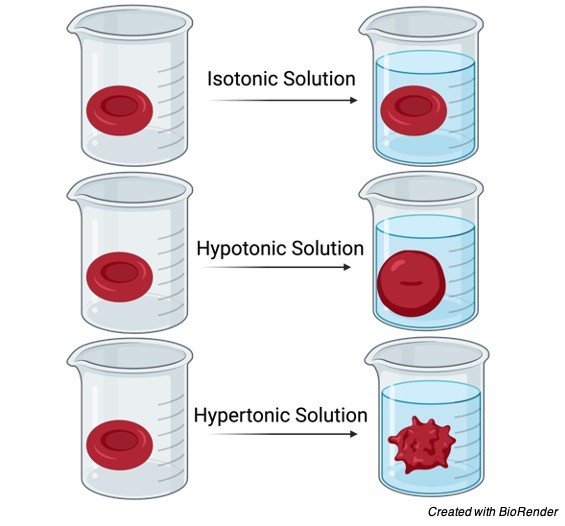
Isotonic Solution
The word tonicity has the prefix ‘iso,’ which means “the same.” As a result, two solutions separated by a semipermeable membrane are said to be isotonic if they contain the same quantity of non-penetrating solutes. A cell will neither shrink nor swell under this circumstance, since there will be no net movement of the solvent molecules due to the lack of an osmotic gradient. With blood serum, any solution with a tonicity of 280 – 300 mOsm/liter is considered isotonic. The typical example of an isotonic solution is 0.9 percent sodium chloride.
Hypotonic Solution
Tonicity (which means “push” or “thrust”) has the prefix “hypo,” which implies “low.” Hypotonic solutions have a low number or concentration of non-penetrating solutes in contrast to the other solutions across a semipermeable barrier, according to biology. When a cell is submerged in a hypotonic solution, the amount of non-penetrating solutes decreases as the water concentration rises.
An osmotic gradient is created as a result of the hypotonic environment, which leads to the flow of the solvent or water into the cell, causing the cell to expand. A cell in a hypotonic solution will expand and finally lyse as a result.
The rupture of the cellular membrane caused by an osmotic gradient, a virus, or enzymes is known as lysis. The activity of lyse is referred to as lysis. The breakdown of big particles into smaller ones is referred to as lyse. As a result, these agents are classified as lytic because they can cause total rupture of the cellular membrane, resulting in cell death.
It is critical to distinguish between the word’s plasmolysis and cytolysis in this section. Plasmolysis is the shrinking of a cell caused by the outflow of water from the cells in a hypertonic environment. The lack of a cell wall causes the cells to shrink in animal cells.
Water efflux from plant cells, on the other hand, causes the cellular membrane to rupture from the cell wall, resulting in gaps or pockets between the cell wall and the cellular membrane. When a cell is put in a hypotonic solution, it undergoes cytolysis. Water molecules will flow inside the cell due to the osmotic gradient, causing the cell to expand and finally rupture and lyse. Only animal cells and protozoa undergo cytolysis.
Cell lysis is classified as “haemolysis” (lysis of red blood cells), “oncolysis” (lysis of malignant cells), and so on, depending on the kind of cell involved.
Plant cells are protected from cytolysis by having a cell wall and vacuoles. The extra water is taken up by the vacuoles, which pushes the cellular membrane towards the cell wall, resulting in turgor pressure.
As a result, plasmolysis and cytolysis occur in opposite directions in hypertonic and hypotonic environments, respectively. The tonicity of a solution refers to the osmotic pressure or tension of a solution, as cells inflate or shrink in response to the environment’s tonicity. It influences the direction in which two solutions diffuse.
The concentration of solutes between liquids separated by a semi-permeable plasma membrane influences tonicity in cells (cell membrane).
Hypotonic, hypertonic, and isotonic solutions are the three kinds of solutions characterised by tonicity. The osmotic pressure of a hypotonic solution is lower than that of the solution being compared to.
Hypotonic solutions have fewer solutes (in terms of concentration) than other solutions. As a result, a hypotonic solution would have more water. A cell in a hypotonic solution, for example, would allow water to enter (diffuse) the cell. The cell would then enlarge as a result of this.
Examples of Hypotonic Solution
Hypotonic solutions are categorised in biology using blood serum as a reference. Hypotonic solutions are defined as those with an osmolarity of less than 280 mOsm/liter in blood serum. Hypotonic saline, such as 0.45 percent sodium chloride or 0.25 percent sodium chloride with or without dextrose, 2.5 percent dextrose solution, and others, are examples of hypotonic intravenous solutions that are hypotonic with regard to blood serum.
Is it true that water is a hypotonic solution?
A hypotonic solution is water, which is the classic example. However, this, too, will be subjective to the solution being compared. Purified distilled water is a solvent, and it is always hypotonic compared to an aqueous solution of a solute containing any quantity of the solute. Because purified distilled water is free of any material, it is hypotonic to any aqueous solution of a solute.
Biological Importance of Hypotonic Solution
Tonicity is required to keep life processes running smoothly. Protists without a cytoskeleton or a cell wall, such as paramecia and amoebae, are able to maintain a rigid structure thanks to tonicity control. Because these protists live in a hypotonic environment, there is a constant input of water. These protists have a specialised organ known as contractile vacuoles that collect an excess of water from the cell and subsequently discharge it out to keep the cell structure and prevent cell lysis.
The production of turgor pressure in plant cells is caused by a constant inflow of water in a hypotonic environment. Turgor pressure is used by plants to impart structure and stiffness to their structures.
Fungi (such as mushrooms) and plants control their environment in order to keep their cells hypotonic. The entry of water will result in the creation of turgor pressure as a result of the hypotonic environment. As a result, cells remain upright, retaining the stiffness of their structure.
This pressure is also used by plants to move water throughout their bodies, from the roots to the top stem. When plants are not watered for an extended period of time, a hypertonic environment forms around them, and their turgor pressure decreases, giving them a wilted appearance.
When such plants are re-watered, turgor pressure is restored, and the plants regain their form and structure. Because of the high salt concentration, marshy regions and mangroves have a hypertonic environment. In such severe hypertonic circumstances, a typical plant will wilt.
Mangroves, for example, have evolved to generate a hypertonic cytosolic state in their root cells because they grow in marshy regions. As a result, a hypotonic external environment around the roots aids in the absorption of water from the environment.
Similarly, all aquatic creatures, whether they live in saltwater or freshwater, have a system called osmoregulation that allows them to manage the effects of osmosis. Salt concentration in the water is important for aquatic life in any water body because of this osmoregulation. With the aid of salt glands, sea turtles have evolved to generate a hypertonic interior environment.
The exterior environment becomes hypotonic for them as a result of the hypertonic internal environment, allowing these marine creatures to survive even in a very hypertonic environment.
Freshwater fish cannot thrive in seawater and vice versa due to tonicity and the osmotic gradient. One example of a hypotonic solution is freshwater. As a result, freshwater fish cells have a greater salt content than the water in the surrounding river or lake. These fish have evolved a mechanism to drain away extra water from their bodies on a regular basis.
Freshwater fish, on the other hand, would have hypotonic cells compared to the external hypertonic environment if they were exposed to saltwater. In such circumstances, their bodies will lose water, causing them to get dehydrated and finally die. As a result, changes in the salt content of water have a significant impact on the fish population in any body of water.
Because of the plasma membrane, not all solutes can enter or leave the cell. The plasma membrane is a crucial component of the cell that controls the flow of ions and molecules into and out of it. As a result, there are variations in solute concentrations between the cytosol of the cell and the fluid surrounding it.
Hypotonic dehydration occurs when the amount of sodium lost exceeds the amount of water lost. This results in a decrease in serum osmolarity, which causes hypotonic dehydration (or hyponatremia). Reduced serum osmolarity causes water to flow from the extracellular to the intracellular region, causing cellular swelling and edema.
Neurological symptoms such as nausea, headache, disorientation, unconsciousness, weakening or absence of tendon reflex, stupor, and lethargy develop as a result of the sodium imbalance, finally leading to coma and death. It’s crucial to realise that hypotonic dehydration involves cellular swelling and edema caused by excessive water retention.
Excessive fluid loss from wounds or burns, persistent diarrhoea, Addison’s illness, renal tubular acidosis, prolonged use of intravenous hypotonic fluids or normal saline in patients, Cystic fibrosis, and chronic use of diuretics can all contribute to this condition.
Treatment for hypotonic dehydration is usually started with 3 percent hypertonic saline or 0.9 percent isotonic saline (depending on the severity of the disease) and constant monitoring of blood sodium levels to prevent myelinolysis, as recommended by doctors.
Hypotonic Solution Citations
- Scientific basis for the use of hypotonic solutions with ultrasonic liposuction. Aesthetic Plast Surg . Mar-Apr 2006;30(2):233-8.
- Administration in a hypotonic solution is preferable to dose escalation in intraperitoneal cisplatin chemotherapy for peritoneal carcinomatosis in rats. Oncology . 1999 Jul;57(1):77-82.
- The effect of hypo- and hypertonic solutions on volume and ion distribution of smooth muscle of guinea-pig taenia coli. J Physiol. 1968 Mar; 195(1): 107–118.
- Isotonic versus hypotonic solutions for maintenance intravenous fluid administration in children. Cochrane Database Syst Rev . 2014 Dec 18;(12):CD009457.
- Maintenance Fluid Therapy: Isotonic Versus Hypotonic Solutions. Vet Clin North Am Small Anim Pract . 2017 Mar;47(2):383-395.
- The effect of hypotonic solutions on the morphology of cells of Mycoplasma gallisepticum. Can J Microbiol . 1971 Sep;17(9):1203-5.
Share
Similar Post:
-

Multiple Alleles: Definition, Types, and Examples
Continue ReadingWhat is Multiple Alleles?
Alleles are genetic pairs that share a locus, which is a specific place on a chromosome. A gene in a diploid organism usually contains just two alleles. Multiple allelism is a condition in which a gene exists in more than two allelic variants. Allelism is the term used to describe any of a gene’s numerous variations. Hereditary variations are caused by these genetic differences, which are generally caused by mutation.
Each gene, according to Gregor Mendel, should contain just two alleles. Alleles are gene variants that come in two or more forms. Each gene is passed down in two forms, one from each parent. As a result, having two distinct alleles for a characteristic is also a possibility.
Heterozygous offspring have genotypes made up of various alleles of a gene, whereas homozygous offspring have genotypes made up of the same alleles (i.e., of a gene for a certain characteristic). Multiple allele characteristics may occur at the population level, despite the fact that humans (and other diploid creatures) can only have two alleles for any particular gene in genetics. As a result, many alleles play an essential role in fostering diversity within a species.
A particular chromosomal locus was inhabited by two unique sorts of gene alternatives in Mendelian inheritance-one dominant and one recessive. Both of these options are alleles of the same gene. However, there are times when there are more than two options (alleles) available to a population. When different versions of the same gene exist in the population, this is referred to as “multiple allelism.” Multiple alleles refer to three or more variations of the same gene.
Multiple Alleles Definition
A gene with three or more alleles. Multiple allelism is a biological term that refers to the existence of multiple alleles. The ABO blood group system in humans is one example. IA, IB, and IO are the three allelic variants of human gene I (I stands for “isohaemagglutinin”). On the cell surface of RBCs, IA and IB generate type A and type B antigens, respectively, but IO (or I) is a recessive allele that does not produce antigens. A child with both IA and IB genes, for example, will have blood type AB, while those with IOIO (or ii) will have blood type O.
Multiple Alleles Examples
Let’s look at some examples of many alleles to help us comprehend the notion better.
i. Cat Coat Color
Domestic cats have been bred for thousands of years to produce a wide range of coat colours. The gene that determines the cat’s coat colour appears to have several variations, since coat colours range from black to orange to brown to white. This indicates that the coat colour is determined by many alleles.

In the population, the coat colour gene has several alleles, and the pigment-producing protein will be determined by the inheritance and expression of these alleles. Curliness, shading, patterning, and even texture is all controlled by the same genes. Because of the numerous potential genotype combinations and expressions resulting from these genes, a broad range of breeds exist. Even when two parents share just four alleles for each gene, the variety may be amazing.
Cats’ coat colour phenotype — patches of black and orange (tortoiseshell pattern), black, grey, white, and patches of white hair — strongly shows multiple allelism, since there appear to be more than two alleles for the coat colour phenotype (piebald spotting).
The genotype of a cat may usually be detected by looking at its coat colouring and pattern. It is typically feasible to estimate the colouring possibilities of kittens if the phenotypes of the parents are known, but the computations would be difficult in most situations.
ii. Multiple Alleles in Fruit Flies
The genomic mapping of the common fruit fly, Drosophila melanogaster, was finished in 2000. Because of its rapid reproductive rate and the simplicity of keeping and analysing large numbers of flies, the fruit fly has been and continues to be a valuable laboratory animal. The DNA of a fruit fly is far smaller than that of a human, with just 165 million base pairs.
Fruit flies have just four chromosomes, whereas humans have 23. Despite this, just four chromosomes contain about 17,000 genes. Each gene controls a different element of the fly’s appearance and is susceptible to mutation and the development of new alleles.
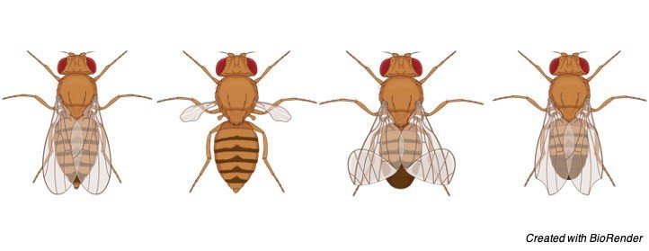
Drosophila melanogaster’s wings are normally fairly long. Two mutations at the same gene happened in different flies, one of which resulted in vestigial (reduced) wings and the other in antlered (less developed) wings.
When a fly with vestigial wings crosses with one with antlered wings, the F1 hybrids have intermediate wing lengths, suggesting that none of the mutant genes is dominant. This hybrid, also known as the vestigial antlered compound, is made up of two mutant genes at the same locus. There is evidence of Mendelian segregation and recombination.
Other phenotypes include nicked wings, strapped wings, and no wings at all, in addition to vestigial and antlered wings. Multiple alleles in this fruit fly population are the gene variations responsible for various characteristics.
iii. Multiple Alleles in Humans
Humans and other organisms have traits with three or more different types of alleles (genes). Multiple allele inheritance refers to the inheritance of three or more different alleles in a characteristic. The ABO blood type alleles/trait in humans is an example of a multiple allele trait. There are three types of alleles: allele A (IA), allele B (IB), and allele i. (IO or i).

Protein A is generated when the allele A is present on the chromosome, and protein A is found on the membranes of the individual’s red blood cells. Protein B will be generated if allele B is present on the chromosome, and protein B will be found in the membranes of red blood cells. Finally, neither protein A nor protein B will be produced if allele I is present on the chromosome. The ABO blood group characteristic is made up of these three alleles.
Allele A and allele B have a codominant inheritance pattern (co-dominance). When neither allele is dominant over the other and a heterozygous individual displays both traits, this is known as co-dominance.
For example, if an individual has allele A on one homologous chromosome and allele B on the other, both proteins are expressed, and red blood cells have both proteins A and B on their cell membranes.
In humans, the ABO blood type genetic system is an example of many alleles blood type. Blood group A, blood group B, blood group AB, and blood group O are the four types of phenotypes. In this example, there are three alleles in the population. When the IA allele is expressed, A molecules are present on red blood cells; when the IB allele is expressed, B molecules are present on red blood cells; and when the IO allele is expressed, no such antigens are present on red blood cells. Not only are the IA and IB alleles codominant, but they are also dominant over the IO allele. Because the IO allele is recessive, it will be expressed even if IA or IB are not present.
Despite the fact that a population has three alleles, each person gets only two of them from their parents. The genotypes and phenotypes shown below are the consequence of this. Consider that there are six genotypes when three alleles are present. The dominance relationships between the three alleles dictate the number of phenotypes that can be produced.
iv. Multiple Alleles in Plants
While it is commonly assumed that the form of a potato tuber is constant, visual traits such as round or long tubers can be distinguished at the diploid level. Although this is the first report of experimental evidence for the occurrence of several allele systems in a potato tuber, this study may be compared to one in maize. The tuber shape recessive allele can be considered a qualitatively acknowledged null or near-null allele.
The difference between dominant alleles is quantifiable. The concept of a null or near-null allele for the (most) recessive allele is compatible with how quantitative effects at a multiallelic locus are characterised. When extra metric characteristics are resolved into Mendelian factors in experimental designs utilising heterozygous parents, inferences regarding the relative significance of various allele traits to numerous loci in explaining quantitative genetic allele variance can be reached.
v. Multiple Alleles in Bacteria
Bacteria carry a combination of genes, and some of them have many alleles. These wild-type alleles are typically linked to different forms of pathogenicity and can be used to categorise subspecies (e.g., housekeeping genes for Multi Locus Sequence Typing, MLST). As a result, identifying not just the target gene but also the appropriate allele is important.
Sequencing-based techniques now available are confined to mapping reads to each known allele reference, which is a time-consuming procedure. More than knowing the species responsible for the infection, it is required to understand and forecast the pathogenic impact and epidemic potential of a bacterial infection.
Bacterial virulence is typically controlled at the sub-species level by a set of genes or even alleles, necessitating the adoption of different treatment methods for infections caused by the same bacterial species.
Minor changes in a gene, for example, might result in a varied array of antibiotic resistance profiles within a single taxonomic group.
Varied alleles of the same gene may be responsible for different adhesion and invasion tactics, immunological responses to the infected organism, and toxin synthesis, among other things.
Identifying alleles of certain genes leads to a more exact categorization of bacteria, in addition to its relevance in understanding pathogenicity.
Multiple Allelism, Pleiotropy and Epistasis
Pleiotropy and epistasis are two additional popular concepts in genetics that might be confused with multiple allelism. Differences between multiple allelism (defined as the presence of many alleles) and other genetic disorders.
Pleiotropy
Pleiotropy is a situation in which more than one gene can affect the phenotype in numerous ways. Pleiotropy entails more than one gene that influences phenotype,
Whereas multiple allelism simply comprises a single gene with numerous variations (referred to as many alleles).
Albino people, for example, are more likely than pigmented ones to have crossed eyes. Albinos may also have a cross-eyed characteristic, in addition to having insufficient colour production in their skin and hair. However, not all albinos have this feature, showing that the two qualities are not connected in these situations. (BioL110F2012 – Confluence, 2012).
The Complex Expression Patterns of Multiple Alleles Another example is the colour of one’s eyes. More than one gene influences the characteristic. OCA2 and HERC2 are two of these genes.
Epistasis
When one gene influences the expression of another, this is known as epistasis. When genes cooperate to generate a certain characteristic, this happens.
One example is the determination of coat colour in some species (such as horses), where the effect of one gene is influenced by the influence of another gene that controls hair pigment deposition. (BioL110F2012 – Confluence, 2012). Multiple Alleles’ Complex Expression Patterns.
Multiple Alleles Citations
- The Involvement of HLA Class II Alleles in Multiple Sclerosis: A Systematic Review with Meta-analysis. Dis Markers . 2019 Nov 6;2019:1409069.
- Schizophrenia: a common disease caused by multiple rare alleles. Br J Psychiatry . 2007 Mar;190:194-9.
- HLA-DRB1 alleles and cervical cancer: A meta-analysis of 36 case-control studies. Cancer Epidemiol . 2020 Aug;67:101748.
Share
Similar Post:
-

Induced Fit Model: Definition, Mechanism, and Examples
Continue ReadingWhat is Induced Fit Model?
A model for enzyme–substrate interaction states that only the right substrate may cause the active site to align properly, allowing the enzyme to execute its catalytic activity.
The induced fit model is a model for the interaction of enzymes and substrates. It states that only the appropriate substrate may cause the active site to align properly, allowing the enzyme to execute its catalytic activity. It also implies that the active site evolves until it is fully bonded to the substrate, at which time the final shape and charge are decided.
Who Proposed Induced Fit Model?
Daniel Koshland proposed the induced fit model in 1958. It is more widely recognised than the lock-and-key paradigm for enzyme-substrate complexes. The interaction of the substrate and the enzyme is compared to a key (the substrate) that is extremely specific to the lock in the lock-and-key paradigm (the active site of the enzyme).
Induced Fit Model Mechanism
Unlike the lock-and-key concept, the induced fit model demonstrates that enzymes are fairly flexible structures, with the active site constantly reshaping as a result of interactions with the substrate until the substrate is entirely bound to it (which is also the point at which the final form and shape of the enzyme is determined).
Induced Fit Model Citations
- Enzyme evolution in natural products biosynthesis: target- or diversity-oriented? Curr Opin Chem Biol . 2020 Dec;59:147-154.
- A thermodynamic and theoretical view for enzyme regulation. Biochemistry (Mosc) . 2015 Jan;80(1):1-7.
- Kinetic Measurements for Enzyme Immobilization. Methods Mol Biol . 2017;1504:215-232.
Share
Similar Post:
-

Stimulus: Definition, Types, and Examples
Continue ReadingWhat is Stimulus?
Our sensitivity to temperature allows us to recognise hot or cold situations. Sweating (perspiration) begins in our bodies when the temperature is too high. Similarly, in a cold environment, our bodies’ small hairs stand up. Piloerection is the term for this occurrence. As a result, we attempt to stay cool in the summer and layer up in the winter to be warm.
So, how can humans and other animals sense and respond to changes in the environment’s temperature? You’ve probably noticed that many plants drop their leaves in the winter. What is the source of this transition? How do plants detect this transition? Snakes, hares, squirrels, and other animals, too, run when they detect human presence. What is it that makes them flee? Understanding the term “stimulus” or its plural “stimuli” is the key to answering all of these issues.
Stimulus Definition
In biology, a stimulus is defined as a “detectable change (physical or chemical) in an organism’s environment that results in some functional activity.” Sunlight, for example, works as a stimulant for plants, causing them to grow or migrate towards it. High temperatures, for example, activate (stimulate) the sweat system in our bodies, causing our bodies to cool down as a result.
Stimulus Examples
The act of nature or the environment on an organism that activates (stimulates) it or a component of it to react in some way is also referred to as a stimulus. It is a frequent observation that frogs come out leaping after rain. As a result, rain serves as a motivator for them.
Human behaviour researchers frequently use the term stimulus (or its plural, stimuli). Stimuli are activities, behaviours, or processes that elicit a response from the human mind in psychology. Visual, auditory, tactile, or a combination of these stimuli may be used. The use of stimuli in the therapy of achluophobia is one example (fear of the dark).
The patient (human) is exposed to darkness after therapy. The trigger for achluophobia is darkness. The patient’s bodily responses are monitored in order to determine the patient’s reaction and stage of healing.
Using their sensory organs, the creatures can sense the changes. The sensory organs can sense changes in the environment (temperature, light, sound, etc.) as well as changes within the body (loss of energy results in hunger).
The sensory system sends signals to the mind that cause it to react. Physical action (moving, running, changing shape, etc.) or an internal response can be used as a response (perspiration). Furthermore, an organism can only perceive a signal if it exceeds an absolute threshold.
A stimulus is an item, event, or circumstance that can cause a physiological reaction. Any of the five senses can be elicited by a stimuli. There are two sorts of stimuli based on the stimuli applied to the sensory organs: (1) homologous stimulus and (2) heterologous stimulus. The origin is Latin stimuli (“goad, prick”).
Science of Stimulus
The method of stimulus recognition in animals is as follows:
• An observable change in the environment is referred to as a stimulus.
• Receptors: Environmental inputs are converted into electrical nerve impulses by the receptor.
• Neurons: Neurons are the carriers of nerve signals to the central nervous system.
• Effectors: As a result of the stimuli, effectors create a reaction. Muscles and glands are examples.
In the stimulus-response circuit, three kinds of neurons are involved.
• Sensory Neurons: These cells carry messages from receptors to the brain and spinal cord.
• Relay Neurons: As part of the decision-making process, they transfer signals within the central nervous system.
• Motor neurons: send signals from the central nervous system to effectors (muscles or glands) to initiate a reaction.
What method does the central nervous system employ to assess stimulus strength?
The stimulus’s intensity determines whether or not a nerve fibre fires. If a particular threshold is achieved, the stimulus will have an effect. The neurons will not communicate any data to the brain if they fall below that threshold.
Types of Stimulus
The exterior stimulus and the interior stimulus are the two primary forms of stimuli. Any sort of response to a stimuli is either learnt or instinctive in nature. A deer, for example, will escape if it sees a predator, but a human response may be anything from hiding to driving away in a car to shooting a gunshot. All of these answers were learnt, whereas the deer’s behaviour was intuitive.
I. External Stimulus
Touch and pain are examples of external stimuli, as are vision, smell, taste, sound, and balance (equilibrium). External alterations activate these sensory stimuli.
i. Touch and Pain
Pain is a stimuli that can elicit a strong reaction from the body. Pain has the ability to alter an organism’s behaviour. If the mind thinks that a response is required in response to pain, a signal will be delivered to the muscles, which will respond appropriately.
The pain receptors known as nociceptors detect the pain signal. Another input that might affect an organism’s behaviour change is touch. Touch stimuli, for example, cause the sensitive plant (also known as touch-me-not) to close its leaves.
ii. Vision
Photoreceptor cells are a specific sort of neuron that detects vision inputs. The creatures use eyesight to assess the state of the environment or space around them. Visual cues might entice an animal to pursue its prey. Visual stimulation, on the other hand, will cause prey to escape in order to survive. Visual stimuli aid us in nearly everything we do as humans, from crossing the street to flying an aeroplane.
iii. Taste
Taste is an external stimulus since it is elicited by the tongue’s contact with an external object (food). Gustatory cells are the cells that line the taste buds. They are in charge of developing a sense of taste. The tongue is stimulated to create several digestive enzymes as a result of this perception of taste. As a result, taste triggers the creation of saliva and digesting enzymes.
iv. Smell
Smell is a stimulant for many creatures that either attracts them to the food or repels them if it is rotten or toxic. Before eating, goats, sheep, and other animals of similar breeds usually smell their food. Because of the fragrance, they will avoid the plants even before tasting them.
The aroma of delicious food stimulates the salivary glands in humans. The scent of a toxic gas like ammonia, on the other hand, causes the body to flee the region. The smell is detected by the olfactory organs in the nose. The olfactory organs are stimulated when volatile molecules come into contact with them.
v. Sound
Sound is a stimulant for a wide range of species. Sound aids in the detection of other animals or things. Bats, for example, send out sound waves at regular intervals to detect potential obstacles in their path.
Predators like lions and tigers are stimulated by the sounds of buffalo, cows, goats, deer, and other animals. When an eagle is soaring about, the hen makes a sound that serves as a warning to her brood. The chicks will run up to the hen and hide behind her feathers. In this scenario, the voice of a hen serves as a stimulant for chicks. Sound has an impact on humans as well.
vi. Balance
Animals require balance to walk and travel from one location to another. If the animal is not balanced, gravity will continually drag it down, which might result in a fall. Balance is simpler for four-legged creatures than it is for two-legged ones. An animal’s orientation is an external element that serves as a stimulus.
The cochlea sends information about direction to the brain through its signals. The brain then processes this information, and impulses are sent to muscles to maintain balance.
II. External Stimulus
Internal stimulation comes from within the organism, as the term indicates. Hunger, for example, is an internal stimulus that indicates a lack of energy in the body. It encourages us to consume food in order to recover lost energy.
i. Blood Pressure
Blood pressure is a mammalian internal sensation that is detected by artery receptors. The arteries stretch when blood pressure is too high, and receptors transmit a signal to the brain. The heart rate will be lowered by the brain. The absence of a signal from the receptor indicates that the blood pressure is low. To maintain normal blood pressure, the brain will raise the heart rate. All of this occurs in the absence of any discernible symptoms.
ii. Homeostasis
Homeostasis is the internal physical and chemical equilibrium of separate circumstances that a mammal’s body maintains in order to stay alive. Blood levels, nutrition levels, temperature, and other factors are examples of homeostasis. A change in internal circumstances that is detected by a multitude of receptors is the stimulus for homeostasis.
Stimulus Examples
The three types of stimulation, i.e., stimulation in plants, animals, and humans, are discussed here.
Stimulus in Plants
Water and sunshine are two of a plant’s most important necessities. Light, gravity, weather, and touch are only some of the environmental cues that the plant responds to. A plant’s reaction is either positive (growth toward the stimulus) or negative (growth away from the stimulus) (growth away from the stimulus).
Phototropism, for example, is a plant’s reaction to a stimulus, such as sunshine. By stimulating growth in a specific region of a stem, the plant hormone “auxin” preserves the plant’s orientation towards the light.
Plants’ gravitropism also responds to the stimulus, which is gravity. To get the nutrients it needs, the plant should be kept upright. When a plant falls, auxin levels in the lower section of the stem rise, encouraging cell elongation and bending the stem upwards.
Stimulus in Animals
The sight of another animal (particularly predators), which causes them to flee or fight, is an example of stimulation in animals. Animals’ instinctive reactions to stimuli account for the majority of their responses. When motivated by danger, an ostrich can sprint at speeds of up to 70 kilometres per hour. Dogs may be taught to respond to commands like sit, stand, eat, and so on. The dog perceives the human voice as a stimulus and behaves appropriately.
Stimulus in Humans
Humans are superior in terms of intellectual capabilities, and as a result, they respond to a variety of stimuli in addition to the fundamental ones, like taste, smell, temperature, sound, and so on. Humans can be stimulated by the sight of a beautiful landscape or the scent of food. The trigger for another person is human action. Kindness and softness have a beneficial influence on people.
Stimulus Citations
- Behavioral studies of stimulus learning in zebrafish larvae. Behav Processes . 2019 Jul;164:150-156.
- Detraining: loss of training-induced physiological and performance adaptations. Part I: short term insufficient training stimulus. Sports Med . 2000 Aug;30(2):79-87.
- Stimulus-Responsive Nanomedicines for Disease Diagnosis and Treatment. Int J Mol Sci . 2020 Sep 2;21(17):6380.
- Stimulus-responsive luminescent hydrogels: Design and applications. Adv Colloid Interface Sci . 2020 Dec;286:102301.
Share
Similar Post:
-

What is Secondary Consumer? Definition, and Examples
Continue ReadingWhat is Secondary Consumer?
Many trophic levels are depicted in a food chain. In a food chain or an ecological pyramid, a trophic level is a position. The species that make up a trophic level all eat in the same way. There are three basic methods in which organisms get their food. Producers, sometimes known as autotrophs, are organisms that eat inorganic food and can synthesize it (through photosynthesis).
Producers are at the bottom of the ecological pyramid (or food chain). Consumers who get their nutrition from organic matter, such as plants and animals, are following them. The decomposers or detritivores are the last group in the food chain or ecological pyramid, and they are at the top.
Secondary Consumer Example
Consumers are organisms that get the majority of their nutrients from organic materials (e.g., animals). Because they are unable to photosynthesise, they must rely on hunting, parasitism, predation, and other means of obtaining food. Consumers in a food chain can be divided into three categories: main consumers, secondary consumers, and tertiary consumers.
Secondary consumers are predominantly carnivores that feed on main consumers or herbivores. Other members of this category are omnivores that eat both main and secondary consumers, as well as producers and autotrophs. One example is a fox devouring a bunny.
Secondary Consumer Citations
Share
Similar Post:
-

True Breeding: Definition, Mechanism, and Examples
Continue ReadingTrue Breeding Definition
A kind of breeding in which parents with a specific trait generate only offspring of that phenotype. True breeding is a form of breeding in which the parents generate children with the same phenotype as the parents. This indicates that both parents are homozygous for all of their children’s traits.
What is True Breeding?
Aberdeen Angus cattle are a good illustration of genuine breeding. The cattle have stayed black for several generations, indicating that a characteristic passed down through the ancestors has remained pure. Another example is when two canines of the same breed mate. As a consequence, the quality of the children would be more predictable.
True Breeding Example
When plants self-pollinate, they produce only offspring of the same variety, which is known as pure breeding. A plant with blue blooms, for example, will only generate seeds that will grow into blue-flowering plants. When genuine breeding is used, the characteristics are handed down through the generations. Parents must be homozygous for a trait in order for this to happen, which implies they must be both dominant and recessive.
True breeding, on the other hand, implies a small gene pool. As a result, there is a high likelihood of a specific characteristic (e.g., genetic diseases) being inherited, which might be harmful to the offspring’s health.
True Breeding Citations
- Ethics in animal breeding. Reprod Domest Anim . 2018 Nov;53 Suppl 3:4-13.
- Implications of the difference between true and predicted breeding values for the study of natural selection and micro-evolution. J Evol Biol . 2006 Mar;19(2):309-20.
- Breeding Buckwheat for Increased Levels and Improved Quality of Protein.Plants (Basel). 2020 Dec 24;10(1):14.
Share
Similar Post: