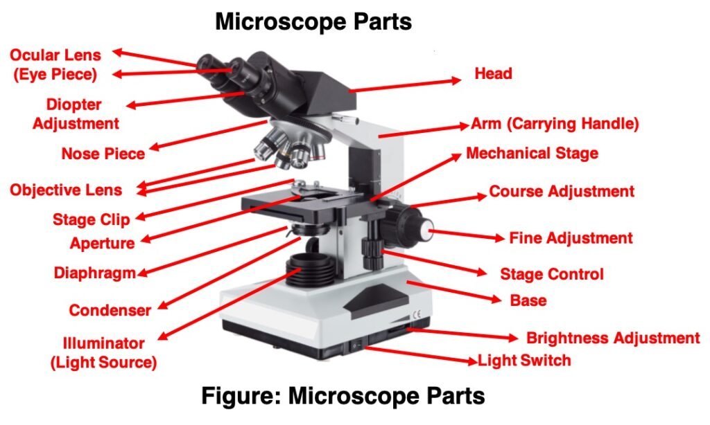
Eyepiece Lens: the lens at the top that you look through, usually 10x or 15x power.
Tube: Connects the eyepiece to the objective lenses.
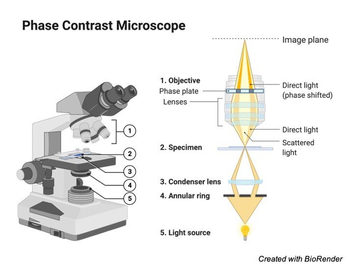
Illuminator: Illuminator is the most important microscope parts and it serve as light source for a microscope during slide specimen visualization. It is a continuous source of light (110 volts) used in place of a mirror. The mirror of microscope is used to reflect light from the external light source up through the bottom of the stage. Usually, the Illuminator located in the base of the microscope. Most light microscopes use low voltage, halogen bulbs with continuous variable lighting control located within the base.
Stage with Stage Clips: The stage of a microscope is a flat platform where you place your subject slides. Stage clips hold the slides in place. The mechanical stage of your microscope will help you to move the slide around by turning two knobs. One knobs moves it left and right, the other knobs moves it up and down.
Revolving Nosepiece or Turret: Turret is the part of the microscope that holds two or multiple objective lenses and helps to rotate objective lenses and also helps to easily change power.
Objective Lenses: Three are 3 or 4 objective lenses on a microscope. The objective lenses almost always consist of 4x, 10x, 40x and 100x powers. The most common eyepiece lens is 10x and when it coupled with others, total magnification is 40x (4x times 10x), 100x , 400x and 1000x. Objectives can be forward or rear-facing.
Microscope Rack Stop Rack Stop: Rack Stop is an important microscope parts that determines how close the objective lens can get to the slide. It keeps the students from damaging the high power objective lens down into the slide. If you cann’t able to focus on the specimen at high power while using very thin slides then slight adjustment helps you to adjust the focus.
Diaphragm or Iris: Most of the laboratory microscopes have a rotating disk under the stage known as diaphragm or iris. Iris Diaphragm controls the amount of light reaching the specimen. The Iris Diaphragm is located above the condenser lens and below the microscope stage.
The different sized holes in the diaphragm helps to vary the size of the cone and intensity of light that is projected upward into the slide. However, there is no set rule regarding which setting to use for a particular power.
The specimen transparency, degree of contrast and particular objective lens in use decide the Diaphragm or Iris setting. Majority of high quality microscopes used in laboratory include an Abbe condenser with an iris diaphragm. When iris diaphragm is combined with Abbe condenser, it control both the quantity of light applied as well as focus on the specimen.
Aperture: It is the hole in the stage through which the base (transmitted) light reaches the stage.
Condenser: Condenser lenses are used to collect and focus the light from the illuminator on to the specimen. Usually the Condenser lenses are located under the stage in conjunction with an iris diaphragm. Condenser lenses helps in ensuring clear and sharp images are produced with a high magnification of 400X and above.
Magnification power of the condenser is directly related to the image clarity. Most of the sophisticated microscopes in the laboratory come with an Abbe condenser that has a high magnification of about 1000X. Condenser Focus Knob moves the condenser up or down to control the lighting focus on the specimen.


