Category: Uncategorized

Category: Uncategorized

  • Protein Synthesis: Definition, Steps, Site, and Diagram

    Protein Synthesis or Protein Translation

    o Protein Translation is the process of protein synthesis directed by mRNA.

    o mRNA is the template which carries the genetic code from the nucleus to the cytosol in the form of codons.

    o The mRNA message is always written in the 5’ → 3’ direction, and the polypeptide chain is synthesized starting with its N-terminal residue.

    o The mRNA is always read in groups of 3 nucleotides.

    o Prokaryotic translation may occur simultaneously with transcription.

    o tRNA contains a set of nucleotides that is complimentary to the codon called the anticodon.

    o tRNA sequesters the amino acid that corresponds to its anticodon.

    o The 5’ base of the anticodon is capable of a “wobble” in its position during translation, allowing it to make alternative hydrogen bonding arrangements with several different codon bases.

    o Identity elements (not on the anticodon) as well as the anticodon determine which amino acid is bound to the tRNA by the aminoacyl-tRNA synthetases.

    o rRNA with protein makes up the ribosome, which provides the site for translation to take place.

    o The ribosome is composed of a small subunit and a large subunit made from rRNA and many separate proteins.

    o The ribosome and its subunits are measured in terms of sedimentation coefficients given in Svedberg units (S).

    o The sedimentation coefficient gives the speed of a particle in a centrifuge, and is proportional to mass, and related to shape and density.

    Protein Synthesis Diagram

    Protein Synthesis, Protein Synthesis Steps, What is Protein Synthesis, Protein Synthesis diagram, site of Protein Synthesis,

    o Prokaryotic ribosomes are smaller than eukaryotic ribosomes.

    o Prokaryotic ribosomes are made from a 30S and a 50S subunit and have a combined sedimentary coefficient of 70S.

    o Eukaryotic ribosomes are made from a 40S and a 60S subunit and have a combined sedimentary coefficient of 80S.

    o The complex structure of ribosomes requires a special organelle called the nucleolus in which to manufacture them.

    o Prokaryotes don’t have a nucleolus, but synthesis of prokaryotic ribosomes is similar to that of eukaryotic ribosomes.

    o Although the ribosome is assembled in the nucleolus, the small and large subunits are exported separately to the cytoplasm.

    o rRNA is synthesized in the nucleolus.

    o After posttranscriptional processing in a eukaryote, mRNA leaves the nucleus through the nuclear pores and enters the cytosol.

    o With the help of initiation factors, the 5’ end attaches to the small subunit of a ribosome.

    o A tRNA possessing the 5’-CAU-3’ anticodon sequesters the amino acid methionine and settles in the P site (peptidyl site).

    o This is the signal for the large subunit to join and form the initiation complex.

    o This process is called initiation. Now elongation of the polypeptide begins.

    "Prokaryotic ribosomes are smaller than eukaryotic ribosomes"

    o A tRNA with its corresponding amino acid attaches to the A site (aminoacyl site) at the expense of two GTPs.

    o The C-terminal of the methionine attaches to the N-terminal of the incoming amino acid at the A site in a dehydration reaction catalyzed by peptidyl transferase, an activity possessed by the ribosome.

    o In an elongation step known as translocation, the ribosome shifts 3 nucleotides along the mRNA toward the 3’ end.

    o The tRNA that carried methionine is carried to the E site where it can exit the ribosome.

    o Then the tRNA that is in the A site is moved to the P site.

    o Translocation requires the expenditure of another GTP.

    E ⇒ EXIT

    P ⇒ PROCESS

    A ⇒ ARRIVE

    Protein Synthesis Steps

    Protein Synthesis, Protein Synthesis Steps, What is Protein Synthesis, Protein Synthesis diagram, site of Protein Synthesis,

    o Translation ends when a stop codon (UAA, UAG, UGA) is reached in a step called termination. When a stop (or nonsense) codon reaches the A site, proteins known as release factors bind to the A site allowing a water molecule to add to the end of the polypeptide chain.

    o The protein is released and the rRNA breaks up into its subunits and waits for it to be used again in another round of translation Even as the polypeptide is being translated, it begins folding.

    o The amino acid sequence determines the folding conformation and the folding process is assisted by proteins called chaperones.

    o In post-translational modification, sugars, lipids, or phosphate groups may be added to the amino acids.

    o The polypeptide may be cleaved in one or more places, or other polypeptides might join to form a quaternary structure.

    o Translation may take place on a free floating ribosome in the cytosol producing proteins that function in the cytosol or a ribosome may attach itself to the rough ER during translation and inject the protein into the ER lumen.

    o Proteins injected into the ER lumen are destined to become membrane bound proteins of the nuclear envelope, ER, Golgi, plasma membrane, or to be secreted from the cell.

    o Free floating ribosomes are identical in structure to the ribosomes that attach to the ER.

    o The growing polypeptide may or may not cause the ribosome to attach to the ER depending upon the polypeptide.

    o A 20 amino acid sequence called a signal peptide near the front of the peptide is recognized by protein-RNA signal- recognition particles (SRPs) that carries the entire ribosome complex to a receptor on the ER.

    o The peptide is actually pulled through the membrane through an ATP driven process.

    o The signal peptide is also usually removed by an enzyme.

    "Signal peptides may also be attached to polypeptides to target them to mitochondria, the nucleus, or other organelles"

    Protein Synthesis Citations

    Share

    Similar Post:

    Continue Reading
  • Genetic Code: Definition, Characteristics, Table, and Facts

    Genetic Code Chart

    o There are four different nucleotides in RNA that together must form an unambiguous code for the 20 common amino acids.

    o The code must be a combination of any three nucleotides.

    o However, any three nucleotides gives 4^3 = 64 possible combinations.

    o These are more possibilities than there are amino acids.

    o Thus more than one series of three nucleotides may code for any amino acid; The code is degenerative.

    o The code is also unambiguous (clear), one codon codes for only one amino acid and never two.

    o In addition the code is almost universal (mitochondria present an exception), nearly every living organism uses the same code.

    Genetic Code Chart

    Genetic Code Chart, Genetic Code, Table, Definition, and Examples Codon- Definition, Function, and Mechanism I Research Tweet

    o Redundancy is usually expressed in the third letter of the codon (3’ position).

    o Three consecutive nucleotides on a strand of mRNA represent a codon.

    o All but three possible codons code for amino acids, so there are only 61 codes for amino acids.

    o The remaining codons UAA, UGA, and UAG are stop codons, which signal an end to the protein synthesis.

    o AUG is a start codon and it also codes for amino acid methionine.

    o Codon changes have been opposed by the most intense selective pressure during evolution.

    o By convention, a sequence of RNA nucleotides is written 5’ → 3’ We must understand probabilities.

    o For instance, you must be able to figure out how many possible codons exist.

    o As discussed above, four possible nucleotides can be placed in 3 spots giving 4^3 = 64.

    o What about this? “A polypeptide contains 100 amino acids. How many possible amino acid sequences are there for this polypeptide? ”

    o Well first we know that there are 20 amino acids and from the information we just received we know that there are 100 spots to place them. So it must be 20^100 possible sequences.

    Genetic Code Chart Citations

    Share

    Similar Post:

    Continue Reading
  • DNA Technology: Definition, Types, and Facts

    DNA Technology: Nucleic Acid Hybridization

    o When heated or immersed in high concentration salt solution or high pH solution, the hydrogen bonds connecting the two strands in a double stranded DNA molecule are disrupted, and the strands separate; the DNA molecule is said to be denatured or melted.

    o Denatured DNA is less viscous (less syrupy), denser, and more able to absorb UV light.

    o Separated strands will spontaneously associate with their original partner or any complementary nucleotide sequence.

    o Thus, the following double stranded combinations can be formed through nucleic acid hybridization.

    o DNA-DNA, DNA-RNA, RNA-RNA

    Restriction Enzymes

    o One method bacteria use to defend themselves from viruses is to cut the viral DNA into fragments with restriction enzymes.

    o The bacteria protect their own DNA from these enzymes by methylation.

    o Methylation is usually, but not always, associated with inactivated genes.

    o Restriction enzymes (also called restriction endonucleases) digest/cut nucleic acid only at certain nucleotide sequences along the chain.

    o Such a sequence is called a restriction sire or recognition sequence.

    o Typically, a restriction site will be a palindromic sequence four to six nucleotides long.

    o Most restriction endonucleases cleave the DNA strand unevenly, leaving complementary single stranded ends.

    o These ends can reconnect through hybridization and are termed sticky ends.

    o Sticky ends are produced by cutting the DNA in a staggered manner within the recognition site producing single-stranded DNA ends.

    restriction enzymes - restriction endonucleases - DNA Technology

    o It can also be cut so that it has blunt ends.

    o Two DNA fragments cleaved by the SAME endonuclease can be joined together regardless of the origin of the DNA.

    o Such DNA is called recombinant DNA; it has been artificially recombined.

    Plasmid

    o A plasmid is an extra-chromosomal DNA molecule separate from the chromosomal DNA which is capable of replicating independently of the chromosomal DNA.

    o In many cases, it is circular and double-stranded.

    o Plasmids usually occur naturally in bacteria, but are sometimes found in eukaryotic organisms.

    o Plasmids used in genetic engineering are called vectors.

    o Plasmids serve as important tools in genetics and biotechnology labs, where they are commonly used to multiply (make many copies of) or express particular genes.

    Plasmid - Plasmid map - Plasmid Vector

    o The gene to be replicated is inserted into copies of a plasmid containing genes that make cells resistant to particular antibiotics and a multiple cloning site, which is a short region containing several commonly used restriction sites allowing the easy insertion of DNA fragments at this location.

    o Next, the plasmids are inserted into bacteria by a process called transformation.

    o Then, the bacteria are exposed to the particular antibiotics.

    o Only bacteria which take up copies of the plasmid survive , since the plasmid makes them resistant.

    o Eukaryotic DNA contains introns.

    o Since bacteria have no mechanism for removing introns, it is useful to clone DNA with no introns.

    o In order to do this, the mRNA produced by the DNA is reverse transcribed using reverse transcriptase.

    o The DNA product is called complementary DNA (cDNA).

    o Adding DNA polymerase to cDNA produces a double strand of the desired DNA fragment (an entire DNA double helix free of introns).

    Polymerase Chain Reaction (PCR)

    o The polymerase chain reaction (PCR) is a technique to amplify a single or few copies of a piece of DNA across several orders of magnitude, generating millions or more copies of a particular DNA sequence.

    o The method relies on thermal cycling, consisting of cycles of repeated heating and cooling of the reaction for DNA melting and enzymatic replication of the DNA.

    o Primers (short DNA fragments) containing sequences complementary to the target region along with a DNA polymerase.

    o Almost all PCR applications employ a heat-stable DNA polymerase, are key components to enable selective and repeated amplification.

    o As PCR progresses, the DNA generated is itself used as a template for replication, setting in motion a chain reaction in which the DNA template is exponentially amplified.

    o Starting with a single fragment, 20 cycles produces 2^20 copies

    Southern Blotting

    o Southern blotting is a technique used to identify target fragments of known DNA sequence in a large population of DNA.

    o It is separated according to size by gel electrophoresis.

    o The anode for Southern blotting is positive!.

    o The cathode for Southern blotting is negative.

    Northern Blotting

    o Northern blotting is just like Southern blotting but it identifies RNA fragments.

    Restriction Fragment Length Polymorphism (RFLP)

    o Restriction fragment length polymorphism (RFLP) analysis identifies individuals as opposed to individual specific genes.

    o The DNA of different individuals possesses different restriction sites and varying distances between restrictions sites.

    o After fragmenting the DNA sample with endonucleases, southern blotting is used and produces bands distinct to the individual.

    o The genome of one human differs from the next at about one nucleotide in every 1000.

    o These differences have been called single nucleotide polymorphisms (SNPs)

    DNA Technology Citations

    Share

    Similar Post:

    Continue Reading
  • Post Transcriptional Modification: Definition, Types, and Mechanism

    Post Transcriptional Modification in Prokaryotes

    o rRNA and tRNA go through posttranscriptional processing.

    o Almost all mRNA is directly translated to protein.

    Post Transcriptional Modification in Eukaryotes ​

    o Each type of RNA undergoes posttranscriptional processing.

    o Posttranscriptional processing allows for additional gene regulation.

    o The initial mRNA nucleotide sequence arrived at through transcription is called the primary transcript (also called pre-mRNA, or heterogeneous nuclear RNA [hnRNA]).

    Type of Post Transcriptional Modification

    The primary transcript is processed in three (3) ways:

    1) addition of nucleotides

    2) deletion of nucleotides

    3) modification of nitrogenous bases

    o Even before the eukaryotic mRNA is completely transcribed, its 5’ end is capped in a process using GTP.

    o The 5’ cap serves as an attachment site in protein synthesis and as a protection against degradation by exonucleases.

    o The 3’ end is polyadenylated with a poly A tail, also to protect it from exonucleases.

    o The primary transcript is much longer than the mRNA that will be translated into a protein.

    o This is due to noncoding regions existing in the primary transcript.

    o These regions are called introns.

    o Introns = noncoding regions of DNA in a gene; generally much longer than exons; Introns stay IN nucleus

    o Exons = coding regions of DNA in a gene; Exons EXIT nucleus

    o Enzyme-RNA complexes called small nuclear ribonucleoproteins (snRNPs “snurps”) recognize nucleotide sequences at the ends of the introns.

    o Several snRNP’s associate with proteins to form a complex called a spliceosome.

    o Inside the spliceosome, the introns are looped bringing the exons together.

    o The introns are then excised by the spliceosome and the exons are spliced together.

    o The exons of some genes may be spliced together in different order allowing them to encode for different polypeptides.

    Citations

    Share

    Similar Post:

    Continue Reading
  • RNA, Types, and RNA vs DNA

    Differences between DNA and RNA (RNA vs DNA)

    ○ Carbon #2 on the pentose is not “deoxygenated” (it has a hydroxyl group attached).

    ○ RNA is single stranded.

    ○ RNA contains uracil instead of thymine.

    ○ RNA can move through the nuclear pores and isn’t confined to the nucleus.

    Types of RNA

    RNA exists in 3 forms:

    1) mRNA

    ○ Delivers the DNA code for amino acids to the cytosol where the proteins are manufactured.

    ○ Has a short half-life in the cytosol, so soon after its transcription is over, the mRNA is degraded.

    ○ Many proteins can be transcribed in a single mRNA.

    2) rRNA

    ○ Combines with proteins to form ribosomes, the cellular complexes that direct the synthesis of proteins.

    ○ rRNA is synthesized in the nucleolus.

    3) tRNA

    ○ Collects amino acids in the cytosol, and transfers them to the ribosomes for incorporation into a protein.

    mRNA → massive

    rRNA → rampant

    tRNA → tiny

    Citations

    Share

    Similar Post:

    Continue Reading
  • RNA Transcription, Diagram, Definition, Process, Steps

    RNA Transcription

    ○ All RNA is manufactured from a DNA template in a process called RNA transcription.

    ○ Transcription requires a promoter; Replication requires a primer.

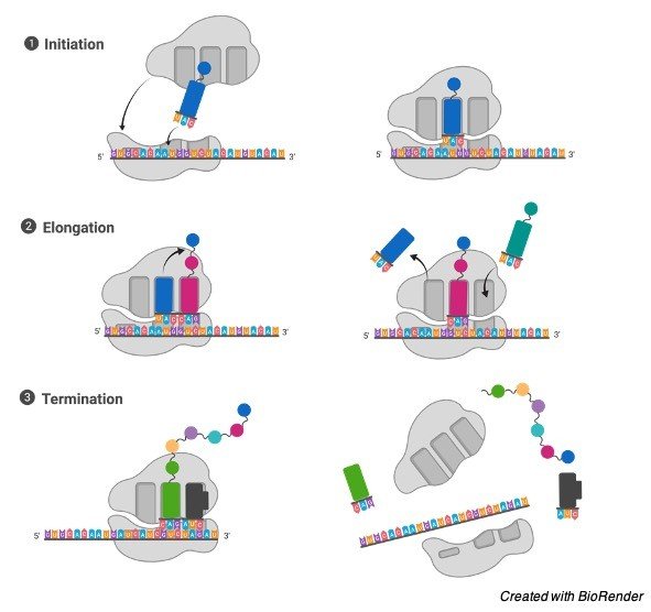
    ○ The beginning of transcription is called initiation.

    ○ In initiation, a group of proteins called initiation factors finds a promoter on the DNA strand, and assembles a transcription initiation complex, which includes RNA polymerase

    ○ Prokaryotes have 1 type of RNA polymerase; Eukaryotes have 3 types of RNA polymerase (one for each type of RNA).

    ○ A promoter is a sequence of DNA nucleotides that designates a beginning point for transcription, and promoter recognition is the rate limiting step in transcription.

    ○ The promoter in prokaryotes is located at the beginning of the gene (said to be upstream).

    ○ The transcription start point is part of the promoter.

    ○ The first base-pair located at the transcription start point is designated +1; base-pairs located before the start point, such as those in the promoter, are designated by negative numbers.

    ○ The most commonly found nucleotide sequence of a promoter recognized by the RNA polymerase of a given species is called the consensus sequence.

    ○ Variation from the consensus sequence causes RNA polymerase to bond less tightly and less often to a given promoter, which leads to those genes being transcribed less frequently.

    ○ After binding to the promoter, RNA polymerase unzips the DNA double helix creating a transcription bubble.

    ○ Next the complex switches to elongation mode.

    ○ In elongation, RNA polymerase transcribes only one strand of the DNA nucleotide sequence into a complementary RNA nucleotide sequence.

    ○ Only one strand in the molecule of double stranded DNA is transcribed.

    ○ This strand is called the template strand or (-) antisense strand.

    ○ The other strand, called the coding strand or (+) sense strand protects its partner from degradation.

    ○ The coding strand/sense strand resembles the universal code sequence of RNA.

    Diagram Representing Regulation of Transcription in Eukaryotes

    Chromatin, What is Chromatin, Chromatin Function, Chromatin Definition,

    ○ Just like DNA polymerase, RNA polymerase reads in the 3’ → 5’ direction and builds in the 5’ → 3’ direction, but it DOESN’T have proofreading ability.

    ○ The end of transcription is called termination, and requires a special termination sequence (high G-C content) and special proteins to dissociate RNA polymerase from DNA.

    ○ Genes are activated or deactivated at the level of transcription.

    ○ For all cells, most regulation of gene expression occurs at the level of transcription via proteins called activators and repressors.

    ○ Activators and repressors bind to DNA close to the promoter, and either activate or repress the activity of RNA polymerase.

    ○ Activators and repressors are often allosterically regulated by small molecules such as cAMP.

    ○ The primary function of gene regulation in prokaryotes is to respond to the environmental changes.

    ○ In contrast, lack of change or homeostasis of the intracellular and extracellular compartments is the hallmark of multicellular organisms.

    ○ The primary function of gene regulation in multicellular organisms is to control the intra- and extracellular environments of the body.

    ○ Prokaryotic mRNA typically contains several genes in a single transcript (polycistronic), whereas eukaryotic mRNA includes only one gene per transcript (monocistronic).

    ○ The genetic unit usually consisting of the operator, promoter, and genes that contribute to a single prokaryotic mRNA is called the operon.

    ○ Genes of an operon are transcribed on one mRNA.

    ○ Genes outside the operon may code for activators and repressors

    ○ An operator is a segment of DNA that a regulatory protein binds to.

    ○ It is classically defined in the lac operon as a segment between the promoter and the genes of the operon.

    ○ A repressor or activator can bind to an operato.

    ○ A good example of an operon is the lac operon.

    ○ The lac operon codes for enzymes that allow E. Coli to import and metabolize lactose when glucose is not present in sufficient quantities.

    ○ Low glucose levels lead to high cAMP levels.

    ○ cAMP binds to and activates catabolite activator protein (CAP).

    ○ The activated CAP protein binds to a CAP site located adjacent and upstream from the promoter to the lac operon.

    ○ The promoter is now activated allowing the formation of an initiation complex and subsequent transcription and translation of the 3 proteins.

    Diagram Representing Regulation of Transcription in Prokaryotes

    Lac Operon- Diagram, Phenotypes, Model, and Regulation 1

    ○ A second regulatory site on the lac operon, called the operator, is located adjacent and downstream to the promoter.

    ○ The operator provides a binding site for a lac repressor protein.

    ○ The lac repressor protein is inactivated by the presence of lactose in the cell.

    ○ The lac repressor protein will bind to the operator unless lactose binds to the lac repressor protein and inactivates it.

    ○ The binding of the lac repressor to the operator in the absence of lactose prevents the transcription of the lac genes.

    ○ Lactose, then, can induce the transcription of the lac operon only when glucose is not present.

    ○ Gene regulation in eukaryotes is more complicated involving the interaction of many genes.

    ○ Thus more room is required than is available near the promoter.

    ○ Enhancers are regulatory proteins commonly used by eukaryotes.

    ○ Their function is similar to activators and repressors, but they act at a much greater distance from the promoter.

    RNA Transcription Citations

    Share

    Similar Post:

    Continue Reading
  • DNA Replication, Replication Fork, DNA Polymerase, Replication...

    DNA Replication

    • DNA replication is semiconservative.

    • This means that when a new double strand is created, it contains one strand from the original DNA, and one newly synthesized strand.

    • It is similar for both Prokaryotes and Eukaryotes.

    • The process of DNA replication is governed by a group of proteins called a replisome.

    DNA Replication Diagram

    DNA Replication - research tweet 2

    • It is made up of a number of subcomponents that each provide a specific function during the process of replication.

    • Replication doesn’t begin at the end of chromosomes, but toward the middle at a site called the origin of replication.

    • A single eukaryotic chromosome contains multiple origins on each chromosome, while replication in prokaryotes usually takes place for a single origin on the circular chromosome.

    • From the origin, two replisomes proceed in opposite directions along the chromosome making replication a bidirectional process.

    • The point where a replisome is attached to the chromosome is called the replication fork.

    DNA Replication Fork

    Replication Fork Definition Structure Diagram and Function - research tweet

    • Each chromosome of eukaryotic DNA is replicated in many discrete segments called replication units or replicons.

    • A replicon is a DNA molecule or RNA molecule, or a region of DNA or RNA, that replicates from a single origin of replication.

    • For most prokaryotic chromosomes, the replicon is the entire chromosome.

    • For eukaryotic chromosomes, there are multiple replicons per chromosome.

    • The definition of replicons is somewhat confused with mitochondria, as they use unidirectional replication with two separate origins.

    "Exonucleases are enzymes that work by cleaving nucleotides one at a time from the end of a polynucleotide chain"

    • DNA helicase uses the energy of ATP hydrolysis to actively unwind the two strands.

    • DNA polymerase, the enzyme that builds the new DNA, cannot initiate a strand from two nucleotides, but can only add nucleotides to an existing strand.

    • Therefore it requires an RNA primer to get started.

    •  It reads the parental strand in the 3’ → 5’ direction.

    • Since it can only add new subunits to the 3’ end of the chain, it must create the new complementary strand in the 5’ → 3’ direction

    • Besides being a polymerase, one of the subunits in DNA polymerase has 3’ → 5’ exonuclease (it removes nucleotides from the strand) capabilities.

    • Exonucleases are enzymes that work by cleaving nucleotides one at a time from the end of a polynucleotide chain

    • There are 3 types of polymerase molecules This enzyme automatically proofreads each new strand, and makes repairs when it discovers any mismatched nucleotides.

    • Primase, an RNA polymerase, creates an RNA primer approximately 10 ribonucleotides long to initiate the strand.

    • Each nucleotide added to the new strand requires the removal of a pyrophosphate group (two phosphates binded together) from a DNTP (deoxynucleotide triphosphate).

    • Some of this energy derived from the hydrolysis of the pyrophosphate is used to drive replication.

    • SSB tetramer proteins (also called helix destabilizer proteins) prevent the two strands from reattaching after the helix is opened.

    • The interrupted strand is called the lagging strand; the continuous new strand is called the leading strand.

    • The lagging strand is made from a series of disconnected strands called Okazaki fragments.

    • Okazaki fragments are about 100-200 nucleotides long in eukaryotes and about 1000-2000 nucleotides long in prokaryotes.

    "SSB tetramer proteins (also called helix destabilizer proteins) prevent the two strands from reattaching after the helix is opened"

    • DNA Ligase moves along the lagging strand and ties the Okazaki fragments together to complete the polymer.

    • Since the formation of one strand is continuous and the other fragmented, the process of replication is said to be semidiscontinuous.

    • The ends of eukaryotic chromosomal DNA possess telomeres.

    • Telomeres are repeated 6 nucleotide units from 100-1000 units long that protect the chromosomes from being eroded through repeated rounds of replication.

    • Telomerase catalyze the lengthening of telomeres at the 3’ ends of DNA strands.

    • Eukaryotic chromosomes contain linear DNA molecules; Prokaryotic chromosomes are usually circular.

    • So telomeres aren’t required by Prokaryotes that have circular DNA.

    DNA Replication Citations

    Share

    Similar Post:

    Continue Reading
  • Chromosomes, Genes, DNA: Definition, Structure, and Functions

    Chromosomes, Genes, DNA

    ○ A gene is a series of DNA nucleotides that generally codes for the production of a single polypeptide or mRNA, tRNA, rRNA.

    ○ The entire DNA sequence of an organism is called the genome.

    ○ Eukaryotes have more than one copy of some genes, while prokaryotes have only one copy of each gene.

    ○ Genes are often referred to as unique sequence DNA; while regions of non-coding DNA found only in eukaryotes are called repetitive sequence DNA.

    ○Generally speaking: One gene; one polypeptide. One exception is posttranscriptional processing of RNA.

    Chromatin Structure

    ○ Negatively charged DNA loops twice around.

    ○Histone octamer (2 each of the positively charged H2A, H2B, H3, and H4) to form nucleosome bead. 

    Gene Nucleotides: Definition, Functions, Types, and Examples

    "Histone octamer (2 each of the positively charged H2A, H2B, H3, and H4) to form nucleosome bead"

    ○Eukaryotic genes that are actively being transcribed by a cell are associated with regions of DNA called euchromatin, while genes not being actively transcribed are associated with tightly packed regions of DNA called heterochromatin.

    ○ Repetitive DNA is found mainly in heterochromatin.

    ○The Central Dogma of gene expression is that DNA is transcribed to RNA, which is translated to amino acids forming a protein.

    DNA

    ○ Four nitrogenous bases exist in DNA:

    ○ Purines: Two ringed structure

    1) Adenine (less C=O)

    2) Guanine (more C=O)

    ○ Pyrimidines: Single ring structure

    3) Cytosine (less C=O)

    4) Thymine (more C=O)

    ○ Deamination of cytosine forms uracil

    ○ In nucleic acids, nucleotides are joined together by phosphodiester bonds between the third carbon of one dexoyribose and the phosphate backbone of a single strand of DNA with a 5 → 3 directionality.

    ○ A phosphodiester bond is a group of strong covalent bonds between the phosphorus atom in a phosphate group and two other molecules over two ester bonds.

    "In DNA, two strands are joined by the hydrogen bonds"

    ○ Nucleotides are written 5’ → 3’

    ○In DNA, two strands are joined by the hydrogen bonds to make the structure called the double helix.

    ○ This model was proposed by Watson and Crick.

    ○ The members of each base pair can fit together within the double helix only if the two strands of the helix are antiparallel.

    ○ By convention the top DNA strand goes 5’ → 3’ and the bottom 3’ → 5’

    ○ Going in the 5’ → 3’ Direction is referred to as going downstream

    ○ Going in the 3’ → 5’ Direction is referred to as going upstream The two strands are complementary strands.

    ○ The double helix contains two distinct grooves called the major groove and minor groove, which moves around once every 10 base-pairs.

    Chromosomes, Genes, DNA Citations

    Share

    Similar Post:

    Continue Reading
  • Electron Transport Chain (ETC): Definition, Steps, and...

    Electron Transport Chain (ETC)

    Electron Transport Chain (ETC) is an aerobic, occurs across the inner cell membrane for prokaryotes, inner mitochondrial membrane for eukaryotes.

    Structure of Electron Transport Chain (ETC)

    electron transport chain - Research Tweet

    • Oxidative phosphorylation oxidizes NADH to NAD+, which creates a proton gradiant, via the pumping out of H+, that propels protons through ATP synthase.

    • So the intermembrane space has a lower pH/higher [H+ concentration] than the matrix.

    • The inter-cristal space or the mitochondrial matrix has a low H+ concentration and a high pH.

    • NADH oxidation back to NAD and FADH2 oxidation back to FAD occur along with ATP production, allowing the earlier stages to continue.

    • As electrons move within the ETC each intermediate carrier is reduced by the preceding molecule and oxidized by the following

    • Oxygen is the last electron acceptor in the ETC

    • 1 NADH produces 3 ATP molecules

    • 1 FADH produces 2 ATP molecules

    Electron Transport Chain (ETC) Summary

    • 36 net ATP produced in eukaryotes, 38 net ATP produced in prokaryotes (because the electrons from the NADH produced from pyruvate decarboxylation do not have to be transported across the mitochondrial membrane in prokaryotes; doing this causes a net loss of two ATP in eukaryotes)

    • Products and Reactants for Respiration:

    Glucose + O2 → CO2 + H2O

    Electron Transport Chain (ETC) Citations

    Share

    Similar Post:

    Continue Reading
  • Krebs Cycle or TCA Cycle: Definition, Steps,...

    Krebs Cycle or TCA Cycle

    Krebs Cycle or TCA Cycle is an aerobic process, occurs in the cytoplasm for prokaryotes, mitochondrial matrix for eukaryotes.

    The process of ATP production in the Krebs cycle is called Substrate-level phosphorylation

    Krebs Cycle or TCA Cycle

    TCA Cycle Krebs Cycle - research tweet 1

    Total:

    ⊛ 4 CO2 produced

    ⊛ 2 GTP/ATP produced

    ⊛ 6 NADH produced (making 18 ATP in ETC)

    ⊛ 2 FADH2 produced (making 4 ATP in ETC)

    "Each turn in the Krebs Cycle produces (1 glucose provides two turns)"

    ⊛ 2 CO2 produced

    ⊛ 1 GTP/ATP produced

    ⊛ 3 NADH produced (making 9 ATP in ETC)

    ⊛ 1 FADH2 produced (making 2 ATP in ETC)

    Krebs Cycle or TCA Cycle Regulation

    ⊛ The cycle is regulated at the three steps that are highly exergonic: those catalyzed by citrate synthase, isocitrate dehydrogenase, and a- ketoglutarate dehydrogenase.

    ⊛ NADH and ATP are both negative regulators; ADP and Ca2+are positive regulators (activators).

    ⊛ Triglycerides can also be catabolized for ATP. Fatty acids are converted to acyl CoA along the outer membrane of the mitochondrion and endoplasmic reticulum at the expense of 1 ATP. Then 2 carbons are cleaved to make acetyl CoA in the matrix. This reaction also produces FADH2 and NADH for every two carbons taken from the original fatty acid. Acetyl CoA then enters into the Krebs cycle as usual. This is called beta oxidation.

    ⊛ The fatty acids are linked to Coenzyme A and carried into the mitochondrial matrix by the g-amino acid L-carnitine. They are then are oxidized TWO carbons at a time in the KREB cycle, yielding an NADH, FADH2, and acetyl CoA.

    ⊛ Amino acids are deaminated in the liver. The deaminated product is either chemically converted to pyruvic acid or acetyl CoA, or it may enter the Kreb cycle at various stages depending upon which amino acid was deaminated.

    Krebs Cycle or TCA Cycle Citations

    Share

    Similar Post:

    Continue Reading