Notochord Definition
An elastic rod, found in all chordate organisms, is known as the notochord. It provides rigid support to the organism. The notochord is replaced by the vertebral column in chordates, called the vertebrates. It is formed by a cartilaginous substance between vertebrae. Lancelets are one of the simplest chordates, which also contain notochord.
What is Notochord?
In anatomy, the notochord is a flexible rod formed of a material similar to cartilage. If a species has a notochord at any stage of its life cycle, it is, by definition, a chordate. The notochord lies along the anteroposterior axis (front to back), is usually closer to the dorsal than the ventral surface of the embryo, and is composed of cells derived from the mesoderm.
The length of the notochord is extended to the length of the organism, and it allows the muscles to attach. Thus, the lancelet can swim in fast bursts. The notochord is developed at some point in all chordate organisms, like lancelets, however, some organisms may lose it later in life.
For instance, some marine organisms like tunicates, live in the bottom of the ocean and filter feed. The adult form of a tunicate does not require a notochord, but in the larval stage, it helps in swimming to potential settling sites, thus they lose notochord at the adult stage.
Other species do not grow a vertebral column and retain the notochord throughout life. These animals are called invertebrate chordates. Lancelets, tunicates, and some large fish such as the sturgeon and coelacanth are included in this group.
The length of the notochord can be around 3-4 feet in these organisms and will be really thick about half an inch. This huge notochord is used as a spine in notochord by these fish. However, how it protects the spinal cord and what it is made of are different in them. In invertebrates, the spinal cord is surrounded by the bony vertebrae and protects it on all sides.
This protection is not found in animals, which have only a notochord, and in these organisms, the spinal cord sits between the notochord and the skin. The spinal cord and notochord are protected by armored plates and thick skin in animals like sturgeon and coelacanth.
Invertebrates, the notochord is converted into cushioning intervertebral discs that provide protection to the vertebrae from smashing together. By the time a human is around 4 years old. However, the spine with other materials entirely replaces the original notochord.
Notochord Structure
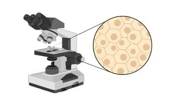
There are several structural molecules such as glycoproteins, form the notochord, which resembles cartilage in many ways. Under a microscope, the cross-section of the notochord appears as a series of concentric rings.
The rings surrounded by each other are layers of the notochord and are made of various molecules, which provide strength and elasticity to the notochord. The glycoproteins and other structural molecules extend from cells and are spaced apart in the notochord.
A large vacuole is found in each cell that can pressurize. The cells push against each other and the surrounding structural materials by pressurization. The notochord becomes extra rigid due to this, which helps the organisms in swimming quickly.
Notochord Function
The notochord becomes a very important and useful structure to attach muscles to due to its strength. To flex properly, muscles need places for attachment. In small invertebrates, the muscles attach down the length of the notochord, due to this they can use muscles all over to swim.
It provides enough support for most of the muscles of the body of some large fishes, which rely on notochord. The cells of the notochord create turgor pressure that makes it extra rigid. While many organisms get enough support from this. In vertebrates, this body plan is taken one step further with the spine.
The spine is made of bone, which increases its rigidity and also protects the spinal cord by fully encompassing it. It is also recorded that during normal vertebrate embryogenesis, the notochord serves important signaling functions.
The proteins secreted by the notochord stimulate the formation of organ systems. This process is known as organogenesis, which starts when the embryo is a hollow ball of cells known as the gastrula. Gastrula has three layers, and the notochord arises from the middle layer, known as mesoderm.
The process of organogenesis is further started with the secretion of several chemical signals from the notochord. Eventually, the spine is created with the formation of bones, and the notochord gets sandwiched between these vertebrae. The vertebrae also protect the notochord from the bones rubbing and smashing together.
Notochord Citations
Share


