The process through which interbreeding animals split into two or more evolutionary groupings is known as divergent evolution. It indicates that these species used to be connected and comparable. However, as time passed, they grew increasingly different.
What is Divergent Evolution?
Divergent evolution may also refer to the process of tracing two or more species back to their common ancestor and determining how they have diverged or diversified. Divergent evolution is one of three types of evolutionary patterns; the other two are convergent and parallel evolution. Environmental variables and predator activities have a big impact on species evolution. One of the most well-known examples of divergent evolution is Galapagos finches deviating from their descendent species.
Divergent Evolution Etymology
The phrase divergent evolution is claimed to have been coined by John Thomas Gulick, an American missionary and naturalist. Divergent is derived from the Latin divergentem, which means “to travel in several ways.” Evolution is derived from the Latin evolutionem, which is derived from the verb evolvere, which means “to unroll.”
Divergent Evolution vs Convergent Evolution
Divergent evolution is a form of evolution in which a species gradually becomes more distinct from its forebears. Unrelated groups of animals acquire comparable structures despite their evolutionary predecessors being very distant or unrelated, which contrasts with convergent evolution.
Analogous structures are biological structures that perform comparable functions but differ in development and anatomical features. Divergent evolution occurs when species with a common ancestor develop comparable anatomical features (called homologous structures) but different roles. Migration is one probable source of diverse evolution. When a species migrates to a new habitat, it is exposed to new environmental circumstances, and, as a result, it is more likely to acquire unique characteristics that help it adapt to its new environment. One example is the development of the so-called Darwin finches.
Environmental pressure is the primary driving force behind convergent evolution. Species evolve characteristics that make them adapt to their particular environments, even if they are unrelated. Insect, bird, and bat wings are examples of this.
Divergent Evolution vs Parallel Evolution
While species in divergent evolution basically diverge and merge, species in parallel evolution tend to evolve structures in tandem with other species in the same environment. Parallel evolution evolved features in animals that were not evolutionarily linked, similar to convergent evolution.
The distinction is that unrelated species acquired a comparable mechanism to adapt to the same environmental circumstances in parallel evolution. Unrelated species do not always dwell in the same habitat in convergent evolution.
Parallel evolution differs from divergent evolution in the same manner that convergent evolution differs from divergent evolution. Species from several evolutionary lineages have been linked to both parallel and convergent evolution. Divergent evolution refers to the evolution of a species away from its origins.
Divergent Evolution vs Adaptive Radiation
Adaptive radiation is the process through which numerous new species emerge from a recent ancestral source. Each of these species has evolved to take advantage of or occupy an open adaptation zone. As a result, this zone provides an ecological chance for some groups of species to diversify into new forms, frequently quickly. Adaptive radiation, like divergent evolution, leads to speciation, as Darwin’s Galapagos finches demonstrate.
Adaptive radiation, on the other hand, is concerned with small-scale evolution during a shorter period of time, whereas divergent evolution examines the development of species diverging from their progenitors over a longer period of time. Adaptive radiation, on the other hand, may result in divergent evolution over time as the species becomes increasingly different from its predecessors.
Importance of Divergent Evolution
Homologous structures are features that indicate a species is diverging from its origin in divergent evolution. These structures do not have to serve the same purpose as those of the species’ forefathers. Human and bat forelimbs, for example, are both homologous structures. Although they are utilised in different ways, they have the same fundamental skeletal structure and are derived from the same embryonic source. Their resemblance in this area might suggest that they evolved from a common ancestor.
This demonstrates how diverse evolution permits species with similar ancestral origins to adapt to their respective environments. As a result, since they acquire characteristics that make them precisely adapted to their environment and biological niche, it is likely to reduce competition among individuals. Divergent evolution is also beneficial to biodiversity. Because it leads to speciation, it has the potential to result in a varied spectrum of creatures flourishing in a variety of environments.
Divergent Evolution Examples
Finches in the Galapagos Islands are an excellent illustration of divergent evolution. The finches separated from their descendent species, according to Charles Darwin’s Beagle trip. While the birds share many characteristics with their forefathers, they finally evolved structures that set them apart from their progeny morphologically.
For example, they created beaks with a variety of shapes and sizes to better adapt to their food.
• The higher the structural differences, the wider the range of species divergence. Here are some more examples of divergent evolution:
• A swarm of newborns is moving to a new island. This group gets increasingly adapted over time as new traits emerge to help them survive in their new environment. As a result, they have become a distinct species from their forefathers, and what was previously a single species has split into two.
• Orchid species have developed different characteristics, resulting in an orchid variety (e.g. floral types).
• About 40 million years ago, peccaries (Tayasuidae) split from real pigs (Suidae).
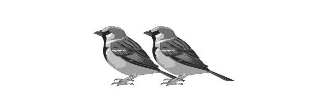
• Humans and apes descended from a shared primate ancestor.
Pattern of genotype-based offspring distribution (i.e. the genetic makeup of an organism that determines its phenotype.)
After a test cross, the genotypic ratio represents the number of times a genotype appears in the progeny. For example, a heterozygous dominant trait test cross between two organisms with the same genotype, Rr, will produce offspring with the genotypes RR, Rr, and rr. The expected genotypic ratio in this case is 1:2:1.
In general, a hybrid is something that has a mixed origin or makeup, or is made up of two or more separate things. A hybrid is the complex generated when two complementary strands of nucleic acids combine in molecular biology.
Reproductive Biology: The offspring of a cross between parents of different species or sub-species.
Molecular Biology: A nucleic acid complex produced by connecting two complementary strands.
What is Hybrid?
A hybrid is an offspring generated by crossing the parents of two distinct species or sub-species in reproductive biology. A mule is an example of a hybrid animal. A hybrid between a horse and a donkey produces the animal. Another animal hybrid is the liger, which is the offspring of a tiger and a lion. Hybridization is quite frequent in plants, and numerous plant hybrids are created naturally or by assisted breeding. Hybridization is one of the methods used in agriculture to create crop varieties with desired characteristics (such as increased disease resistance).
Hybrid Types
• The first generation of offspring produced by a cross between purebred parents is known as a single cross hybrid.
• Double cross hybrids are the progeny of a cross between two single cross hybrids.
• A three-way cross hybrid is the result of crossing a single cross hybrid with an inbred line.
• The child of crossing two distinct three-way cross hybrids is known as a triple cross hybrid.
• Population hybrids are offspring produced by crossing plants or animals from one population with those from another. For example, a hybrid of several races.
Hybrid Etymology
The word hybrid came from the Latin words hybrida and hibrida, which mean “crossbred animal.”
Hunter and Markert (1957) defined isozymes as distinct variations of the same enzyme that have identical functions and are present in the same person. (1) enzyme variations that are the product of separate genes and hence represent various loci (described as isozymes) and (2) enzymes that are the result of different alleles of the same gene are included in this definition (described as allozymes).
What are Isozymes?
Isozymes (also known as isoenzymes) are enzymes with varied amino acid sequences that catalyse the same chemical process. Various kinetic parameters (i.e. different KM values) or regulatory characteristics are frequently displayed by these enzymes.
Isozymes allow metabolism to be fine-tuned to fit the specific demands of a certain tissue or developmental stage (for example, lactate dehydrogenase (LDH)). Isozymes (or isoenzymes) are enzyme isoforms (closely related variations) in biochemistry. They are coded for by homologous genes that have diverged through time in many situations. Although the terms allozymes and isozymes technically refer to distinct alleles of the same gene and separate genes whose products catalyse the same process, the two terms are frequently interchanged.
Isozymes Features
Isozymes are most commonly caused by gene duplication, although they can also be caused by polyploidization or hybridization. If the function of the new variation remains the same as the original over time, one or the other will most likely be lost as mutations accumulate, resulting in a pseudogene. If, on the other hand, the mutations do not instantly stop the enzyme from working, but instead change its function or gene expression pattern, the two versions may be favoured by natural selection and specialise in distinct roles. They may, for example, be expressed at various phases of development or in various tissues.
Point mutations or insertion-deletion (indel) events that alter the gene’s DNA coding sequence can result in allozymes. A new allozyme might do one of three things, just like any other new mutation:
a. The new allele is most likely to be non-functional, in which case it will cause poor fitness and be eliminated from the population by natural selection.
b. If the altered amino acid residue is in a relatively insignificant region of the enzyme, such as a long distance from the active site, the mutation may be selectively neutral and vulnerable to genetic drift.
c. In rare circumstances, the mutation may produce a more efficient enzyme or one that can catalyse a slightly different chemical reaction, in which case the mutation may enhance fitness and be favoured by natural selection.
Isozymes Examples
Glucokinase, a form of hexokinase that is not inhibited by glucose 6-phosphate, is an example of an isozyme. Its unique regulatory characteristics and reduced affinity for glucose (as compared to other hexokinases) allow it to perform a variety of activities in cells of certain organs, such as regulating insulin release in pancreatic beta cells or initiating glycogen synthesis in liver cells. Both of these procedures must take place only when glucose is plentiful or when difficulties arise.
In biology, a non living things are any form that lacks life, such as an inanimate body or item. A non-living object lacks the characteristics that define a living thing as compared to a living creature. A non-living object, for example, lacks the essential component of life: a live cell that develops, metabolises, responds to external stimuli, reproduces, and adapts. A non-living object is made up of components or compounds that arise as a result of chemical processes rather than cells. Rocks, water, and air are examples of non-living objects.
non Living Things Etymology
The words non-living and living stem from the Middle English non-, which means “not,” “lack of,” or “failure to,” and lifende, which means “to live” or “to have a life. “Thing” is derived from the Old English word ing, which can signify “entity,” “being,” “body,” or “material.”
Living and non Living Things
A living creature is one that exhibits the characteristics of life; it is alive. A living creature is defined as one that moves, grows, and reproduces. We interpret the item as inanimate or non-living if it is not otherwise. A non-living object is shown in two ways. On the one hand, a non-living object is an entity that once had life but has since died. A non-living item, on the other hand, is something that has never had, never will have, and never will have life. The stricter definition of a non-living entity is the latter. The tighter definition of a non-living object is more accurate in a biological sense. In biological terms, a living item includes both alive and extinct species.
The fundamental resemblance between living and non-living objects is that they both refer to anything that exists. A separate entity, person, matter, or object is referred to as a thing. It’s possible to classify something depending on whether or not it has life. In contrast to a non-living item that lacks life, a living thing possesses or shows life.
Characteristics of non Living Things
i. Lack of Cell Organization
A non-living entity does not have cells, yet a living creature does. Although both non-living and living things are made up of molecules of elements and compounds, living things are made up of biomolecules that are arranged into cell structures. A living thing’s fundamental biological unit is the cell. It orchestrates and systemsatizes different biological activities. It is in charge of keeping the organism alive by conducting different metabolic activities such as cell development, cell respiration, reacting to stimuli, reproduction, nourishment, biomolecular syntheses, waste removal, and other homeostatic functions.
The protoplasm of the cell is enclosed by a plasma membrane. In the cytosol, many cytoplasmic entities are suspended. The nucleus is one of the most visible cytoplasmic components. Living organisms are categorised as prokaryotes or eukaryotes based on the existence of membrane-bound organelles such as the nucleus. Membrane-bound organelles are absent in prokaryotes, but they are present in eukaryotes.
ii. Growth by Accretion
A non-living organism does not develop in the same manner as a living organism does. Although certain non-living items appear to expand, this growth is caused by accretion rather than metabolic processes.
The cellular level of growth in living things is indicated by an increase in the number of cells or an increase in cell size. Cell division increases the number of cells (e.g. mitosis). The increase in cell size is frequently attributed to an increase in cytoplasmic mass. Some creatures have the ability to regenerate portions that have been lost. Plants, for example, can produce new shoots where they’ve been cut as long as the meristematic tissue isn’t damaged. Salamanders have the ability to regenerate new eyes and limbs. Humans have a limited ability to regenerate. They have the ability to repair skin and portions of the liver.
iii. non Motile
The majority of living things are motile, whereas non-living objects are not. Non-living objects might appear to move. The movement, however, is due to an external factor. Many living creatures have the ability to move about on their own. Animals have locomotory organs, which allow them to move around.
They move with them, particularly in reaction to stimuli. Animals, for example, flee their predators by fleeing away when they notice them. Animals may also travel in order to feed, find a better environment, or find a prospective partner. Plants, unlike most animals, are unable to move on their own volition. Plants are classified as living even though they are not as mobile as most animals. They exhibit many other traits of life.
iv. Lack of Reproduction
Non-living creatures can’t make duplicates of themselves in the natural world; living things can, through reproduction. A living object has the ability to create life. A living organism can reproduce in two ways: sexual reproduction and asexual reproduction. In sexual reproduction, the two parents’ sex cells combine to produce a zygote, which will ultimately grow into a creature of their own species. Sex cells are engaged in asexual reproduction, and the child comes from just one parent.
v. Lack of Metabolism
A living entity metabolises, but a non-living object does not. The numerous processes that allow the cell to stay alive are referred to as metabolism. Catabolism and anabolism are the 2 kinds of metabolism. Catabolism is a process in which a living organism performs degradative chemical processes that break down complex molecules into smaller components and receives energy from the process. Energy-driven chemical processes in anabolism create compounds from smaller components. As a result, a living organism needs energy to power these processes. Non-living objects, on the other hand, do not go through such metabolic processes and do not require energy to maintain their life.
vi. Not Responding to Stimuli
A non-living creature can not notice or respond to changes in its surroundings, but a living entity can. Non-living things lack the specific sensors that allow living things to perceive environmental changes. Sight, hearing, smell, touch, and taste are all senses that humans and other animals have. Plants and other creatures lack the complex sense organs seen in mammals, yet they can nevertheless notice changes in their surroundings and respond to stimuli. It’s possible that the answer will be favourable or negative. A positive reaction is one that is directed toward the stimulus source, whereas a negative response is one that is directed away from the stimulus source.
vi. Not Capable of Adaption
A non-living object does not adapt; a living object does so by adapting to changing circumstances. Living things are distinguished from non-living objects by their ability to adapt to changes in the environment. They have the ability to change in order to better adapt to their surroundings. In this context, it’s also worth noting that a non-living object doesn’t mutate, but a living thing does and so diversifies.
vii. Lack of Life Cycle
Because a non-living item has no life, it does not die. A non-living object decomposes rather than dies. The decomposition of a material by chemical or physical processes is referred to as abiotic decomposition. A living object, on the other hand, dies and decays. Death happens when a person’s life comes to an end. Organs, tissues, and cells stop working when a live entity dies. The breakdown process is referred to as decay in biology. Biodegradation is the process through which a deceased organism decomposes. Microbes, for example, degrade organic molecules into simpler forms.
non Living Things Examples
The term “non-living” refers to something that lacks the qualities of life. Non-living objects, according to that description, include rocks, water, sand, glass, and the sun. None of them exhibit any of the hallmarks of life. Others describe a non-living item as anything that once belonged to a live creature. Coal, wood, rubber, and paper, for example. Despite the fact that they were formerly a part of a live tree, they are now considered non-living.
Non-living objects are part of the abiotic components (abiotic elements) of the environment, such as soil and the atmosphere, in ecology. They have an impact on the growth, reproduction, and maintenance of living organisms. The biotic components or biotic elements are the living organisms themselves.
The part of a prokaryotic cell that contains the genophore (genetic material).
What is Nucleoid?
The lack of a “true” nucleus distinguishes a prokaryotic cell. Instead of a nucleus, the cell contains a nucleoid, which is an area where the cell’s genetic material is stored.
Characteristics of Nucleoid
The nucleoid, which literally means “nucleus-like,” is an irregularly shaped region that houses the prokaryotic cell’s genetic material. The genetic material is not encased in a membrane to separate it from the cytoplasm, as it is in the nucleus of a eukaryotic cell.
DNA makes up the majority of the genetic components found in the nucleoid. They account for roughly 60% of the total. The remaining percentages come from RNAs (mRNAs, for example) and proteins (e.g. transcription factor proteins and nucleoid-associated proteins).
Nucleoid Function and Importance
The genophore, or genetic material of a prokaryotic cell, is found in the nucleoid. The prokaryote’s DNA is circular and double-stranded. At any given time, a single cell may have several copies of DNA. And, just like any other cell, the genetic material inside the cell must be compressed to accommodate everything inside.
Nucleoid Examples
NAPs are nucleoid proteins such as HU, H-NS, and Fis, which are also known as nucleoid-associated proteins (or simply nucleoid proteins). These proteins are involved in nucleoid condensation, which is the process of genetic material being compacted into a nucleoid region. Unlike eukaryotic histones, which form nucleosomes to facilitate DNA compaction, NAPs promote compaction by DNA looping (i.e. DNA bending, bridging, and aggregation).
One of the most common varieties of NAP is HU. It is usually 20 kDa and a heterodimer (for example, in Escherichia coli, it is made up of HU and HU). In contrast to H-NS, Hu compacts DNA by limiting toroidal DNA supercoils while not inhibiting transcription. H-NS (histone-like nucleoid structuring protein) is a type of NAP that helps to compact DNA by building complexes with one another and then binding to distinct portions of DNA to bring them together.
H-NS blocks gene expression while doing so (by binding to AT rich DNA). Fis is a NAP (National Association of Professionals) (as well as a global regulatory protein in E. coli). It’s thought to play a part in defining nucleoid shape, regulating bacterial chromatin structure, and beginning DNA replication as a NAP.
Nucleoid Visualization
An electron microscope can be used to see the nucleoid. When the specimen is stained with the Fuelgen stain, which makes the DNA visible, it can also be seen with a light microscope. Fluorescence microscopy and staining procedures with DAPI and ethidium bromide are another option.
Population can also be defined as the one which consist of a similar group of species in a particular habitat and who have the ability to produce offspring is called as Population. There are various termed related to population such as population size, decline, population ecology, population biology and bottleneck. Animal population study and its biological relation such as evolution, population size, growth, traits and regulation is called as Population biology.
What is Population?
The word Population originates from a Latin word “populus” which means people. There are various definition of population according to various topics and they are: in taxonomy it signifies lowest rank of taxonomy. In statistic population means a group of data from which samples are taken. In genetics, when a huge bunch of organism reside in a particular habitat, it is called as Population. Population could also mean the number of citizens residing in a place.
The interaction between the species in the population and the environment is called as Population ecology. N.A is the term denoted for the population size i.e the number of organism in a population in the Population ecology and population biology. The fall out in the number of organism is called as Population decline. Drop in the number of the organism within a population for a small amount of time is called as Bottleneck population, which is due to environmental factors. Overpopulation occurs when the number of species in a population increases more than the capacity the ecosystem or the environment could hold is Over-population.
Protein synthesis is the process by which cells produce protein molecules using DNA, RNA, and different enzymes. Transcription and translation are the two steps that make up protein synthesis.
Meanwhile, in eukaryotic cells, the process of making transcripts starts in the nucleus (mRNA). This step of protein synthesis in cells proceeds when mRNA is translated into polypeptide protein molecules by ribosomes.
Protein Synthesis Steps
Amino acids are required for the stages of protein synthesis to take place. Some amino acids may be generated by the body from carbon sources such as glucose through a series of metabolic reactions. You can get some of the other amino acids from the food you consume. Transcription takes place in the nucleus of eukaryotic cells, whereas translation takes place on ribosomes in the cytoplasm. DNA RNA Protein are two processes that may be condensed into one.
Protein Synthesis Diagram
Amino acids are required for the stages of protein synthesis to take place. Some amino acids may be generated by the body from carbon sources such as glucose through a series of metabolic reactions. You can get some of the other amino acids from the food you consume.
I. Transcription Process
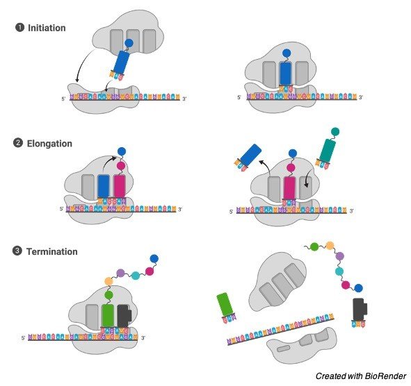
The initiation, elongation, and termination of the mRNA chain are the three steps of the transcription process in protein synthesis. Transcription factors are a class of proteins. Transcription factors attach to particular DNA regions known as enhancers and promoters in order to direct RNA polymerase to the correct transcription site.
The initiation, elongation, and termination of the mRNA chain are the three steps of the transcription process in protein synthesis.
a). Initiation
A transcription initiation complex is made up of transcription factors and RNA polymerase. This complex will start transcription, after which RNA polymerase will start mRNA synthesis by matching complementary bases with the original DNA strand.
b). Elongation
The RNA travels along the DNA and untwists the DNA double helix to produce an elongated RNA molecule during the elongation process.
c). Termination
Transcription is terminated once the mRNA strand is entirely produced, and the mRNA is detached from the DNA template. During the translation step, the newly generated mRNA copy of the gene will exit the nucleus and serve as a template for protein synthesis. This is a sequence that signals the end of the transcription process.
Transcription is terminated once the mRNA strand is entirely produced, and the mRNA is detached from the DNA template. During the translation step, the newly generated mRNA copy of the gene will exit the nucleus and serve as a template for protein synthesis.
II. Translation Process
The genetic code is a collection of principles that govern how an mRNA sequence is converted into a 20-letter amino acid code. These atoms and molecules
Codons are three-letter nucleotide pairings that make up the genetic code. Each of these codons will either match a certain type of amino acid or a process stop signal. The building blocks of protein synthesis are these amino acids.
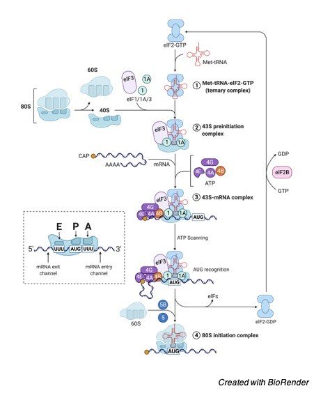
The start, elongation, and termination phases of translation are similar to those of transcription. in the ribosome, which serves as a protein production factory. Ribosomes are complicated entities made up of multiple ribosomal RNA molecules and a number of proteins with tiny and big subunits.
The start, elongation, and termination phases of translation are similar to those of transcription.
a). Initiation
The AUG sequence is found in the start codon of all mRNA molecules, and it codes for methionine. The big ribosomal subunit then binds to start forming the whole initiation complex.
b). Elongation
The ribosome will continue to translate each codon in turn throughout the elongation step. The relevant amino acids are added to the elongated chain and peptide bonds are used to bind them together. The process of elongation continues until all codons have been read.
c). Termination
This is the order in which protein synthesis takes place in the nucleus and on ribosomes. The translation complex is split once the new protein produced as a result of the translation process is released.
A carrier protein is a type of protein in biology that transports a specific item across intracellular compartments, into the extracellular fluid, or between cells, as opposed to channel proteins, which are another form of membrane transport protein that transports molecules less selectively. Carrier proteins, like other membrane transport proteins, are present in lipid bilayer cell structures such as cell membranes, mitochondria, and chloroplasts.
What is Carrier Protein?
Carrier protein is a type of cell membrane protein that helps things leave or enter the cell by facilitating diffusion and active transport. Carrier proteins are in charge of sugar, amino acid, and nucleoside transport. They are also the proteins that pick-up glucose molecules and transport them, as well as other molecules (such as salts, amino acids, and other nutrients), throughout the cell.
For example, carrier proteins embedded in the cell membrane, such as integral transmembrane proteins, would have a high affinity for certain chemicals on the cell exterior and then undergo a conformational shift to allow these substances to flow through the membrane barriers into the cell interior.
Carrier Protein vs Channel-Formers
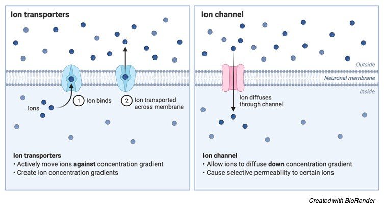
Carrier proteins, like channel proteins, are membrane transport proteins. Membrane transport proteins are proteins that move molecules from one site to another in biological membranes. However, there are some differences between these transporters. As the name indicates, channel proteins produce a “channel” that allows molecules to flow through. Their hydrophobic regions interface with the membrane’s lipids and keep them securely embedded in the plasma membrane.
Pores are channels that stay open to both the inside and exterior of the cell. Aquaporin is an example of a channel protein that permits water molecules to pass across the cell membrane. Carrier proteins, on the other hand, do not create channels. Rather, they have binding sites where molecules can attach themselves. The molecules are then shuttled to their final destination, which is either the inside or exterior of the membrane.
Carrier proteins with binding sites are more discriminating when it comes to the chemicals they carry. Furthermore, they are not open to both the inside and outside of the cell at the same time, unlike certain channel proteins, such as porins, which are open on both sides at the same time. As a result, unlike porin channels, carrier proteins may actively transport molecules across a concentration gradient.
Types of Carrier Protein
Carrier proteins engaged in the active transport of molecules or substances can be categorised according to the type of transport activity they perform. Carrier proteins that are accelerated by a concentration gradient rather than ATP hydrolysis are known as carrier-mediated diffusion proteins. They move molecules from a high-concentration location to a low-concentration area. Sugars, amino acids, and nucleosides, for example, are transported across the majority of cell membranes by carrier proteins.
Carrier proteins that carry molecules across a concentration gradient consume a lot of energy. The carrier proteins can be categorised as (1) ATP-driven, (2) electrochemical potential-driven, or (3) light-driven, depending on the energy source. ATP-driven carrier proteins transport molecules utilizing ATP, whereas electrochemical potential-driven proteins transport molecules using electrochemical potential. Pumps that are powered by photons are known as light-driven pumps. In bacterial cells, these pumps are widely encountered.
ATP-driven Carrier Proteins
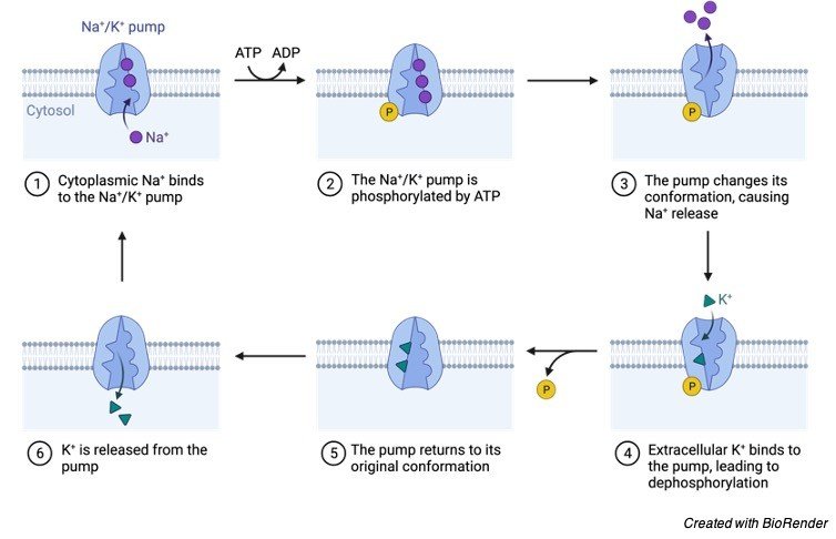
Carrier proteins that require ATP coupling to transport molecules are known as ATP-driven. An ATP-driven carrier is the sodium-potassium pump in the plasma membrane of animal cells. The sodium and potassium ions are selectively bound by the pump. This pump maintains adequate amounts of such ions in order to preserve homeostasis. For each ATP molecule it consumes, the pump actively transfers three sodium ions (Na+) from the interior of the cell and replaces them with two potassium ions (K+) from the outside. Primary active transport is a form of active transport in which the process is powered by chemical energy (ATP).
Electrochemical potential-driven carrier proteins are those whose transport activity is fuelled by an electrochemical potential gradient. Secondary active transportation is the name given to this type of active transportation. Because two molecules are carried through a membrane at the same time, it is also known as linked transport.
A symporter is a carrier protein that transports two molecules in the same direction. An antiporter is a carrier protein that transports two molecules in opposing directions. Nonetheless, certain porters are capable of transporting a single molecule from one side to the other of the membrane. Uniporters are what they’re called. Look for the figure illustrating the three types of carrier-mediated transport in this material to get schematic depictions of the three types of porters.
Function of Carrier Protein
Carrier proteins are engaged in biological transport mechanisms that are both passive and active. Molecules are moved downhill via passive transport, from a higher to a lower concentration. A concentration gradient is created by the difference in concentrations between two areas, which is sufficient to initiate passive transport. However, due to the cell membrane’s lipid bilayer structure, not all molecules will be able to flow out or into the cell in accordance with their concentration gradient.
Polar molecules and ions are unable to pass across the membrane easily. Membrane transport proteins, similar to carriers, are required to assist their movement. If a carrier protein is used in the process, the molecule “sits” on the carrier protein on one side of the membrane before being transported to the other side and released. Facilitated diffusion is a type of diffusion (or passive transport) that uses a membrane protein to move molecules along a concentration gradient.
While certain membrane proteins are unable to participate in active transport, carrier proteins do. Molecules attached to carrier proteins have the ability to migrate upwards, from a lower concentration location to a higher concentration area. Active transport occurs when molecules travel against the concentration gradient, i.e., in the opposite direction of where they would normally go, since the region is already concentrated. As a result, an energy source (such as ATP) is required to power the process. This is what happens when Na+ and K+, as well as NADH, are actively transported through the inner mitochondrial membrane, where ATP is linked to their transport.
Transport Mechanism
The carrier proteins in both passive and active transport move molecules by attaching to them and then changing their conformation. As they transport molecules from one side of the membrane to the other, they alter their form. However, chemical energy is necessary for active transport. When ATPases catalyse the breakdown of ATP to ADP, energy is released by ATP hydrolysis.
The release of one inorganic phosphate from ATP results in the simultaneous release of energy. Direct ATP coupling does not power all active transport activities. Instead of ATP, an electrochemical gradient is used in another kind of active transport. Cations travelling passively, for example, produce entropy that can be used to power the active transport of another set of ions.
Carrier Proteins Examples
1. Glucose Transporters
When the cell contains less glucose than the outside, “glucose transporters” in the cell membrane of animal cells take up glucose molecules without using ATP. Glucose is an important biomolecule since it provides a source of energy. There are 14 glucose transporters in human cells. They are uniporters, meaning they only bind to and transport glucose molecules. GLUT1 is a glucose transporter that is found in virtually all cell types. It is most highly expressed in red blood cells in adults.
ii. Sodium-Potassium Pump (Na+/K+ Pump)
An antiporter is the Na+/K+ pump. It has Na+ and K+ ion binding sites. The pump requires an energy source since the flow of these ions is against their concentration gradients. As a result, it attaches to ATP and hydrolyses it into ADP, releasing energy. This energy is used by the pump to change its form. The ions dissolve from the pump after conformational change, but they are released in opposing directions. Na+ ions are pushed out of the cell, whereas K+ ions are pumped in. The function of the Na+/K+ pump is critical because it is involved in nerve impulse transmission and cell membrane potential maintenance. The function of motor neurons, and hence the target muscles, can be disturbed if there are insufficient K+ ions.
iii. Glucose-Sodium Transport Proteins
Glucose-sodium transport proteins are active glucose transporters that function as symport carriers. The glucose-sodium transporter is used when a cell has a lot of glucose within but still needs to take in more. Glucose and two Na+ ions have binding sites on this transporter. Because there are fewer Na+ ions in the cell at first, the Na+ ions diffuse passively. As a result, an electrochemical potential gradient is created, which causes the glucose transporter to actively transfer the glucose molecule into the cell.
FAQ About Carrier Protein
A membrane transport protein known as a carrier protein is a kind of membrane transport protein. A channel protein is another form of membrane transport protein. The binding site that chooses molecules to transport is one method of distinguishing a carrier protein from a channel protein. When a molecule or solute attaches to this location, the carrier protein transports it across the membrane to the opposite side. Some carriers will require an energy source (e.g., ATP or an electrochemical potential gradient) or a photon to cause them to change shape, allowing the attached molecule or solute to be released.
When all of a carrier protein’s binding sites are occupied, it is said to be saturated. As a result, the transport rate will be at its maximum. The transport rate, also known as Vmax, is a characteristic of a particular carrier that indicates how quickly it may shift between its two conformational states. The binding constant of a specific transporter for its solute (Km) is equal to the concentration of the solute when the transport rate is half of its highest value.
Endosymbiotic theory suggests that the eukaryotic cell’s organelles, such as mitochondria and chloroplasts, evolved as a result of early endosymbiosis between prokaryotic endosymbionts and the eukaryotic host cell.
The Endosymbiotic hypothesis is one of the oldest evolutionary hypotheses still in use today. It is assumed that the early living forms formed an endosymbiotic relationship. This type of symbiosis entails a bigger cell acting as the host and a smaller cell acting as the endosymbiont. The bigger cell absorbed or took in the smaller one, according to the endosymbiotic hypothesis. The bigger cell is a modern eukaryotic cell, whereas the smaller one is a prokaryotic cell.
The presence of membrane-bound cellular components termed organelles distinguishes a eukaryotic cell from a prokaryotic cell. The organelles mitochondria and chloroplasts, according to this view, are the early prokaryotic endosymbionts that were taken in. They spent so much time within the host cell that they evolved into the semi-autonomous organelles we know today.
Endosymbiosis
Endosymbiosis is one of many different types of symbiotic connections (symbioses) that may exist between or among organisms. The endosymbiont dwells inside the host’s body in endosymbiosis. Endosymbiosis still occurs in nature today. The biological relationship between Rhizobium and plant legumes is one example. Rhizobium is an endosymbiont found in the roots of legumes that fixes atmospheric nitrogen into a form that the beans can use. Rhizobium metabolites like malate and succinate are produced by photosynthesis in the legume.
Endosymbiosis formed the basis of the Endosymbiotic Theory in evolutionary biology, which was initially conceived by the botanist Konstantin Mereschkowski (4 August 1855 – 9 January 1921) and then supported by empirical data by Lynn Margulis, 1938–2011.
Endosymbiosis became the mechanism by which organelles such as mitochondria and chloroplasts within eukaryotic cells arose, according to the Endosymbiotic Theory.
This hypothesis proposes that 1.5 billion years ago, a bigger cell took in tiny free-living prokaryotes (bacteria), and the prokaryotes lived as endosymbionts inside the cell.
The mitochondria evolved from proteobacteria (such as the SAR11 clade), whereas the chloroplasts arose from cyanobacteria, according to research findings (particularly the nitrogen-fixing cyanobacteria).
The same characteristics shared by these organelles and their prokaryotic predecessors provide evidence that this idea is realistic.
The following are some of the qualities they share:
• Mitochondria and plastids are both capable of self-replication via a mechanism similar to prokaryotic binary fission.
• Both mitochondria and plastids have a single circular DNA that is comparable in size and structure to that of bacteria but differs from that of the cell’s nucleus.
• Porins in mitochondrial and chloroplast outer membranes are comparable to porins in bacterial cell membranes. Cardiolipin is a membrane lipid found solely in the membranes of bacteria and the inner mitochondrial membrane.
Other Evolutionary Theory
i. Miller-Urey Experiment
The Earth’s age is believed to be about 4.54 billion years, with life appearing around 3.5 billion years ago or earlier. According to the current view of abiogenesis, life on Earth began when the first living organisms ingested non-living elements. They utilised these organic chemicals to make biomolecules and other life-supporting components. Self-replication, self-assembly, autocatalysis, and cell membrane creation are likely biochemical mechanisms that lead to the development of living organisms. These processes were thought to be gradual and made up of a series of occurrences.
The findings of the Miller-Urey experiment showed that the simulated-primitive Earth promoted chemical syntheses of the cell membrane’s basic structures. Amino acids are created by combining the gases methane, ammonia, hydrogen, and water, then electrically electrifying them.
ii. Prebiotic Soup
The Earth was unfriendly to life some four billion years ago. Due to the severe circumstances, no living forms could exist. Simple organic compounds eventually developed. The prebiotic (primordial) soup is a hypothesised model of the early Earth with circumstances that led to the synthesis of simple organic molecules. The notion of the heterotrophic origin of life hypothesis was conceived by Alexander Oparin (1894–1980) and John Burdon Sanderson Haldane (1892–1964), who separately created hypotheses that formed the heterotrophic origin of life theory. They both hypothesised that the early Earth’s atmosphere was chemically decreasing. It helped in the creation of organic molecules.
These chemicals are collected and create a so-called prebiotic soup as they are generated. These simple chemical molecules have evolved into increasingly sophisticated organic polymers throughout time. Life came into being in the end. To flourish and live in the prebiotic soup, the earliest living forms ingested and utilised organic materials. They hypothesised that the first forms of life were heterotrophic in nature. However, new evidence shows that autotrophs were the earliest creatures.
iii. RNA World Hypothesis
Nucleic acids (RNA, DNA), carbohydrates (a variety of sugars), lipids (fats), and amino acids are the four main macromolecules required for life (constituents of proteins). Because RNA may act as both a genetic material and a catalyst, it is thought that primordial life was RNA-based. The evolution of primordial life forms into single-celled living beings happened over millions of years.