Category: Uncategorized

Category: Uncategorized

  • Chick Embryo Fibroblasts Cells: Growth and Maintenance

    Fundamentals of Chick Embryo Fibroblasts

    This in vitro system enables to study various stages in embryonic development of chick.

    It has the advantage of providing easy access to the embryo and extra-embryonic membranes for the observation of morphogenesis and growth as well as for application of different agents under study and inoculation of viruses in specific extra embryonic membranes.

    The technique does not require any sophisticated equipments, media or sera and could be used not only for research purposes but also for teaching of embryology in schools, colleges and universities.

    The present model may serve as an in-vitro model suitable for the fields of developmental biology, toxicology, teratology, virology and several other aspects of biomedical research.

    Materials Required for Chick Embryo Fibroblasts Culture

    Freshly fertilized hen’s eggs

    Unfertilized hen’s eggs

    Transparent glass bowl (surface diameter 8.5 cm, bottom diameter 4.5 cm and depth 4.5 cm)

    Glass petri dish (9.5 cm diameter)

    70% alcohol, scalpel, incubator

    Procedure for Chick Embryo Fibroblasts Culture

    Wash freshly fertilized hen’s eggs with water, allow to air dry and incubate at 37.5 C with a relative humidity of 70-80% for 24 h before culturing.

    Allow incubated eggs to cool for 25-30 mins, wipe with 70% alcohol under laminar flow to minimize the chances of contamination from shell surface.

    Keep the eggs in the horizontal position to assure that the embryo is properly positioned.

    Thin albumin from unfertilized eggs was poured into a sterile bowl.

    This albumin acted as a shock absorber, provided a cushion for the culture and also limited desiccation.

    Crack the fertilized, incubated egg from above with the help of a scalpel at about 3-3.5 cm from the narrow end and gently release the contents of the egg over the albumin cushion in the bowl.

    Transferring of the egg contents without damage to embryo and yolk and upright position of the embryo is necessary for better survival of the embryo.

    Cover the bowl with a glass petridish and incubate cultures at 37.5 C and 80% humidity.

    Observe the cultures after different duration of time to obtain the embryos of various Hamburger Hamilton stages.

    Embryo culture can be continued for 17-19 days.

    This system has been developed and used to demonstrate the glucose induced malformations in developing chick embryos.

    Chick Embryo Fibroblasts Culture Citations:

    Share

    Similar Research Position:

    Continue Reading
  • Comet Assay Full Protocol to Assess DNA...

    About Comet Assay

    Several man-made chemicals find their way into the environment and pose health risk to human population.

    These chemicals have been found to interact with the vital tissue macromolecules regulating the cellular functions leading to long lasting health disorders.

    Acute and chronic exposure to environmental chemicals such as pesticide, metals, polycyclic aromatic hydrocarbons (PAHs), radiation and others have been shown to produce marked toxicity at the target sites.

    Some of these chemicals affect the DNA, which is the carrier of inherited information and any gross change in its structure potentiates serious biological changes.

    Hence there is a need to test the chemicals for their genotoxic potential before being released into the environment.

    The conventional methods for evaluating genetic damage include chromosomal aberration, micronucleus assay, sister chromatid exchanges.

    However, these are time consuming, resource intensive and require proliferating cell population.

    Hence, newer and more sensitive test systems have now been introduced for assessing the genotoxicity of chemicals.

    Fundamental Of Comet Assay

    The comet assay and microgel electrophoresis (MGE) were first introduced by Ostling and Johanson in 1984.

    This was a neutral assay in which the lysis and electrophoresis were done under neutral conditions.

    The image obtained looks like a “comet” with a distinct head, comprising of intact DNA and a tail, consisting of damaged or broken pieces of DNA hence the name “Comet” Assay was given.

    The extent of DNA liberated from the head of the comet was the function of the dose of damage.

    The above neutral assay was modified by two groups, Singh and co-workers (1988) and Olive et al (1989). Singh et al used microgels, for electrophoresis under highly alkaline conditions (pH>13).

    This enables the DNA super coils to get relaxed and unwind, which are then pulled out during application of electric-current which made possible detection of single strand breaks in DNA and alkali labile sites expressed as frank single strand breaks in individual cells.

    This method was developed to measure low levels of strand breaks with high sensitivity.

    Olive and co-workers conducted the electrophoresis under neutral or mild alkaline condition (pH 12.3) to detect single stranded breaks.

    This method is optimized to detect a subpopulation of cells with varying sensitivity to drug or radiation.

    Importance of Comet Assay

    The single cell gel electrophoresis or comet assay is one such state-of-the-art technique for the measurement of DNA damage and repair in vitro in any eukaryotic cell and some prokaryotic cells.

    This technique is rapid, non-invasive, sensitive, visual and inexpensive as compared to the conventional techniques and is a powerful tool to study factors modifying mutagenicity and carcinogenicity.

    In addition, it combines the simplicity of biochemical techniques for detecting DNA single strand breaks (strand breaks and incomplete excision repair sites), alkali-labile sites and cross-linking with the single cell approach typical of cytogenetic assays.

    Comet assay measures, double strand breaks (DBSs), single strand breaks (SSBs), alkali labile sites, oxidative DNA base damage, DNA-DNA/ DNA-protein/ DNA-Drug cross linking and DNA repair.

    Comet Assay Principle

    The assay works upon the principle that strand breakage of the super coiled duplex DNA leads to the reduction of the size of the large molecule and these strands can be stretched out by electrophoresis.

    Comets form as the broken ends of the negatively charged DNA molecule become free to migrate in the electric field towards the anode.

    Two principles in the formation of the comet are:

    1. DNA migration is a function of both size and the number of broken ends of the DNA

    2. Tail length increases with damage initially and then reaches a maximum

    Advantage of Comet Assay

    1. It is a non-invasive technique.

    2. It requires <10,000 cells and collection of data at the level of the individual cell, allowing for more robust types of statistical analyses.

    3. Counting of 50-100 cells per individual/ treatment group through computerized image analysis software gives a complete analysis.

    4. Any eukaryotic cell population (nasal and buccal mucosal cells, epithelial cells, male germ cells, fine needle biopsy) is amenable to analysis.

    5. Duration of this experiment is few hours only whereas conventional cytogenetic assays require few days.

    6. Single strand breaks (SSBs) and alkali labile lesions (capable of being transformed into SSBs under alkaline conditions) in the DNA of individual cells can be assessed.

    Materials Required for Comet Assay

    Frosted slides 

    Coverslips 

    Lymphocyte Separation Medium

    Hydrogen peroxide (H2O2): 25 μM and 50 μM

    Phosphate Buffer Saline (PBS) (i.e. 137mM NaCl, 2.7mM KCl, 10mM Na2HPO4.2H2O and 2 mM KH2PO4, pH 7.4).

    Dulbecco’s modified Eagle Medium (DMEM)

    Fetal Bovine Serum (FBS)

    0.4% Trypan blue dye

    Normal melting point agarose (NMPA)

    Low melting point agarose (LMPA)

    Propidium iodide

    Lysis buffer (2.5 M NaCl, 100 mM EDTA, 10 mM Trizma base). 

    Neutralising Buffer (Add 0.4 M Tris to ~800 ml distilled water. Adjust the pH to 7.5 using conc. HCl to 1000 ml with distilled water).

    Electrophoresis buffer (Add 30 ml 10N NaOH and 5 ml 200 mM EDTA to 1000 ml with distilled water, mix well. Ensure pH > 13 before use).

    IX-51 inverted research microscope

    Horizontal gel electrophoresis units

    Sorvall Centrifuge

    Comet Assay Procedure

    Isolation of lymphocytes:

    Blood was collected in microfuge tubes containing heparin (10 IU heparin per ml of blood)

    Blood was diluted with 1X PBS in the ratio of 1:3 (Blood: PBS) and layered on lymphocyte separation medium (LSM) (1 ml of LSM per 3 ml of diluted blood)

    Tubes were centrifuged at 400 g for 15 minutes at 15oC.

    Lymphocyte layer was collected and diluted with 1X PBS in the ratio of 1:1.

    Again it was centrifuged at 400 g for 10 mins at 15oC to get the lymphocyte pellet.

    Pellet was resuspended in 1X PBS.

    Estimation of cell viability using trypan blue exclusion assay. The standard trypan blue assay was carried out on isolated lymphocytes as mentioned in earlier protocol for cell counting.

    Chemical treatment of cells:

    An aliquot of cells were treated with two different concentrations of H2O2 (25 μM and 50 μM) for 15 minutes and the other aliquot of untreated cells were used as negative control.

    A layer of 1% normal melting point agarose (NMPA) was prepared on frosted-end slides and slides were kept overnight at 37oC.

    After chemical treatment, cells (1 x 10⁶ cells/ml) were mixed with 1ml of 2% low melting point agarose (LMPA).

    The suspension of 200 μl LMPA and cells was layered on to the precoated slides and covered with cover slips.

    The layer was allowed to solidify at 4oC for 15 mins.

    After solidification of the layer, cover slips were removed.

    Slides were immersed in cold lysis solution (pH 10) and kept at 4C for 3 hours.

    Slides were washed gently with alkaline electrophoresis buffer (pH 13) for 15 mins, twice.

    This helps in allowing denaturation of DNA.

    Subsequently, slides were transferred to an electrophoresis tank with fresh alkaline electrophoresis buffer and electrophoresis was performed at field strength of 20 V/ 250 mA for 25 min at 4C.

    Slides were neutralized in chilled distilled water for 5 min and stained with 5 μg/ml propidium iodide for 20 mins.

    DNA damage Analysis in Comet Assay

    For visualization of DNA damage, observations are made with PI stained DNA using a 10X/20X objective on a fluorescent microscope.

    Any image analysis system may be suitable for the quantification of SCGE data.

    CASP, freely downloadable software, is used to assess the quantitative extent of DNA damage in the cells by measuring the length of DNA migration and the percentage of migrated DNA.

    The software calculates the tails moment and olive tail moment.

    Generally, 50 to 100 randomly selected cells are analyzed per sample.

    The softwares are designed to differentiate comet head from tail and to measure a variety of parameters including tail length; % of total fluorescence in head and tail; and ‘tail moment’, calculated in different ways but essentially representing the product of tail length and relative tail intensity.

    Percent DNA in tail is linearly related to DNA break frequency up to about 80% in tail, and this defines the useful range of the assay.

    Tail length tends to increase rapidly with dose at low levels of damage, but soon reaches its maximum.

    It is therefore the most sensitive parameter at near-background levels of damage.

    Tail moment is an attempt to combine the information of tail length and tail intensity, but suffers from lack of linearity.

    DNA damage Analysis with CASP Software in Comet Assay

    The CASP software can be downloaded from http://www.casplab.com

    The software CASP generates the frame to limit the comet area, the line to limit the tail length, the circle to identify the comet head and the cross to identify the head centre.

    The small rectangle at the bottom of each picture is generated by CASP as background reference.

    Comet Assay Parameters

    1. % Head DNA = (Head optical intensity/ (Head optical intensity + Tail optical intensity)) X 100

    2. % Tail DNA = 100- % Head DNA.

    3. Tail length = Tail length is the distance of DNA migration from the body of the nuclear core and it is used to evaluate the extent of DNA damage.

    4. Tail length = Tail extent (Tail from centre) + Head extent / 2

    5. Olive tail moment = Tail moment is defined as the product of the tail length and the fraction of total DNA in the tail.

    Tail moment incorporates a measure of both the smallest detectable size of migrating DNA (reflected in the comet tail length) and the number of relaxed/ broken pieces (represented by the intensity of DNA in the tail).

    6. Olive Tail Moment = (mean Tail- mean Head) X % Tail DNA/ 100 7. Extent Tail Moment = Tail length X % Tail DNA / 100

    Comet Assay Citations:

    Share

    Similar Post:

    Continue Reading
  • Cell Plating, Media Change, and Culture Maintenance...

    About Cell Plating or Cell Passaging

    Most types of cells stop or slow down their growth at a characteristic population density in any particular medium.

    Cell to cell contact, localized or generalized depletion of nutrients or production of cell generated inhibitors leads to density dependent inhibition or sometimes called as contact inhibition of cell growth.

    Cells at this stage are said to be confluent.

    This is important to avoid Contact Inhibition of Proliferation (CIP), exhibited by most cancerous cell lines.

    The phenomenon of a cell ceasing to proliferate after contact with other cells is called as CIP.

    Adherent cells cover the entire growth surface available to them is referred to as confluence.

    Hence they need to be subcultured, passaging, cell plating (indicated by passage number).

    The passage number is the number of times that the culture has been subcultured.

    Why Cell Plating or Cell Passaging

    Cells are often passaged at semiconfluency when they are in log phase.

    Failure to subculture or cell plating, the proliferating cells results in reduced mitotic index and eventually cell death.

    The most important step in subculturing or cell plating of monolayers is to detach cells from the surface of the primary culture vessel by either enzymatic means such as trypsinization or mechanical means such as scraping of monolayer cells.

    The resultant cell suspension is then reseeded, into fresh cultures.

    Secondary cultures are checked for growth, fed periodically, and may be subsequently subcultured to produce tertiary cultures, etc.

    The time between passaging cells depends on the growth rate and varies from cell line to cell line.

    Detachment of cells by mechanical means is accomplished by scraping monolayer of cells with commercially available silicon rubber spatula known as a scrapper.

    Whereas proteases such as trypsin, collagenase and pronase etc. are used for enzymatic dissociation.

    Prolonged enzymatic dissociation may damage cell membrane.

    Thus, it is wise to decrease the incubation period and physical injury when in enzyme solution.

    Cell Plating - Research Tweet 1

    Cell Plating or Cell Passaging Overview. Images are created with BioRender

    Cell Plating or Cell Passaging Requirements

    1. Confluent culture of cells

    2. Appropriate culture media

    3. Antibiotics and antifungal agent

    4. Sterile PBS (Phosphate buffer saline)

    5. Trypsin/EDTA solution (0.25% Trypsin in 1mM EDTA in PBS)

    6. Sterile pipettes, culture vessels, sterile beakers

    7. Cotton swabs, 70% IPA,

    8. 15 ml & 50 ml Centrifuge tubes

    Cell Plating or Passaging of Adherent Cells

    Check all the cell lines under the microscope for contamination, cell morphology, density and presence of clumps or detachment.

    1. The protocol described here is for a typical confluent culture in a 25 cm2 flask

    2. Remove medium with a sterile pipette without disturbing the cells.

    3. Wash adhering cell layer with sterile PBS (Phosphate buffer saline) to remove traces of serum that can inhibit trypsin.

    4. Add 2 ml trypsin/EDTA at 37ºC to cover the cell layer.

    5. Incubate for 2 minutes at 37ºC in CO2 incubator. Tap occasionally to verify that the cells are releasing. Check in microscope to visualize detachment of cells.

    6. Remove trypsin-EDTA. Add fresh 2 ml of medium and rinse cell layer two or three times to dissociate cells and to dislodge any remaining adherent cells.

    7. Transfer the required number of cells to a new labelled flask containing pre-warmed medium. Maintain the split ratio as recommended.

    8. Incubate cells in humidified incubator at 37ºC, 5% CO2.

    Cell Plating or Passaging of Suspension Cells

    In suspension culture cells are suspended in complete growth medium rather than attached to a culture flask surface, it is not necessary to disperse them enzymatically before passaging.

    However, before passaging, cells must be maintained in culture by feeding every 2 to 3 days until they reach confluency (i.e. until the cells clump together in the suspension and medium appears turbid when the flask is swirled).

    1. With a pipette, mix the cells thoroughly by pipetting up and down and rinsing the side of the flask.

    2. Decant the cell suspension into the centrifuge tube, spin at 1300 rpm for 7 minutes at 4 °C temperature to pellet down all the cells.

    3. Add 1 ml of fresh media.

    4. Split into two flasks with 0.5 ml of cell suspension and add 6.5 ml of fresh media.

    5. Label new passage number on both the flasks.

    6. Incubate cells in humidified incubator at 37 C, 5% CO2.

    Precautions During Cell Plating or Passaging

    Cell degeneration may lead to change in morphology i.e. rounding up of cells and their detachment from the surface of the culture vessel.

    The most frequent reasons for rapid cell degeneration include use of too high seeding density, use of poor quality or too high concentration of fetal calf serum when preparing cultures.

    Whenever rapidly growing, continuous cell lines are maintained in a laboratory there is a risk of cell line cross-contamination.

    The problem of cell degeneration can usually be overcome by appropriate adjustment of the cell count or foetal calf serum concentration.

    Only one cell line should be used in at any one time.

    After removal of the cell cultures from the cabinet, the cabinet should be swabbed down with a suitable disinfectant and the cabinet UV run for five minutes before introduction of another cell line.

    Bottles or aliquots of medium should be dedicated for use with only one cell line.

    Regularly return to frozen stocks — never grow a cell line for more than three months 15 passages from stock passage level, whichever is the shorter period.

    All culture vessels must be carefully and correctly labelled. (including name of cell line, passage number and date of transfer)

    Some cultures whilst growing as attached lines adhere only lightly to the flask called as fragile cells, thus it is important to ensure that the growth medium is retained and the flasks are handled with care to prevent any cells detachment prematurely.

    Although majority of cells will detach in the presence of trypsin alone the EDTA is added to enhance the activity of the enzyme.

    Presence of serum inactivate Trypsin activity. Therefore, it is essential to wash the monolayer of cells with PBS without calcium and magnesium in order to remove all traces of serum from the culture medium.

    Cells should only be exposed to trypsin/EDTA long enough to detach cells.

    Prolonged exposure could damage cell surface receptors.

    In case of cells lost in trypsin removal, trypsin should be neutralised with serum prior to seeding cells into new flasks otherwise cells will not attach.

    Trypsin may also be neutralised by the addition of soyabean trypsin inhibitor, where an equal volume of inhibitor at a concentration of 1mg/ml is added to the trypsinised cells.

    The cells are then centrifuged, resuspended in fresh culture medium and counted as above.

    This is especially necessary for serum-free cell cultures.

    If the cells harvested are at too low a cell density to re-seed at the appropriate cell density into fresh flasks it may be necessary to centrifuge the cells e.g. 5 mins at 1300 rpm, and resuspend in a smaller volume of medium.

    Precautions During Cell Plating or Passaging Ciations:

    Share

    Similar Post:

    Continue Reading
  • Micronucleus Assay: a Widely Used Assay for...

    About Micronucleus Assay

    Genotoxicity is a word in genetics defined as a destructive or adverse effect on a cell’s genetic material either DNA or RNA, affecting its integrity.

    Genotoxins are mutagens (compounds that changes nucleotide); they can cause mutations.

    Genotoxins include both radiation and chemical genotoxins.

    A substance (radiation and chemical compounds) that has the property of genotoxicity is known as a genotoxin.

    Micronucleus Assay Principle

    The purpose of in vitro testing is to determine whether an environmental factor, substrate, or any product induces damage to genetic material.

    One technique entails cytogenetic assays using different animals cells.

    The aberrations detected in cells affected by a genotoxic substance are chromosome gaps, chromatid, complex rearrangements, chromosome breaks, fragmentation, translocation, chromatid deletions, and many more.

    One such example is testing for the formation of micronuclei.

    The micronucleus test is used as a tool for genotoxicity assessment of various chemicals.

    Chromosomal aberration test is easy to conduct in order to evaluate genotoxicity.

    A micronucleus is the erratic (third) nucleus that is formed during the anaphase of mitosis or meiosis.

    Micronuclei (the name means ‘small nucleus’) are cytoplasmic bodies having a portion of acentric chromosome or whole chromosome which was not carried to the opposite poles during the anaphase.

    Micronuclei formation results in the daughter cell lacking a part or all of a chromosome.

    These fragments of chromosome or whole chromosomes normally develop nuclear membranes and form as micronuclei as a third nucleus.

    After cytokinesis, one daughter cell ends up with one nucleus and the other ends up with one large and one small nucleus, i.e., micronuclei.

    There is a chance of more than one micronucleus forming when more genetic damage has happened.

    Micronucleus Assay Requirements

    1. Cancer cell line

    2. Media (DMEM or alpha MEM)

    3. FBS

    4. 12 well or 24 well or 4 well plate or 6 well plate

    5. Coverslips

    6. 1X PBS (pH-7.2)

    7. Triton-X 100- 0.05%

    8. Ethanol-70%

    9. Paraformaldehyde (PFA)-4%

    10. Dapi stain (0.5 ug/ml)

    11. Bright field microscope

    Procedure of Micronucleus Assay

    1. MDA-MB-468 cells were grown on coverslips in 12 well plates.

    2. After treatment period (24 hour or 48 hour), media was removed and cells were washed twice with PBS. Further the cells were fixed in 4% PFA (1 hour at RT or overnight to 1 week at 4oC).

    3. After fixation PFA was removed and the cells were washed with PBS (pH 7.2)

    4. After washing add 0.05% triton –X 100 (it should cover the cells grown on coverslip) and incubate for 1 hour.

    5. After incubation triton-X 100 was removed and the cells were washed gently with PBS (pH-7.2).

    6. After washing Dapi stain was added (it should cover the surface and make sure it should not get dried during incubation) and incubate for 30 minutes. Observe under fluorescence microscope.

    Note: For better result cells should be 70%-80% confluent.

    Micronucleus Assay Citations:

    Share

    Similar Post:

    Continue Reading
  • Transition Words And Phrases: Why And How...

    Transition Words

    If you are writing an assay that is full of idea and perfectly written but what if its not clear to your readers.

    That’s where transition words come in. Transitions are words, phrases, sentences, that link one idea to another showing the connection between them.

    What are a Transition Words?

    A transition means a change from one idea to another. There are many transitions in English.

    We use transitions to add information, to show contrast, to show condition, to show conclusion, etc.

    There are a lot of transition words out there, and they play very important role in English because they can join idea within the sentences and more importantly they can join two sentences.

    transition words - Research Tweet 1

    Why Transition Words are so Important?

    They create flow in your writing.

    Flow makes easier to understand your assay or writing and also makes your content a little bit more interesting to your reader.

    Transition words also act as a connecting bridge within the sentences and also between the sentences.

    Categories of Transition Words

    Causation: These words and phrases are used to connecting instigator(s) to consequence(s) and used after a sentence or paragraph that describes a cause.

    • Accordingly
    • As a result
    • And
    • So
    • Because
    • Consequently
    • For that reason
    • Hence
    • On account of
    • Since
    • Therefore
    • Thus

    The plant is growing very fast. [Accordingly / Consequently / As such] it is necessary to cut new branches.

    Because he failed to respond, John and Steve offered coffee.

    The speaker’s flight has been cancelled. [Therefore / As such / As a result] we will reschedule the conference lecture.

    Monday is a national holiday. [As a result / Accordingly / Thus / Hence / Consequently] classes will be canceled.

    transition words - Research Tweet 3

    Chronology:It is widely used to connect what issues in regard to when they occur.

    • After
    • Afterwards
    • Always,
    • At length
    • During
    • Earlier
    • Following
    • Immediately
    • In the meantime
    • Later
    • Never
    • Next
    • Now
    • Once
    • Simultaneously
    • So far
    • Sometimes
    • Soon
    • Subsequently
    • Then
    • This time
    • Until now
    • When
    • Whenever
    • While

    I went to the school after he gave me bike.

    He almost always wins lottery.

    During the morning, you can play.

    First, we are going fishing. Then, we will get ready for movie.

    transition words - Research Tweet 4

    Combinations: It connects multiple events or numerous elements that create long sentences.

    • Additionally
    • Again
    • Also
    • And
    • Or
    • Not
    • As a result
    • Besides
    • Even more
    • Finally
    • First,
    • Firstly
    • Further
    • Furthermore
    • In addition
    • In the first place
    • In the second place
    • Last
    • Lastly
    • Moreover
    • Next
    • Second
    • Secondly
    • Too

    Steve promised to respect, love and also obey his wife.

    Moreover, he will do all the cleaning and cooking while he cooks at home.

    Furthermore, he is grateful that his girlfriend earns enough money his groceries.

    transition words - Research Tweet 6

    Contrast: It connect two sentences by focusing on their differences.

    • After all
    • Although
    • And yet
    • At the same time
    • But
    • Despite
    • However
    • In contrast
    • Nevertheless
    • Nonetheless
    • Notwithstanding
    • On the contrary
    • On the other hand
    • Otherwise
    • Though
    • Yet

    Jones argues for the Oxford comma, although everyone doesn’t agree.

    I ran home, but I was still late.

    Despite the last fight, it is also very important to think about dogs.

    During my Chicago trip, I went to see my friend. However, I wasn’t sure.

    transition words - Research Tweet 5

    Example: It connects a general idea to a particular instance of this idea.

    • As an illustration
    • e.g., (from a Latin abbreviation for “for example”)
    • For example
    • For instance
    • Specifically
    • That is
    • To demonstrate
    • To illustrate

    Importance: It connect what is critical to what is more inconsequential.

    • Chiefly
    • Critically
    • Foundationally
    • Most importantly
    • Of less importance
    • Primarily

    Location: It connect elements according to where they are placed in relationship to each other.

    • Above
    • Adjacent to
    • Below
    • Beyond
    • Centrally
    • Here
    • Nearby
    • Neighboring on
    • Opposite to
    • Peripherally
    • There
    • Wherever
    transition words - Research Tweet 7

    Similarity: It connect to things by suggesting that they are in some way alike.

    • By the same token
    • In like manner in Similar fashion
    • Here
    • In the same way
    • Likewise
    • Wherever

    Share

    Continue Reading
  • qPCR: Real-Time Polymerase Chain Reaction (RT-PCR)

    qPCR Objective

    To estimate the copy number of a target gene

    About qPCR

    Polymerase chain reaction (PCR) is a biochemical process that copies and amplifies DNA using a thermally stable DNA polymerase.

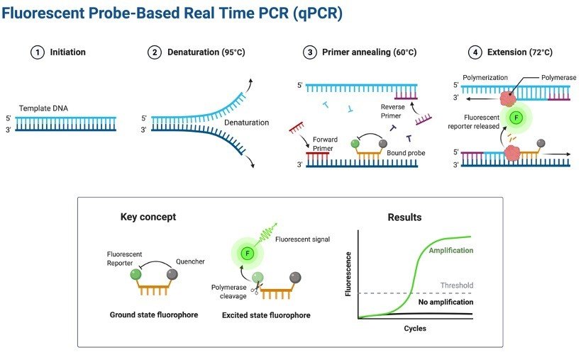
    Real Time Quantitative PCR (qPCR) is a major development of PCR technology that enables reliable detection and measurement of products during each cycle of PCR reaction which are directly proportional to the amount of template prior to start of PCR process.

    qPCR Principle

    PCR is a technique for amplifying DNA. Generally, the PCR reaction consists of a series of temperature changes that are repeated 25 – 45 times.

    These PCR cycles normally consist of three stages: the first, at around 95 °C, allows the separation of the nucleic acid’s double strands; the second, at a temperature of around 50-60 °C, allows the primer binding with the DNA template; the third, at between 68 – 72 °C, facilitates the polymerization carried out by the DNA polymerase.

    This principle of amplification is utilized in real-time PCR also widely known as qPCR (quantitative PCR) but instead of looking at bands on a gel at the end of the reaction, the continuous reaction is monitored in “real-time”.

    The reaction is placed in to a PCR machine that watches the reaction in real-time manner with a camera or fluorescence detector.

    There are a lot of techniques out there that are used to allow the progress of a PCR process to be monitored but they all have one thing in common.

    The DNA amplification to the generation of fluorescence which can be detected with a camera or sensor during each PCR cycle.

    Hence, as the number of PCR copies increases during the reaction, so the fluorescence increases.

    qPCR - research tweet 1

    Adopted from BioRender

    SYBR Green (or other intercalating dye)

    SYBR® Green is by far the most commonly used intercalating dye. This dye operate via a simple mechanism.

    The dye is fluorescent in its own but in the presence of double stranded DNA, the dye intercalates with the DNA double helix and thus alters the structure of the dye and causes it to fluoresce more.

    An increase in DNA product during PCR therefore leads to an increase in fluorescence intensity measured at each cycle.

    Nomenclature Commonly Used in qPCR

    Baseline is defined as PCR cycles in which a reporter fluorescent signal is accumulating but is beneath the limits of detection of the instrument.

    Threshold is an arbitrary level of fluorescence chosen on the basis of baseline variability.

    A signal that is detected above the threshold is considered a real signal that can be used to define the threshold cycle (Ct) for a sample.

    Threshold can be adjusted for each experiment so that it is in the region of exponential amplification across plots.

    Ct is defined as the fractional PCR cycle number at which the reporter fluorescence is greater than the threshold.

    The Ct is a basic principle of real time PCR and is essential component in producing accurate and reproducible data.

    qPCR Applications

    1. Quantitative mRNA expression studies.

    2. DNA copy number measurements in genomic or viral DNAs.

    3. Allelic discrimination assays or SNP genotyping.

    4. Verification of microarray results.

    5. Drug therapy efficacy.

    6. DNA damage measurement.

    Requirements for qPCR Assay

    1. Genomic DNA (2-20 ng per reaction)

    2. Primers for endogenous control and target gene

    3. SYBR green

    4. DNAse free Milli-Q water 5. Plasticware

    6. Real time instrument

    qPCR Procedure

    1. Make a mastermix for 10 reactions.

    ComponentsVol. per Reaction (ul)Vol. for 10 reaction (ul)
    SYBR Green550
    Forward Primer0.3 (300 nM-500 nM)3
    Reverse Primer0.3 (300 nM-500 nM)3
    cDNA1 (100 ng-100 fg)10
    Water3.434
    Total10100

    2. Mix well and centrifuge briefly

    3. Aliquote 9 μl of the mastermix in the wells of the 96-well plate

    4. Add 1 μl of the DNA ( 50 ng/ μl) in each well and then seal the 96-well plate with

    5. Setup the PCR reaction in the STEP ONE real-time PCR

    6. Cycling condition 

    7. Total number of cycle varies but usually 40.

    StageTemperature (C) Min
    Stage 1 (Denaturing)953:00
    Stage 2 (Annealing)950:10
    Stage 3 (Melt Curve)55-600:30
    55-950:05

    8. Introduce a hold at 4 ºC at the end of cycling if you need to store samples for some time (max. overnight) before the plate read.

    9. Take reading in STEP ONE.

    Formula for Calculating Relative Level of mRNA Expression

    The relative level of mRNA Expression were calculated using the cycle threshold (Ct) method:

    2−ΔΔCt = 2{ΔCt (treated samples) − ΔCt (untreated control)} where ΔCt = Ct (Gene of interest) − Ct (Endogenous Gene)

    qPCR Citations

    Share

    Similar Post:

    Continue Reading
  • How To Embed A Video In Powerpoint?...

    Don’t know how to embed a video in powerpoint ? The most…

    Continue Reading
  • How To Add Music To Powerpoint ?...

    Don’t know how to add Music in powerpoint ? The most easy…

    Continue Reading
  • Do All Birds Migrate? Everything You Need...

    Do All Birds Migrate? It’s no wonder that bird migration is stunning,…

    Continue Reading
  • Top 6 Smartest Birds: Everything You Need...

    Top 6 Smartest Birds in the Worlds Owning a pet is a…

    Continue Reading